Adductor hiatus

Last updated
Adductor hiatus
Gray433.png
Deep muscles of the medial femoral region. (Adductor hiatus visible as hole in adductor magnus at lower left.)
Gray544.png
The arteries of the gluteal and posterior femoral regions. (Adductor hiatus is not labeled, but popliteal artery is visible at bottom center.)
Details
Identifiers
Latin hiatus adductorius
TA98 A04.7.03.008
TA2 2634
FMA 58784
Anatomical terminology

In human anatomy, the adductor hiatus also known as hiatus magnus is a hiatus (gap) between the adductor magnus muscle and the femur that allows the passage of the femoral vessels from the anterior thigh to the posterior thigh and then the popliteal fossa. [1] It is the termination of the adductor canal and lies about 8–13.5 cm (3.1–5.3 in) superior to the adductor tubercle. [2]

Contents

Structure

Kale et al. classified the adductor hiatus according to its shape and the structures surrounding. [3] An adductor hiatus is described as oval or bridging depending on the shape of the upper boundary. It can also be described as muscular or fibrous depending on whether the structure surrounding is the muscular part or the tendinous part of the adductor magnus muscle. For example, the top drawing on the right shows an oval fibrous type of adductor hiatus, and the bottom one shows a bridging muscular adductor hiatus.

Four structures are associated with the adductor hiatus. However, only two structures enter and then leave through the hiatus; namely the femoral artery and femoral vein. Those vessels become the popliteal vessels (popliteal artery and popliteal vein) immediately after they leave the hiatus, [4] where they form a network of anastomoses called the genicular arteries. The genicular arteries supply the knee joint.

The other two structures that are associated with the adductor hiatus are the descending genicular artery and the saphenous nerve. The saphenous nerve does not leave through the adductor hiatus but penetrates superficially halfway through the adductor canal.

Clinical significance

Fracture of distal femur

Fracture at the supracondylar area of femur, where the adductor part of the adductor magnus attaches, will most likely cause damage to the femoral artery and may cause impairment of the blood supply to the lower leg. [5] Popliteal artery can also be damaged by the fracture of distal femur. [6]

Popliteal artery entrapment syndrome

Abnormality in the relationship between the adductor hiatus and the popliteal artery can also contribute to a condition called popliteal artery entrapment syndrome. [7]

Additional images

Related Research Articles

<span class="mw-page-title-main">Human leg</span> Lower extremity or limb of the human body (foot, lower leg, thigh and hip)

The human leg is the entire lower limb of the human body, including the foot, thigh or sometimes even the hip or buttock region. The major bones of the leg are the femur, tibia, and adjacent fibula. The thigh is between the hip and knee, while the calf (rear) and shin (front) are between the knee and foot.

<span class="mw-page-title-main">Thigh</span> Area between the pelvis and the knee; upper leg

In human anatomy, the thigh is the area between the hip (pelvis) and the knee. Anatomically, it is part of the lower limb.

<span class="mw-page-title-main">Sartorius muscle</span> Longest muscle in the human body

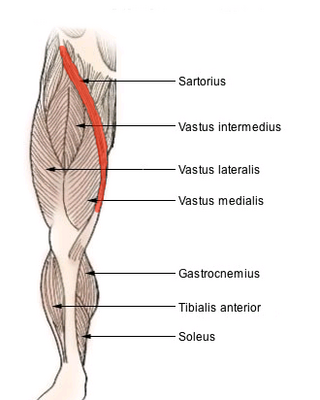
The sartorius muscle is the longest muscle in the human body. It is a long, thin, superficial muscle that runs down the length of the thigh in the anterior compartment.

<span class="mw-page-title-main">Femoral artery</span> Large artery in the thigh

The femoral artery is a large artery in the thigh and the main arterial supply to the thigh and leg. The femoral artery gives off the deep femoral artery and descends along the anteromedial part of the thigh in the femoral triangle. It enters and passes through the adductor canal, and becomes the popliteal artery as it passes through the adductor hiatus in the adductor magnus near the junction of the middle and distal thirds of the thigh.

<span class="mw-page-title-main">Deep femoral artery</span> Large branch of the femoral artery that supplies the thigh

The deep femoral artery also known as the deep artery of the thigh, or profunda femoris artery, is a large branch of the femoral artery. It travels more deeply ("profoundly") than the rest of the femoral artery. It gives rise to the lateral circumflex femoral artery and medial circumflex femoral artery, and the perforating arteries, terminating within the thigh.

<span class="mw-page-title-main">Popliteal artery</span> Continuation of the femoral artery that supplies the lower leg

The popliteal artery is a deeply placed continuation of the femoral artery opening in the distal portion of the adductor magnus muscle. It courses through the popliteal fossa and ends at the lower border of the popliteus muscle, where it branches into the anterior and posterior tibial arteries.

<span class="mw-page-title-main">Popliteal vein</span> Large leg vein

The popliteal vein is a vein of the lower limb. It is formed from the anterior tibial vein and the posterior tibial vein. It travels medial to the popliteal artery, and becomes the femoral vein. It drains blood from the leg. It can be assessed using medical ultrasound. It can be affected by popliteal vein entrapment.

<span class="mw-page-title-main">Hip</span> Anatomical region between the torso and the legs, holding the buttocks and genital region

In vertebrate anatomy, hip refers to either an anatomical region or a joint.

<span class="mw-page-title-main">Adductor magnus muscle</span> Muscle in the thigh

The adductor magnus is a large triangular muscle, situated on the medial side of the thigh.

<span class="mw-page-title-main">Popliteal fossa</span> Depression at back of knee joint

The popliteal fossa is a shallow depression located at the back of the knee joint. The bones of the popliteal fossa are the femur and the tibia. Like other flexion surfaces of large joints, it is an area where blood vessels and nerves pass relatively superficially, and with an increased number of lymph nodes.

The articularis genus is a small skeletal muscle located anteriorly on the thigh just above the knee.

The middle genicular artery is a small branch of the popliteal artery. It supplies parts of the knee joint.

<span class="mw-page-title-main">Medial circumflex femoral artery</span>

The medial circumflex femoral artery is an artery in the upper thigh that arises from the profunda femoris artery. It supplies arterial blood to several muscles in the region, as well as the femoral head and neck.

<span class="mw-page-title-main">Adductor canal</span> Aponeurotic tunnel in the middle third of the thigh

The adductor canal is an aponeurotic tunnel in the middle third of the thigh giving passage to parts of the femoral artery, vein, and nerve. It extends from the apex of the femoral triangle to the adductor hiatus.

The cruciate anastomosis is a circulatory anastomosis in the upper thigh formed by the inferior gluteal artery, the lateral and medial circumflex femoral arteries, the first perforating artery of the deep femoral artery, and the anastomotic branch of the posterior branch of the obturator artery.

<span class="mw-page-title-main">Femoral sheath</span> Anatomical structure of the upper thigh

The femoral sheath is a funnel-shaped downward extension of abdominal fascia within which the femoral artery and femoral vein pass between the abdomen and the thigh. The femoral sheath is subdivided by two vertical partitions to form three compartments ; the medial compartment is known as the femoral canal and contains lymphatic vessels and a lymph node, whereas the intermediate canal and the lateral canal accommodate the femoral vein and the femoral artery (respectively). Some neurovascular structures perforate the femoral sheath. Topographically, the femoral sheath is contained within the femoral triangle.

<span class="mw-page-title-main">Perforating arteries</span>

The perforating arteries are branches of the deep artery of the thigh, usually three in number, so named because they perforate the tendon of the adductor magnus to reach the back of the thigh. They pass backward near the linea aspera of the femur underneath the small tendinous arches of the adductor magnus muscle.

<span class="mw-page-title-main">Descending genicular artery</span>

The descending genicular artery arises from the femoral artery just before its passage through the adductor hiatus.

The saphenous nerve is the largest cutaneous branch of the femoral nerve. It is derived from the lumbar plexus (L3-L4). It is a strictly sensory nerve, and has no motor function. It commences in the proximal (upper) thigh and travels along the adductor canal. Upon exiting the adductor canal, the saphenous nerve terminates by splitting into two terminal branches: the sartorial nerve, and the infrapatellar nerve. The saphenous nerve is responsible for providing sensory innervation to the skin of the anteromedial leg.

<span class="mw-page-title-main">Patellar network</span> Network of blood vessels around the knee

The patellar network is an intricate network of blood vessels around and above the patella, and on the contiguous ends of the femur and tibia, forming a superficial and a deep plexus.

References

  1. Moore, Keith L.; Dalley, Arthur F.; Agur, A. M. R. (2013-02-13). Clinically Oriented Anatomy. Lippincott Williams & Wilkins. ISBN   9781451119459.
  2. Olson, S. A.; Holt, B. T. (Feb 1995). "Anatomy of the medial distal femur: a study of the adductor hiatus". Journal of Orthopaedic Trauma. 9 (1): 63–65. doi:10.1097/00005131-199502000-00010. ISSN   0890-5339. PMID   7714656.
  3. Kale, Ayşin; Gayretli, Ozcan; Oztürk, Adnan; Gürses, Ilke Ali; Dikici, Fatih; Usta, Ahmet; Sahinoğlu, Kayıhan (Dec 2012). "Classification and localization of the adductor hiatus: a cadaver study". Balkan Medical Journal. 29 (4): 395–400. doi:10.5152/balkanmedj.2012.030. ISSN   2146-3123. PMC   4115878 . PMID   25207041.
  4. Agur, A. M. R.; Dalley, Arthur F.; Grant, John Charles Boileau (2013). Grant's Atlas of Anatomy. Lippincott Williams & Wilkins. ISBN   9781608317561.
  5. Copley, L. A.; Dormans, J. P.; Davidson, R. S. (1996). "Vascular injuries and their sequelae in pediatric supracondylar humeral fractures: toward a goal of prevention". Journal of Pediatric Orthopedics. 16 (1): 99–103. doi:10.1097/00004694-199601000-00020. ISSN   0271-6798. PMID   8747364.
  6. Green, Neil E.; Swiontkowski, Marc F. (2009). Skeletal Trauma in Children. Elsevier Health Sciences. ISBN   978-1416049005.
  7. Wright, Lonnie B.; Matchett, W. Jean; Cruz, Carlos P.; James, Charles A.; Culp, William C.; Eidt, John F.; McCowan, Timothy C. (March 1, 2004). "Popliteal Artery Disease: Diagnosis and Treatment". RadioGraphics. 24 (2): 467–479. doi:10.1148/rg.242035117. ISSN   0271-5333. PMID   15026594.