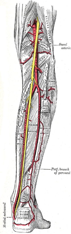
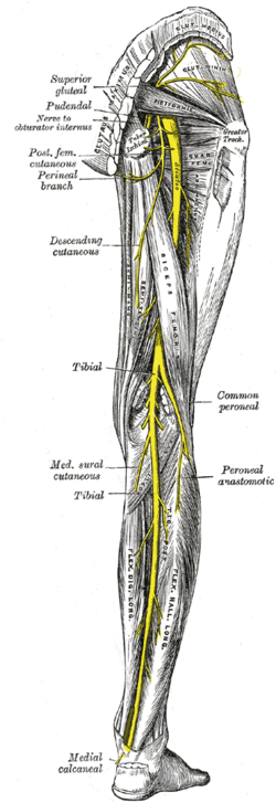
Popliteal fossa

Last updated
Popliteal fossa
Sobo 1909 718.png
Popliteal fossa of the right leg.
Details
Identifiers
Latin fossa poplitea
TA98 A01.2.08.013
TA2 324
FMA 22525
Anatomical terminology

The popliteal fossa (also referred to as hough [1] or kneepit in analogy to the cubital fossa) is a shallow depression located at the back of the knee joint. The bones of the popliteal fossa are the femur and the tibia. Like other flexion surfaces of large joints (groin, armpit, cubital fossa and essentially the anterior part of the neck), it is an area where blood vessels and nerves pass relatively superficially, and with an increased number of lymph nodes.

Contents

Structure

Boundaries

The boundaries of the fossa are: [1]

 MedialLateral
Superiorthe semimembranosus & semitendinosus muscles [2] the biceps femoris muscle [2]
Inferiorthe medial head of the gastrocnemius muscle [2] the lateral head of the gastrocnemius muscle and plantaris muscle [2]

Roof

Moving from superficial to deep structures, the roof is formed by:

  1. the skin. [1]
  2. the superficial fascia. [1] This contains the small saphenous vein, the terminal branch of the posterior cutaneous nerve of the thigh, posterior division of the medial cutaneous nerve, lateral sural cutaneous nerve, and medial sural cutaneous nerve. [1]
  3. the popliteal fascia. [1] [2]

Floor

The floor is formed by:

  1. the popliteal surface of the femur. [2]
  2. the capsule of the knee joint and the oblique popliteal ligament. [2]
  3. strong fascia covering the popliteus muscle. [2]

Contents

Structures within the popliteal fossa include, (from superficial to deep): [1]

It is of note that the common fibular nerve also begins at the superior angle of the popliteal fossa. [4]

Additional images

See also

Related Research Articles

<span class="mw-page-title-main">Human leg</span> Lower extremity or limb of the human body (foot, lower leg, thigh and hip)

The leg is the entire lower limb of the human body, including the foot, thigh or sometimes even the hip or buttock region. The major bones of the leg are the femur, tibia, and adjacent fibula. The thigh is between the hip and knee, while the calf (rear) and shin (front) are between the knee and foot.

Articles related to anatomy include:

<span class="mw-page-title-main">Femoral triangle</span> Anatomical region of the thigh

The femoral triangle is an anatomical region of the upper third of the thigh. It is a subfascial space which appears as a triangular depression below the inguinal ligament when the thigh is flexed, abducted and laterally rotated.

<span class="mw-page-title-main">Great saphenous vein</span> Large, subcutaneous, superficial vein of the leg

The great saphenous vein (GSV) or long saphenous vein is a large, subcutaneous, superficial vein of the leg. It is the longest vein in the body, running along the length of the lower limb, returning blood from the foot, leg and thigh to the deep femoral vein at the femoral triangle.

<span class="mw-page-title-main">Popliteal artery</span> Continuation of the femoral artery that supplies the lower leg

The popliteal artery is a deeply placed continuation of the femoral artery opening in the distal portion of the adductor magnus muscle. It courses through the popliteal fossa and ends at the lower border of the popliteus muscle, where it branches into the anterior and posterior tibial arteries.

<span class="mw-page-title-main">Popliteal vein</span> Large leg vein

The popliteal vein is a vein of the lower limb. It is formed from the anterior tibial vein and the posterior tibial vein. It travels medial to the popliteal artery, and becomes the femoral vein. It drains blood from the leg. It can be assessed using medical ultrasound. It can be affected by popliteal vein entrapment.

<span class="mw-page-title-main">Cubital fossa</span> The human elbow pit

The cubital fossa, chelidon or inside of elbow is the area on the anterior side of the upper part between the arm and forearm of a human or other hormid animals. It lies anteriorly to the elbow when in standard anatomical position.

<span class="mw-page-title-main">Tibial nerve</span> Branch of the sciatic nerve

The tibial nerve is a branch of the sciatic nerve. The tibial nerve passes through the popliteal fossa to pass below the arch of soleus.

The biceps femoris is a muscle of the thigh located to the posterior, or back. As its name implies, it consists of two heads; the long head is considered part of the hamstring muscle group, while the short head is sometimes excluded from this characterization, as it only causes knee flexion and is activated by a separate nerve.

<span class="mw-page-title-main">Gracilis muscle</span> Most superficial muscle on the medial side of the thigh

The gracilis muscle is the most superficial muscle on the medial side of the thigh. It is thin and flattened, broad above, narrow and tapering below.

The semimembranosus muscle is the most medial of the three hamstring muscles in the thigh. It is so named because it has a flat tendon of origin. It lies posteromedially in the thigh, deep to the semitendinosus muscle. It extends the hip joint and flexes the knee joint.

The common fibular nerve is a nerve in the lower leg that provides sensation over the posterolateral part of the leg and the knee joint. It divides at the knee into two terminal branches: the superficial fibular nerve and deep fibular nerve, which innervate the muscles of the lateral and anterior compartments of the leg respectively. When the common fibular nerve is damaged or compressed, foot drop can ensue.

<span class="mw-page-title-main">Posterior cutaneous nerve of thigh</span> Sensory nerve that supplies the back of the thigh, leg, buttock, and perineum

The posterior cutaneous nerve of the thigh is a sensory nerve of the thigh. It is a branch of the sacral plexus. It supplies the skin of the posterior surface of the thigh, leg, buttock, and also the perineum.

<span class="mw-page-title-main">Lateral cutaneous nerve of thigh</span> Nerve of the thigh

The lateral cutaneous nerve of the thigh is a cutaneous nerve of the thigh. It originates from the dorsal divisions of the second and third lumbar nerves from the lumbar plexus. It passes under the inguinal ligament to reach the thigh. It supplies sensation to the skin on the lateral part of the thigh by an anterior branch and a posterior branch.

The saphenous nerve is the largest cutaneous branch of the femoral nerve. It is derived from the lumbar plexus (L3-L4). It is a strictly sensory nerve, and has no motor function. It commences in the proximal (upper) thigh and travels along the adductor canal. Upon exiting the adductor canal, the saphenous nerve terminates by splitting into two terminal branches: the sartorial nerve, and the infrapatellar nerve. The saphenous nerve is responsible for providing sensory innervation to the skin of the anteromedial leg.

<span class="mw-page-title-main">Popliteal lymph nodes</span>

The popliteal lymph nodes, small in size and some six or seven in number, are embedded in the fat contained in the popliteal fossa, sometimes referred to as the 'knee pit'. One lies immediately beneath the popliteal fascia, near the terminal part of the small saphenous vein, and drains the region from which this vein derives its tributaries, such as superficial regions of the posterolateral aspect of the leg and the plantar aspect of the foot.

<span class="mw-page-title-main">Posterior compartment of thigh</span> One of the fascial compartments that contains the knee flexors and hip extensors

The posterior compartment of the thigh is one of the fascial compartments that contains the knee flexors and hip extensors known as the hamstring muscles, as well as vascular and nervous elements, particularly the sciatic nerve.

<span class="mw-page-title-main">Anterior cutaneous branches of the femoral nerve</span>

The anterior cutaneous branches of the femoral nerve consist of the following nerves: intermediate cutaneous nerve and medial cutaneous nerve.

<span class="mw-page-title-main">Cutaneous innervation of the lower limbs</span> Area of the skin

Cutaneous innervation of the lower limbs is the nerve supply to areas of the skin of the lower limbs which are supplied by specific cutaneous nerves.

<span class="mw-page-title-main">Outline of human anatomy</span> Overview of and topical guide to human anatomy

The following outline is provided as an overview of and topical guide to human anatomy:

References

  1. 1 2 3 4 5 6 Buckenmaier III C; Bleckner L (2008). "Chapter 20: Popliteal nerve block". The Military Advanced Regional Anesthesia and Analgesia Handbook. Rockville, Maryland: Defense & Veterans Pain Management Initiative (DVPMI). Archived from the original (PDF) on 2016-02-20. Retrieved 2011-06-08.
  2. 1 2 3 4 5 6 7 8 Nichols, Jennifer S.; Ashford, Robert U. (2013-04-01). "Surgical anatomy & pathology of the popliteal fossa". Orthopaedics and Trauma. 27 (2): 113–117. doi:10.1016/j.mporth.2013.02.011. ISSN   1877-1327.
  3. 1 2 3 Clinically Oriented Anatomy by Moore, 6th edition
  4. "The Popliteal Fossa - Borders - Contents - TeachMeAnatomy".