Urogenital triangle

Last updated
Urogenital triangle
1116 Muscle of the Female Perineum.png
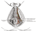
Muscles of the female perineum. (Urogenital triangle is roughly equal to top half of diagram.)
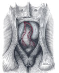
1116 Muscle of the Male Perineum.png
Muscles of male perineum. (Urogenital triangle is roughly equal to top half of diagram.)
Details
Identifiers
Latin regio urogenitalis
TA98 A01.2.06.003
TA2 279
FMA 20348
Anatomical terminology

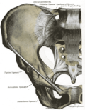
The urogenital triangle is the anterior part of the perineum. In female mammals, it contains the vulva, while in male mammals, it contains the penis and scrotum.

Contents

Structure

The urogenital triangle is the area bound by a triangle with one vertex at the pubic symphysis and the two other vertices at the iliac tuberosities of the pelvic bone.

Components

As might be expected, the contents of the urogenital triangle differ greatly between the male and the female. Some of the components include: [1]

Additional images

See also

Related Research Articles

<span class="mw-page-title-main">Perineum</span> Region of the body between the genitals and anus

The perineum in placental mammals is the space between the anus and the genitals. The human perineum is between the anus and scrotum in the male or between the anus and vulva in the female. The perineum is the region of the body between the pubic symphysis and the coccyx, including the perineal body and surrounding structures. The perineal raphe is visible and pronounced to varying degrees. The perineum is an erogenous zone. This area is also known as the taint or gooch in American slang; it is further known as the chad or grundle in British slang.

<span class="mw-page-title-main">Bulbospongiosus muscle</span> Superficial muscle of the perineum

The bulbospongiosus muscles are a subgroup of the superficial muscles of the perineum. They have a slightly different origin, insertion and function in males and females. In males, these muscles cover the bulb of the penis, while in females, they cover the vestibular bulbs.

<span class="mw-page-title-main">Bulb of vestibule</span> One of two elongated masses of erectile tissue in the human clitoris

In female anatomy, the vestibular bulbs, bulbs of the vestibule or clitoral bulbs are two elongated masses of erectile tissue typically described as being situated on either side of the vaginal opening. They are united to each other in front by a narrow median band. Some research indicates that they do not surround the vaginal opening, and are more closely related to the clitoris than to the vestibule. They constitute the root of the clitoris along with the crura.

<span class="mw-page-title-main">Perineal nerve</span> Nerve of the perineum

The perineal nerve is a nerve of the pelvis. It arises from the pudendal nerve in the pudendal canal. It gives superficial branches to the skin, and a deep branch to muscles. It supplies the skin and muscles of the perineum. Its latency is tested with electrodes.

<span class="mw-page-title-main">Fascia of Colles</span>

The membranous layer of the superficial fascia of the perineum is the deeper layer of the superficial perineal fascia. It is thin, aponeurotic in structure, and of considerable strength, serving to bind down the muscles of the root of the penis. Colles' fascia emerges from the perineal membrane, which divides the base of the penis from the prostate. Colles' fascia emerges from the inferior side of the perineal membrane and continues along the ventral (inferior) penis without covering the scrotum. It separates the skin and subcutaneous fat from the superficial perineal pouch.

<span class="mw-page-title-main">Fascia of Scarpa</span> Deep membranous layer of the superficial fascia of the abdomen

The fascia of Scarpa is the deep membranous layer (stratum membranosum) of the superficial fascia of the abdomen. It is a layer of the anterior abdominal wall. It is found deep to the fascia of Camper and superficial to the external oblique muscle.

<span class="mw-page-title-main">Posterior scrotal nerves</span>

The posterior scrotal branches are two in number, medial and lateral. They are branches of the perineal nerve, which is itself a branch of the pudendal nerve. The pudendal nerve arises from spinal roots S2 through S4, travels through the pudendal canal on the fascia of the obturator internus muscle, and gives off the perineal nerve in the perineum. The major branch of the perineal nerve is the posterior scrotal/posterior labial.

The perineal raphe is a visible line or ridge of tissue on the body that extends from the anus through the perineum to the scrotum (male) or the vulva (female). It is found in both males and females, arises from the fusion of the urogenital folds, and is visible running medial through anteroposterior, to the anus where it resolves in a small knot of skin of varying size.

<span class="mw-page-title-main">Perineal membrane</span> Anatomical term

The perineal membrane is an anatomical term for a fibrous membrane in the perineum. The term "inferior fascia of urogenital diaphragm", used in older texts, is considered equivalent to the perineal membrane.

<span class="mw-page-title-main">Superficial perineal pouch</span>

The superficial perineal pouch is a compartment of the perineum.

<span class="mw-page-title-main">Deep perineal pouch</span> Anatomic space enclosed partly by the perineum

The deep perineal pouch is the anatomic space enclosed in part by the perineum and located superior to the perineal membrane.

<span class="mw-page-title-main">Anal triangle</span> Posterior part of the perineum

The anal triangle is the posterior part of the perineum. It contains the anus in mammals.

<span class="mw-page-title-main">Posterior labial nerves</span>

The posterior labial nerves are branches of the pudendal nerve. They supply the female labia majora.

<span class="mw-page-title-main">Crus of penis</span> Internal base of the human biological male sex organ

The two crura of penis constitute the root of penis along with the bulb of penis. The two crura flank the bulb - one to each side of the bulb. Each crus is attached at the angle between the perineal membrane and ischiopubic ramus. The deep artery of the penis enters the anterior portion of the crus. Distally, each crus transitions into either corpus spongiosum of the body of the penis.

<span class="mw-page-title-main">Bulb of penis</span> Flare of the urethra tissue at the base of the human biological male sex organ

The bulb of penis is the proximal/posterior bulged end of the corpus spongiosum penis. Together with the two crura, it constitutes the root of the penis. It is covered by the bulbospongiosus.

<span class="mw-page-title-main">Perineal branches of posterior femoral cutaneous nerve</span>

The perineal branches of the posterior femoral cutaneous nerve are distributed to the skin at the upper and medial side of the thigh.

<span class="mw-page-title-main">Outline of human anatomy</span> Overview of and topical guide to human anatomy

The following outline is provided as an overview of and topical guide to human anatomy:

<span class="mw-page-title-main">Root of penis</span> Internal portion of the human penis

In human male anatomy, the radix or root of the penis is the internal and most proximal portion of the human penis that lies in the perineum. Unlike the pendulous body of the penis, which is suspended from the pubic symphysis, the root is attached to the pubic arch of the pelvis and is not visible externally. It is triradiate in form, consisting of three masses of erectile tissue; the two diverging crura, one on either side, and the median bulb of the penis or urethral bulb. Approximately one third to one half of the penis is embedded in the pelvis and can be felt through the scrotum and in the perineum.

<span class="mw-page-title-main">Muscular branches of perineal nerve</span> Nerve of the perineum

The deep branch of the perineal nerve is a nerve of the perineum. It is a branch of the perineal nerve, from the pudendal nerve. It supplies the superficial transverse perineal muscle, bulbospongiosus muscle, ischiocavernosus muscle, the bulb of penis, levator ani, and the external anal sphincter.

<span class="mw-page-title-main">Vaginal support structures</span> Structures that maintain the position of the vagina within the pelvic cavity

The vaginal support structures are those muscles, bones, ligaments, tendons, membranes and fascia, of the pelvic floor that maintain the position of the vagina within the pelvic cavity and allow the normal functioning of the vagina and other reproductive structures in the female. Defects or injuries to these support structures in the pelvic floor leads to pelvic organ prolapse. Anatomical and congenital variations of vaginal support structures can predispose a woman to further dysfunction and prolapse later in life. The urethra is part of the anterior wall of the vagina and damage to the support structures there can lead to incontinence and urinary retention.

References

  1. Daftary, Shirish; Chakravarti, Sudip (2011). Manual of Obstetrics, 3rd Edition. Elsevier. pp. 1-16. ISBN   9788131225561.