
The perineum in humans is the space between the anus and scrotum in the male, or between the anus and the vulva in the female. The perineum is the region of the body between the pubic symphysis and the coccyx, including the perineal body and surrounding structures. The perineal raphe is visible and pronounced to varying degrees. The perineum is an erogenous zone. This area is also known as the taint or chode in American slang.

The levator ani is a broad, thin muscle group, situated on either side of the pelvis. It is formed from three muscle components: the pubococcygeus, the iliococcygeus, and the puborectalis.
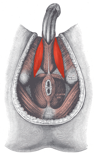
The ischiocavernosus muscle is a muscle just below the surface of the perineum, present in both men and women.

The bulbospongiosus muscle is one of the superficial muscles of the perineum. It has a slightly different origin, insertion and function in males and females. In males, it covers the bulb of the penis. In females, it covers the vestibular bulb.

The external anal sphincter is an oval tube skeletal muscle fibers. Distally, it is adherent to the skin surrounding the margin of the anus. The sphincter exhibits a resting state of tonical contraction.

The posterior cutaneous nerve of the thigh is a sensory nerve of the thigh. It is a branch of the sacral plexus. It supplies the skin of the posterior surface of the thigh, leg, buttock, and also the perineum.

The ischium forms the lower and back region of the hip bone.
In vertebrates, the pubis or pubic bone forms the lower and anterior part of each side of the hip bone. The pubis is the most forward-facing of the three bones that make up the hip bone. The left and right pubic bones are each made up of three sections, a superior ramus, inferior ramus, and a body.

The sacrotuberous ligament is situated at the lower and back part of the pelvis. It is flat, and triangular in form; narrower in the middle than at the ends.

The fascia of Scarpa is the deep membranous layer (stratum membranosum) of the superficial fascia of the abdomen. It is a layer of the anterior abdominal wall. It is found deep to the fascia of Camper and superficial to the external oblique muscle.
The fascia of Camper is a thick superficial layer of the anterior abdominal wall.

The perineal membrane is an anatomical term for a fibrous membrane in the perineum. The term "inferior fascia of urogenital diaphragm", used in older texts, is considered equivalent to the perineal membrane.
The superficial perineal pouch is a compartment of the perineum.
The deep perineal pouch is the anatomic space enclosed in part by the perineum, and located superior to the perineal membrane.
The superior fascia of the urogenital diaphragm is continuous with the obturator fascia and stretches across the pubic arch.

Buck's fascia is a layer of deep fascia covering the three erectile bodies of the penis.

The following outline is provided as an overview of and topical guide to human anatomy:

In human male anatomy, the radix or root of the penis is the internal and most proximal portion of the human penis that lies in the perineum. Unlike the pendulous body of the penis which is suspended from the pubic symphysis, the root is attached to the pubic arch of the pelvis and is not visible externally. It is triradiate in form, consisting of three masses of erectile tissue; the two diverging crura, one on either side, and the median bulb of the penis or urethral bulb. Approximately one third to one half of the penis is embedded in the pelvis and can be felt through the scrotum and in the perineum.
The subcutaneous tissue of perineum is a layer of subcutaneous tissue surrounding the region of the perineal body.

The vaginal support structures are those muscles, bones, ligaments, tendons, membranes and fascia, of the pelvic floor that maintain the position of the vagina within the pelvic cavity and allow the normal functioning of the vagina and other reproductive structures in the female. Defects or injuries to these support structures in the pelvic floor leads to pelvic organ prolapse. Anatomical and congenital variations of vaginal support structures can predispose a woman to further dysfunction and prolapse later in life. The urethra is part of the anterior wall of the vagina and damage to the support structures there can lead to incontinence and urinary retention.

