
The urethra is the tube that connects the mammalian urinary bladder to the urinary meatus. In placental mammals, the urethra transports urine through the penis or vulva during urination and semen through the penis during ejaculation.

The corpus spongiosum is the mass of spongy tissue surrounding the male urethra within the penis. It is also called the corpus cavernosum urethrae in older texts.

The bulbospongiosus muscles are a subgroup of the superficial muscles of the perineum. They have a slightly different origin, insertion and function in males and females. In males, these muscles cover the bulb of the penis, while in females, they cover the vestibular bulbs.

Older texts have asserted the existence of a urogenital diaphragm, also called the triangular ligament, which was described as a layer of the pelvis that separates the deep perineal sac from the upper pelvis, lying between the inferior fascia of the urogenital diaphragm and superior fascia of the urogenital diaphragm.
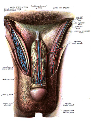
The fundiform ligament of the penis is a structure of the external genitalia whose descriptions differ according to the source consulted.
The external sphincter muscle of the male urethra, also sphincter urethrae membranaceae, sphincter urethrae externus, surrounds the whole length of the membranous urethra, and is enclosed in the fascia of the urogenital diaphragm.

The membranous layer of the superficial fascia of the perineum is the deeper layer of the superficial perineal fascia. It is thin, aponeurotic in structure, and of considerable strength, serving to bind down the muscles of the root of the penis. Colles' fascia emerges from the perineal membrane, which divides the base of the penis from the prostate. Colles' fascia emerges from the inferior side of the perineal membrane and continues along the ventral (inferior) penis without covering the scrotum. It separates the skin and subcutaneous fat from the superficial perineal pouch.

The dorsal artery of the penis is a bilaterally paired terminal branch of the internal pudendal artery which passes upon the dorsum of the penis to the base of the glans penis, where it unites with its contralateral partner and supply the glans and foreskin.
The perineal membrane is an anatomical term for a fibrous membrane in the perineum. The term "inferior fascia of urogenital diaphragm", used in older texts, is considered equivalent to the perineal membrane.
The superficial perineal pouch is a compartment of the perineum.
The deep perineal pouch is the anatomic space enclosed in part by the perineum and located superior to the perineal membrane.

The urogenital triangle is the anterior part of the perineum. In female mammals, it contains the vulva, while in male mammals, it contains the penis and scrotum.
The prostatic urethra, the widest and most dilatable part of the urethra canal, is about 3 cm long.

The spongy urethra is the longest part of the male urethra, and is contained in the corpus spongiosum of the penis.

The internal urethral orifice is the opening of the urinary bladder into the urethra.
The navicular fossa is a short dilated portion of the male urethra within the glans penis just proximal to the external urethral meatus. The roof of the fossa is especially dilated, forming a lacuna; medical instruments being inserted into the male urethra should initially be directed towards the floor of the fossa so as not to get snagged at the fossa. It is one of three dilations of the male urethra.
The superior fascia of the urogenital diaphragm is continuous with the obturator fascia and stretches across the pubic arch.

The urethral sphincters are two muscles used to control the exit of urine in the urinary bladder through the urethra. The two muscles are either the male or female external urethral sphincter and the internal urethral sphincter. When either of these muscles contracts, the urethra is sealed shut.

In human male anatomy, the radix or root of the penis is the internal and most proximal portion of the human penis that lies in the perineum. Unlike the pendulous body of the penis, which is suspended from the pubic symphysis, the root is attached to the pubic arch of the pelvis and is not visible externally. It is triradiate in form, consisting of three masses of erectile tissue; the two diverging crura, one on either side, and the median bulb of the penis or urethral bulb. Approximately one third to one half of the penis is embedded in the pelvis and can be felt through the scrotum and in the perineum.
The vaginal support structures are those muscles, bones, ligaments, tendons, membranes and fascia, of the pelvic floor that maintain the position of the vagina within the pelvic cavity and allow the normal functioning of the vagina and other reproductive structures in the female. Defects or injuries to these support structures in the pelvic floor leads to pelvic organ prolapse. Anatomical and congenital variations of vaginal support structures can predispose a woman to further dysfunction and prolapse later in life. The urethra is part of the anterior wall of the vagina and damage to the support structures there can lead to incontinence and urinary retention.


