
The clitoris is a female sex organ present in mammals, ostriches and a limited number of other animals. In humans, the visible portion – the glans – is at the front junction of the labia minora, above the opening of the urethra. Unlike the penis, the male homologue (equivalent) to the clitoris, it usually does not contain the distal portion of the urethra and is therefore not used for urination. In most species, the clitoris lacks any reproductive function. While few animals urinate through the clitoris or use it reproductively, the spotted hyena, which has an especially large clitoris, urinates, mates, and gives birth via the organ. Some other mammals, such as lemurs and spider monkeys, also have a large clitoris.

The urethra is a tube that connects the urinary bladder to the urinary meatus for the removal of urine from the body of both females and males. In human females and other primates, the urethra connects to the urinary meatus above the vagina, whereas in marsupials, the female's urethra empties into the urogenital sinus.

In male human anatomy, the glans penis, commonly referred to as the glans, is the bulbous structure at the distal end of the human penis that is the human male's most sensitive erogenous zone and primary anatomical source of sexual pleasure. The glans penis is present in the male reproductive organs of humans and most mammals where it may appear smooth, spiny, elongated or divided. It is externally lined with mucosal tissue, which creates a smooth texture and glossy appearance. In humans, the glans is located over the distal ends of the corpora cavernosa and is a continuation of the corpus spongiosum of the penis. At the summit appears the urinary meatus and at the base forms the corona glandis. An elastic band of tissue, known as the frenulum, runs on its ventral surface. In men who are not circumcised, it is completely or partially covered by a fold of skin called the foreskin. In adults, the foreskin can generally be retracted over and past the glans manually or sometimes automatically during an erection.

The corpus spongiosum is the mass of spongy tissue surrounding the male urethra within the penis. It is also called the corpus cavernosum urethrae in older texts.

The palang, or ampallang is a male genital piercing that penetrates horizontally through the entire glans of the penis.

The male reproductive system consists of a number of sex organs that play a role in the process of human reproduction. These organs are located on the outside of the body and within the pelvis.

The urethral artery arises from the internal pudendal artery a branch of the internal iliac artery. The internal pudendal artery has numerous branches including the artery of the bulb of the penis immediately before the urethral and the dorsal artery of the penis more distally.
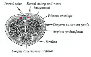
A corpus cavernosum penis (singular) is one of a pair of sponge-like regions of erectile tissue, which contain most of the blood in the penis during an erection.
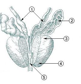
The prostatic urethra, the widest and most dilatable part of the urethra canal, is about 3 cm long.

The spongy urethra is the longest part of the male urethra, and is contained in the corpus spongiosum of the penis.

The membranous urethra or intermediate part of male urethra is the shortest, least dilatable, and, with the exception of the urinary meatus, the narrowest part of the urethra.

In human male anatomy, the dorsal veins of the penis are blood vessels that drain the shaft, the skin and the glans of the human penis. They are typically located in the midline on the dorsal aspect of the penis and they comprise the superficial dorsal veinof the penis, that lies in the subcutaneous tissue of the shaft, and the deep dorsal veinof the penis, that lies beneath the deep fascia.

The two crura of penis constitute the root of penis along with the bulb of penis. The two crura flank the bulb - one to each side of the bulb. Each crus is attached at the angle between the perineal membrane and ischiopubic ramus. The deep artery of the penis enters the anterior portion of the crus. Distally, each crus transitions into either corpus spongiosum of the body of penis.
In male anatomy, the lacuna magna is the largest of several recesses in the roof of the navicular fossa of the male urethra.

In human anatomy, the penis is an external male intromittent organ that additionally serves as the urinary duct. The main parts are the root (radix); the body (corpus); and the epithelium of the penis including the shaft skin and the foreskin (prepuce) covering the glans penis. The body of the penis is made up of three columns of tissue: two corpora cavernosa on the dorsal side and corpus spongiosum between them on the ventral side. The human male urethra passes through the prostate gland, where it is joined by the ejaculatory duct, and then through the penis. The urethra traverses the corpus spongiosum, and its opening, the meatus, lies on the tip of the glans penis. It is a passage both for urination and ejaculation of semen.
The development of the reproductive system is the part of embryonic growth that results in the sex organs and contributes to sexual differentiation. Due to its large overlap with development of the urinary system, the two systems are typically described together as the urogenital or genitourinary system.
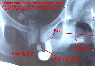
A retrograde urethrography is a routine radiologic procedure used to image the integrity of the urethra. Hence a retrograde urethrogram is essential for diagnosis of urethral injury, or urethral stricture.

The corona of glans penis or penis crown refers to the rounded projecting border or flare that forms at the base of the glans in human males. The corona overhangs a mucosal surface, known as the neck of the penis, which separates the shaft and the glans. The deep retro-glandular coronal sulcus forms between the corona and the neck of the penis. The two sides of the corona merge on the ventral midline forming the septum glandis. The circumference of the corona is richly innervated and is described as a highly erogenous area of the glans.

In male human anatomy, the foreskin, also known as the prepuce, is the double-layered fold of skin, mucosal and muscular tissue at the distal end of the human penis that covers the glans and the urinary meatus. The foreskin is attached to the glans by an elastic band of tissue, known as the frenulum. The outer skin of the foreskin meets with the inner preputial mucosa at the area of the mucocutaneous junction. The foreskin is mobile, fairly stretchable and sustains the glans in a moist environment. Except for humans, a similar structure, known as penile sheath, appears in the male sexual organs of all primates and the vast majority of mammals.

The septum glandis, also septum of the glans, refers to the fibrous partition of the ventral aspect of the glans penis that separates the two glans wings in the ventral midline. The septum extends from the urethral meatus through the glanular urethra and ends in the tunica albuginea of the human penis. Externally it is attached to the frenulum which extends lower on the neck of the penis.
