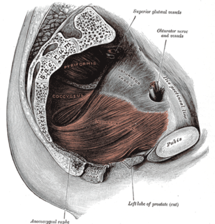
The levator ani is a broad, thin muscle, situated on either side of the pelvis. It is formed from three muscle components: the pubococcygeus, the iliococcygeus, and the puborectalis.

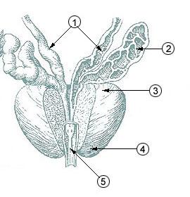
The seminal vesicles, vesicular glands, or seminal glands, are a pair of simple tubular glands posteroinferior to the urinary bladder of some male mammals. Seminal vesicles are located within the pelvis. They secrete fluid that partly composes the semen.
The vas deferens, also called ductus deferens, is part of the male reproductive system of many vertebrates; these ducts transport sperm from the epididymis to the ejaculatory ducts in anticipation of ejaculation. It is a partially coiled tube which exits the abdominal cavity through the inguinal canal.

The ejaculatory ducts are paired structures in male anatomy. Each ejaculatory duct is formed by the union of the vas deferens with the duct of the seminal vesicle. They pass through the prostate, and open into the urethra at the seminal colliculus. During ejaculation, semen passes through the prostate gland, enters the urethra and exits the body via the urinary meatus.

The prostatic utricle is a small indentation in the prostatic urethra, at the apex of the urethral crest, on the seminal colliculus (verumontanum), laterally flanked by openings of the ejaculatory ducts. It is also known as the vagina masculina or uterus masculinus or vesicula prostatica.

The middle rectal artery is an artery in the pelvis that supplies blood to the rectum.

The inferior vesical artery or inferior vesicle artery is an artery in the pelvis that supplies the lower part of the bladder.

The external iliac lymph nodes are lymph nodes, from eight to ten in number, that lie along the external iliac vessels.

The internal iliac vein begins near the upper part of the greater sciatic foramen, passes upward behind and slightly medial to the internal iliac artery and, at the brim of the pelvis, joins with the external iliac vein to form the common iliac vein.

The inferior hypogastric plexus is a plexus of nerves that supplies the viscera of the pelvic cavity. The inferior hypogastric plexus gives rise to the prostatic plexus in males and the uterovaginal plexus in females.

The deep perineal pouch is the anatomic space enclosed in part by the perineum, and located superior to the perineal membrane.
The prostatic urethra, the widest and most dilatable part of the urethra canal, is about 3 cm long.

The membranous urethra or intermediate part of male urethra is the shortest, least dilatable, and, with the exception of the urinary meatus, the narrowest part of the urethra.

In human anatomy, the dorsal veins of the penis comprise the superficial dorsal vein of the penis and the deep dorsal vein of the penis.

The internal urethral orifice is the opening of the urinary bladder into the urethra. It is placed at the apex of the trigonum vesicae, in the most dependent part of the bladder, and is usually somewhat crescent-shaped; the mucous membrane immediately behind it presents a slight elevation in males, the uvula vesicae, caused by the middle lobe of the prostate.
The seminal colliculus, or verumontanum, of the prostatic urethra is a landmark near the entrance of the ejaculatory ducts. Verumontanum is translated from Latin to mean 'mountain ridge', a reference to the distinctive median elevation of urothelium that characterizes the landmark on magnified views. Embryologically, it is derived from the uterovaginal primordium. The landmark is important in classification of several urethral developmental disorders. The margins of seminal colliculus are the following:

The prostatic veins form a well-marked prostatic plexus which lies partly in the fascial sheath of the prostate and partly between the sheath and the prostatic capsule. It communicates with the pudendal and vesical plexuses.
The superior fascia of the urogenital diaphragm is continuous with the obturator fascia and stretches across the pubic arch.
The public domain consists of all the creative works to which no exclusive intellectual property rights apply. Those rights may have expired, been forfeited, expressly waived, or may be inapplicable.

Gray's Anatomy is an English language textbook of human anatomy originally written by Henry Gray and illustrated by Henry Vandyke Carter. Earlier editions were called Anatomy: Descriptive and Surgical, Anatomy of the Human Body and Gray's Anatomy: Descriptive and Applied, but the book's name is commonly shortened to, and later editions are titled, Gray's Anatomy. The book is widely regarded as an extremely influential work on the subject, and has continued to be revised and republished from its initial publication in 1858 to the present day. The latest edition of the book, the 41st, was published in September 2015.

