Related Research Articles

The urethra is a tube that connects the mammalian urinary bladder to the urinary meatus in the glans penis or vulval vestibule. Male and female placental mammals release urine through the urethra during urination, but males also release semen through the urethra during ejaculation.

The bladder is a hollow organ in humans and other vertebrates that stores urine from the kidneys before disposal by urination. In placental mammals, urine enters the bladder via the ureters and exits via the urethra. In humans, the bladder is a distensible organ that sits on the pelvic floor. The typical adult human bladder will hold between 300 and 500 ml before the urge to empty occurs, but can hold considerably more.

The urogenital sinus is a part of the human body only present in the development of the urinary and reproductive organs. It is the ventral part of the cloaca, formed after the cloaca separates from the anal canal during the fourth to seventh weeks of development.
Sinus of Morgagni may refer to:
The development of the urinary system begins during prenatal development, and relates to the development of the urogenital system – both the organs of the urinary system and the sex organs of the reproductive system. The development continues as a part of sexual differentiation.

The pyramid-shaped maxillary sinus is the largest of the paranasal sinuses, located in the maxilla. It drains into the middle meatus of the nose through the semilunar hiatus. It is located to the side of the nasal cavity, and below the orbit.

The agger nasi is a small ridge on the lateral side of the nasal cavity. It is located midway at the anterior edge of the middle nasal concha, directly above the atrium of the middle meatus. It is formed by a mucous membrane that is covering the ethmoidal crest of the maxilla.

Oral mucocele is a condition caused by two related phenomena - mucus extravasation phenomenon and mucous retention cyst.

The superior sagittal sinus, within the human head, is an unpaired area along the attached margin of the falx cerebri. It allows blood to drain from the lateral aspects of anterior cerebral hemispheres to the confluence of sinuses. Cerebrospinal fluid drains through arachnoid granulations into the superior sagittal sinus and is returned to venous circulation.

The mastoid part of the temporal bone is the posterior (back) part of the temporal bone, one of the bones of the skull. Its rough surface gives attachment to various muscles and it has openings for blood vessels. From its borders, the mastoid part articulates with two other bones.
Morgagni can refer to:

Anal columns are a number of vertical folds, produced by an infolding of the mucous membrane and some of the muscular tissue in the upper half of the lumen of the anal canal. They are named after Giovanni Battista Morgagni, who has several other eponyms named after him.

The laryngeal ventricle, is a fusiform fossa, situated between the vestibular and vocal folds on either side, and extending nearly their entire length. There is also a sinus of Morgagni in the pharynx.
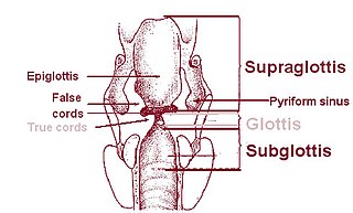
The pyriform sinus is a small recess on either side of the laryngeal inlet. It is bounded medially by the aryepiglottic fold, and laterally by the thyroid cartilage and thyrohyoid membrane. The fossae are involved in speech.
A sinus is a sac or cavity in any organ or tissue, or an abnormal cavity or passage. In common usage, "sinus" usually refers to the paranasal sinuses, which are air cavities in the cranial bones, especially those near the nose and connecting to it. Most individuals have four paired cavities located in the cranial bone or skull.

In male anatomy, the lacuna magna is the largest of several recesses in the roof of the navicular fossa of the male urethra.

Alphonse François Marie Guérin was a French surgeon who was a native of Ploërmel.
The development of the reproductive system is the part of embryonic growth that results in the sex organs and contributes to sexual differentiation. Due to its large overlap with development of the urinary system, the two systems are typically described together as the genitourinary system.
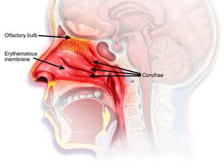
The nasal mucosa lines the nasal cavity. It is part of the respiratory mucosa, the mucous membrane lining the respiratory tract. The nasal mucosa is intimately adherent to the periosteum or perichondrium of the nasal conchae. It is continuous with the skin through the nostrils, and with the mucous membrane of the nasal part of the pharynx through the choanae. From the nasal cavity its continuity with the conjunctiva may be traced, through the nasolacrimal and lacrimal ducts; and with the frontal, ethmoidal, sphenoidal, and maxillary sinuses, through the several openings in the nasal meatuses. The mucous membrane is thickest, and most vascular, over the nasal conchae. It is also thick over the nasal septum where increased numbers of goblet cells produce a greater amount of nasal mucus. It is very thin in the meatuses on the floor of the nasal cavities, and in the various sinuses. It is one of the most commonly infected tissues in adults and children. Inflammation of this tissue may cause significant impairment of daily activities, with symptoms such as stuffy nose, headache, mouth breathing, etc.
A vulvar disease is a particular abnormal, pathological condition that affects part or all of the vulva. Several pathologies are defined. Some can be prevented by vulvovaginal health maintenance.
References
| | This article related to the genitourinary system is a stub. You can help Wikipedia by expanding it. |