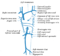
Testicular vein

Last updated
Testicular vein
Gray590.png
Spermatic veins.
Details
Source Pampiniform plexus
Drains to Inferior vena cava, left renal vein
Artery Testicular artery
Identifiers
Latin vena testicularis,
vena spermatica
TA98 A12.3.09.014M
A12.3.09.012M
TA2 5015, 5018
FMA 14344
Anatomical terminology

The testicular vein (or spermatic vein), the male gonadal vein, carries deoxygenated blood from its corresponding testis to the inferior vena cava or one of its tributaries. It is the male equivalent of the ovarian vein, and is the venous counterpart of the testicular artery.

Contents

Structure

It is a paired vein, with one supplying each testis:

The veins emerge from the back of the testis, and receive tributaries from the epididymis. [1] They unite and form a convoluted plexus, called the pampiniform plexus, which constitutes the greater mass of the spermatic cord; the vessels composing this plexus are very numerous, and ascend along the cord, in front of the ductus deferens.

Below the subcutaneous inguinal ring, they unite to form three or four veins, which pass along the inguinal canal, and, entering the abdomen through the abdominal inguinal ring, coalesce to form two veins, which ascend on the Psoas major, behind the peritoneum, lying one on either side of the internal spermatic artery.

These unite to form a single vein, which opens, on the right side, into the inferior vena cava (at an acute angle), on the left side into the left renal vein (at a right angle).

The left spermatic vein passes behind the iliac colon and is thus exposed to pressure from the contents of that part of the bowel.

Variation

The testicular veins usually have valves. [1] However, in post-mortem examinations it was found that up to 40% of left testicular veins lack valves, and up to 23% of right testicular veins lack valves. [1]

Clinical significance

Varicocele

Valveless testicular veins are a major contributing factor to varicocele. [1] Since the left testicular vein goes all the way up to the left renal vein before it empties, this results in a higher tendency for the left testicle to develop varicocele because of the gravity working on the column of blood in this vein, compared to the right internal spermatic vein. One reason for this susceptibility is that the left internal testicular vein often drains into the left renal vein without a right-angle entry like it is with the right testicular vein, which can create increased pressure and possibly disrupt the normal valve function, leading to poor venous return and eventually varicocele development. [2]

The testicular vein may be ligated in part (a branch) or completely to treat varicocele. [3] This is typically very safe. [3] There is debate about whether the testicular artery should also be ligated simultaneously. [3] Affected testicular veins can also be removed completely to further reduce recurrence rates.[ citation needed ]

Compression

The left renal vein passes between the abdominal aorta and the superior mesenteric artery en route to the inferior vena cava, and is often compressed by an enlarged superior mesenteric artery—this is called the "Nutcracker effect". [4]

Additional images

Related Research Articles

Articles related to anatomy include:

<span class="mw-page-title-main">Inferior vena cava</span> One of two veinous trunks bringing deoxygenated blood back to the heart

The inferior vena cava is a large vein that carries the deoxygenated blood from the lower and middle body into the right atrium of the heart. It is formed by the joining of the right and the left common iliac veins, usually at the level of the fifth lumbar vertebra.

<span class="mw-page-title-main">Vas deferens</span> Part of the male reproductive system of many vertebrates

The vas deferens, with the more modern name ductus deferens, is part of the male reproductive system of many vertebrates. The ducts transport sperm from the epididymis to the ejaculatory ducts in anticipation of ejaculation. The vas deferens is a partially coiled tube which exits the abdominal cavity through the inguinal canal.

<span class="mw-page-title-main">Spermatic cord</span> Structure in the human male reproductive system

The spermatic cord is the cord-like structure in males formed by the vas deferens and surrounding tissue that runs from the deep inguinal ring down to each testicle. Its serosal covering, the tunica vaginalis, is an extension of the peritoneum that passes through the transversalis fascia. Each testicle develops in the lower thoracic and upper lumbar region and migrates into the scrotum. During its descent it carries along with it the vas deferens, its vessels, nerves etc. There is one on each side.

<span class="mw-page-title-main">Inguinal canal</span> Human abdominal anatomy

The inguinal canal is a passage in the anterior abdominal wall on each side of the body which in males convey the spermatic cords and in females the round ligament of the uterus. The inguinal canals are larger and more prominent in males.

<span class="mw-page-title-main">Orchiopexy</span> Surgery to fix a testicle into the scrotum

Orchiopexy is a surgery to move and/or permanently fix a testicle into the scrotum. While orchiopexy typically describes the operation to surgically correct an undescended testicle, it is also used to resolve testicular torsion.

<span class="mw-page-title-main">Abdominal aorta</span> Largest artery in the abdomen

In human anatomy, the abdominal aorta is the largest artery in the abdominal cavity. As part of the aorta, it is a direct continuation of the descending aorta.

<span class="mw-page-title-main">Varicocele</span> Medical condition

A varicocele is an abnormal enlargement of the pampiniform venous plexus in the male scrotum; the female equivalent of painful swelling to the embryologically identical pampiniform venous plexus is called pelvic compression syndrome. This plexus of veins drains blood from the testicles back to the heart. The vessels originate in the abdomen and course down through the inguinal canal as part of the spermatic cord on their way to the testis. Varicoceles occur in around 15% to 20% of all men. The incidence of varicocele increase with age.

<span class="mw-page-title-main">Renal artery</span> Vessel supplying blood to kidney

The renal arteries are paired arteries that supply the kidneys with blood. Each is directed across the crus of the diaphragm, so as to form nearly a right angle.

<span class="mw-page-title-main">Periaortic lymph nodes</span> Human lymph nodes

The periaortic lymph nodes are a group of lymph nodes that lie in front of the lumbar vertebrae near the aorta. These lymph nodes receive drainage from the gastrointestinal tract and the abdominal organs.

<span class="mw-page-title-main">Renal vein</span> Short thick veins which return blood from the kidneys to the vena cava

The renal veins in the renal circulation, are large-calibre veins that drain blood filtered by the kidneys into the inferior vena cava. There is one renal vein draining each kidney. Each renal vein is formed by the convergence of the interlobar veins of one kidney.

<span class="mw-page-title-main">Superior mesenteric artery</span> Artery which supplies blood to the intestines and pancreas

In human anatomy, the superior mesenteric artery (SMA) is an artery which arises from the anterior surface of the abdominal aorta, just inferior to the origin of the celiac trunk, and supplies blood to the intestine from the lower part of the duodenum through two-thirds of the transverse colon, as well as the pancreas.

<span class="mw-page-title-main">Abdominal aortic plexus</span>

The abdominal aortic plexus is formed by branches derived, on either side, from the celiac plexus and ganglia, and receives filaments from some of the lumbar ganglia.

<span class="mw-page-title-main">Testicular artery</span>

The testicular artery is a branch of the abdominal aorta that supplies blood to the testis. It is a paired artery, with one for each of the testes.

<span class="mw-page-title-main">Ovarian vein</span>

The ovarian vein, the female gonadal vein, carries deoxygenated blood from its corresponding ovary to inferior vena cava or one of its tributaries. It is the female equivalent of the testicular vein, and is the venous counterpart of the ovarian artery. It can be found in the suspensory ligament of the ovary.

<span class="mw-page-title-main">Lumbar veins</span> Veins that drain the posterior abdominal wall

The lumbar veins are four pairs of veins running along the inside of the posterior abdominal wall, and drain venous blood from parts of the abdominal wall. Each lumbar vein accompanies a single lumbar artery. The lower two pairs of lumbar veins all drain directly into the inferior vena cava, whereas the fate of the upper two pairs is more variable.

<span class="mw-page-title-main">Pampiniform plexus</span> Vein network in human males

The pampiniform plexus is a venous plexus – a network of many small veins found in the human male spermatic cord, and the suspensory ligament of the ovary. In the male, it is formed by the union of multiple testicular veins from the back of the testis and tributaries from the epididymis.

<span class="mw-page-title-main">Outline of human anatomy</span> Overview of and topical guide to human anatomy

The following outline is provided as an overview of and topical guide to human anatomy:

<span class="mw-page-title-main">Scrotal ultrasound</span> Medical ultrasound examination of the scrotum.

Scrotalultrasound is a medical ultrasound examination of the scrotum. It is used in the evaluation of testicular pain, and can help identify solid masses.

Male genital examination is a physical examination of the genital in males to detect ailments and to assess sexual development and is normally included in an annual physical examination. The examination includes checking the penis, scrotum, and urethral meatus. A comprehensive assessment of the male genitals assesses the pubic hair based on Sexual Maturity Rating and the size of the testicles and penis. The exam is conducted to detect various ailments, measure sexual development, or verify a person's age and biological sex. The genitourinary system can also be assessed as part of the male genital examination. During a genital examination, the doctor can detect any of the following: structural abnormalities, urethral opening abnormalities, problems related to uncircumcision, lumps, tumors, redness, excoriation, edema, lesions, swelling, cancer, hair-related issues and many others. In some instances where a physical examination of the male genitals is not sufficient to diagnose an individual than an internal genital examination using imaging or ultrasounds will be needed for further evaluation.

References

PD-icon.svgThis article incorporates text in the public domain from page 678 of the 20th edition of Gray's Anatomy (1918)

  1. 1 2 3 4 Hutson, John M. (2012-01-01), Coran, Arnold G. (ed.), "Chapter 77 - Undescended Testis, Torsion, and Varicocele", Pediatric Surgery (Seventh Edition), Philadelphia: Mosby, pp. 1003–1019, doi:10.1016/b978-0-323-07255-7.00077-5, ISBN   978-0-323-07255-7 , retrieved 2021-02-04
  2. Hutson, John M. (2012), "Undescended Testis, Torsion, and Varicocele", Pediatric Surgery, Elsevier, pp. 1003–1019, doi:10.1016/b978-0-323-07255-7.00077-5, ISBN   978-0-323-07255-7 , retrieved 2023-10-04
  3. 1 2 3 Zelkovic, Paul; Kogan, Stanley J. (2010-01-01), Gearhart, John P.; Rink, Richard C.; Mouriquand, Pierre D. E. (eds.), "The Pediatric Varicocele", Pediatric Urology (Second Edition), Philadelphia: W.B. Saunders, pp. 585–594, doi:10.1016/b978-1-4160-3204-5.00045-1, ISBN   978-1-4160-3204-5 , retrieved 2021-02-04
  4. Ahmed K, Sampath R, Khan MS (April 2006). "Current trends in the diagnosis and management of renal nutcracker syndrome: a review". European Journal of Vascular and Endovascular Surgery . European Society for Vascular Surgery. 31 (4): 410–416. doi: 10.1016/j.ejvs.2005.05.045 . PMID   16431142.