Peritoneum

Last updated
Peritoneum
Gray1040.png
The peritoneum, colored in blue
Gray1035.png
The epiploic foramen, greater sac or general cavity (red) and lesser sac, or omental bursa (blue)
Details
Pronunciation /ˌpɛrɪtəˈnəm/
Part of Abdomen
Identifiers
Latin peritoneum
MeSH D010537
TA98 A10.1.02.002
A10.1.02.005
A10.1.02.006
TA2 3729
TH H3.04.08.0.00001
FMA 9584
Anatomical terminology

The peritoneum is the serous membrane forming the lining of the abdominal cavity or coelom in amniotes and some invertebrates, such as annelids. It covers most of the intra-abdominal (or coelomic) organs, and is composed of a layer of mesothelium supported by a thin layer of connective tissue. This peritoneal lining of the cavity supports many of the abdominal organs and serves as a conduit for their blood vessels, lymphatic vessels, and nerves.

Contents

The abdominal cavity (the space bounded by the vertebrae, abdominal muscles, diaphragm, and pelvic floor) is different from the intraperitoneal space (located within the abdominal cavity but wrapped in peritoneum). The structures within the intraperitoneal space are called "intraperitoneal" (e.g., the stomach and intestines), the structures in the abdominal cavity that are located behind the intraperitoneal space are called "retroperitoneal" (e.g., the kidneys), and those structures below the intraperitoneal space are called "subperitoneal" or "infraperitoneal" (e.g., the bladder).

Structure

Layers

The peritoneum is one continuous sheet, forming two layers and a potential space between them: the peritoneal cavity.

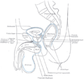
The outer layer, the parietal peritoneum, is attached to the abdominal wall and the pelvic walls. [1] The tunica vaginalis, the serous membrane covering the male testis, is derived from the vaginal process, an outpouching of the parietal peritoneum.

The inner layer, the visceral peritoneum, is wrapped around the visceral organs, located inside the intraperitoneal space for protection. It is thinner than the parietal peritoneum. The mesentery is a double layer of visceral peritoneum that attaches to the gastrointestinal tract. There are often blood vessels, nerves, and other structures between these layers. The space between these two layers is technically outside of the peritoneal sac, and thus not in the peritoneal cavity.

The potential space between these two layers is the peritoneal cavity, filled with a small amount (about 50 mL) of slippery serous fluid that allows the two layers to slide freely over each other.

The right paracolic gutter is continuous with the right and left subhepatic spaces. The epiploic foramen allows communication between the greater sac and the lesser sac. [2] The peritoneal space in males is closed, while the peritoneal space in females is continuous with the extraperitoneal pelvis through openings of the fallopian tubes, the uterus, and the vagina. [3]

Subdivisions

Peritoneal folds are omentums, mesenteries and ligaments; they connect organs to each other or to the abdominal wall. [4] There are two main regions of the peritoneal cavity, connected by the omental foramen.

The mesentery is the part of the peritoneum through which most abdominal organs are attached to the abdominal wall and supplied with blood and lymph vessels and nerves.

Omenta

SourcesStructureFromToContains
Dorsal mesentery Greater omentumGreater curvature of stomach (and spleen)Transverse colonright and left gastroepiploic vessels and fat
Gastrosplenic ligament StomachSpleen Short gastric artery, Left gastroepiploic artery
Gastrophrenic ligament Stomach Diaphragm Left inferior phrenic artery
Gastrocolic ligament Stomach Transverse colon Right gastroepiploic artery
Splenorenal ligament SpleenKidney Splenic artery, Tail of pancreas
Ventral mesentery Lesser omentumLesser curvature of the stomach (and duodenum)LiverThe right free margin-hepatic artery, portal vein, and bile duct,lymph nodes and the lymph vessels,hepatic plexus of nerve,all enclosed in perivascular fibrous sheath. Along the lesser curvature of the stomach-left and right gastric artery,gastric group of lymph nodes and lyphatics, branches from gastric nerve.
Hepatogastric ligament StomachLiver Right and left gastric artery
Hepatoduodenal ligament DuodenumLiver Hepatic artery proper, hepatic portal vein, bile duct, autonomic nerves

Mesenteries

SourcesStructureFromToContains
Dorsal mesenteryMesentery proper Small intestine (jejunum and ileum)Posterior abdominal wall Superior mesenteric artery, accompanying veins, autonomic nerve plexuses, lymphatics, 100–200 lymph nodes and connective tissue with fat
Transverse mesocolon Transverse colon Posterior abdominal wall Middle colic
Sigmoid mesocolon Sigmoid colon Pelvic wall Sigmoid arteries and superior rectal artery
Mesoappendix Mesentery of ileum Appendix Appendicular artery

Other ligaments and folds

SourcesStructureFromToContains
Ventral mesentery Falciform ligament Liver Thoracic diaphragm, anterior abdominal wall Round ligament of liver, paraumbilical vein
Left umbilical vein Round ligament of liver Liver Umbilicus
Ventral mesentery Coronary ligament LiverThoracic diaphragm
Ductus venosus Ligamentum venosum LiverLiver
Phrenicocolic ligament Left colic flexure Thoracic diaphragm
Ventral mesentery Left triangular ligament, right triangular ligament Liver
Umbilical folds Urinary bladder
Ileocecal fold Ileum Cecum
Broad ligament of the uterus Uterus Pelvic wall Mesovarium, mesosalpinx, mesometrium
Round ligament of uterus Uterus Inguinal canal
Suspensory ligament of the ovary Ovary Pelvic wall Ovarian artery

In addition, in the pelvic cavity there are several structures that are usually named not for the peritoneum, but for the areas defined by the peritoneal folds:

NameLocationSexes possessing structure
Rectovesical pouch Between rectum and urinary bladderMale only
Rectouterine pouch Between rectum and uterusFemale only
Vesicouterine pouch Between urinary bladder and uterusFemale only
Pararectal fossa Surrounding rectumMale and female
Paravesical fossa Surrounding urinary bladderMale and female

Classification of abdominal structures

The structures in the abdomen are classified as intraperitoneal, mesoperitoneal, retroperitoneal or infraperitoneal depending on whether they are covered with visceral peritoneum and whether they are attached by mesenteries (mensentery, mesocolon).

IntraperitonealMesoperitonealRetroperitoneal ( or Extraperitoneal )Infraperitoneal / Subperitoneal
Stomach, half of the first part of the duodenum [2.2 cm], jejunum, ileum, cecum, appendix, transverse colon, sigmoid colon, rectum (upper 1/3)The rest of the duodenum, ascending colon, descending colon, rectum (middle 1/3)Rectum (lower 1/3)
Spleen, pancreas (only tail)LiverPancreas (except tail)
Kidneys, adrenal glands, proximal ureters, renal vessels Urinary bladder, distal ureters
In women: ovariesGonadal blood vessels, Uterus, Fallopian Tubes
Inferior vena cava, aorta

Structures that are intraperitoneal are generally mobile, while those that are retroperitoneal are relatively fixed in their location.

Some structures, such as the kidneys, are "primarily retroperitoneal", while others such as the majority of the duodenum, are "secondarily retroperitoneal", meaning that structure developed intraperitoneally but lost its mesentery and thus became retroperitoneal.

Development

The peritoneum develops ultimately from the mesoderm of the trilaminar embryo. As the mesoderm differentiates, one region known as the lateral plate mesoderm splits to form two layers separated by an intraembryonic coelom. These two layers develop later into the visceral and parietal layers found in all serous cavities, including the peritoneum.

As an embryo develops, the various abdominal organs grow into the abdominal cavity from structures in the abdominal wall. In this process they become enveloped in a layer of peritoneum. The growing organs "take their blood vessels with them" from the abdominal wall, and these blood vessels become covered by peritoneum, forming a mesentery. [6]

Peritoneal folds develop from the ventral and dorsal mesentery of the embryo. [4]

Clinical significance

Imaging assessment

CT scan is a fast (15 seconds) and efficient way in visualising the peritoneal spaces. Although ultrasound is good at visualizing peritoneal collections and ascites, without ionising radiation, it does not provide a good overall assessment of all the peritoneal cavities. MRI scan is also increasingly used to visualise peritoneal diseases, but requires long scan time (30 to 45 minutes) and prone to motion artifacts due to respiration and peristalsis and chemical shift artifacts at the bowel-mesentery interface. Those with peritoneal carcinomatosis, acute pancreatitis, and intraabdominal sepsis may not tolerate prolonged MRI scan. [3]

Peritoneal dialysis

In one form of dialysis, called peritoneal dialysis , a glucose solution is sent through a tube into the peritoneal cavity. The fluid is left there for a prescribed amount of time to absorb waste products, and then removed through the tube. The reason for this effect is the high number of arteries and veins in the peritoneal cavity. Through the mechanism of diffusion, waste products are removed from the blood.

Peritonitis

Peritonitis is the inflammation of the peritoneum. It is more commonly associated to infection from a punctured organ of the abdominal cavity. It can also be provoked by the presence of fluids that produce chemical irritation, such as gastric acid or pancreatic juice. Peritonitis causes fever, tenderness, and pain in the abdominal area, which can be localized or diffuse. The treatment involves rehydration, administration of antibiotics, and surgical correction of the underlying cause. Mortality is higher in the elderly and if present for a prolonged time. [7]

Primary peritoneal carcinoma

Primary peritoneal cancer is a cancer of the cells lining the peritoneum.

Etymology

"Peritoneum" is derived from Greek : περιτόναιον, romanized: peritonaion, lit. 'peritoneum, abdominal membrane' [8] via Latin. In Greek, περί, peri means "around", while τείνω, teino means "to stretch"; thus, "peritoneum" means "stretched over". [8]

Additional images

See also

Related Research Articles

<span class="mw-page-title-main">Abdominal cavity</span> Body cavity in the abdominal area

The abdominal cavity is a large body cavity in humans and many other animals that contains many organs. It is a part of the abdominopelvic cavity. It is located below the thoracic cavity, and above the pelvic cavity. Its dome-shaped roof is the thoracic diaphragm, a thin sheet of muscle under the lungs, and its floor is the pelvic inlet, opening into the pelvis.

<span class="mw-page-title-main">Body cavity</span> Internal space within a multicellular organism

A body cavity is any space or compartment, or potential space, in an animal body. Cavities accommodate organs and other structures; cavities as potential spaces contain fluid.

<span class="mw-page-title-main">Coelom</span> The main body cavity in many animals

The coelom is the main body cavity in many animals and is positioned inside the body to surround and contain the digestive tract and other organs. In some animals, it is lined with mesothelium. In other animals, such as molluscs, it remains undifferentiated. In the past, and for practical purposes, coelom characteristics have been used to classify bilaterian animal phyla into informal groups.

<span class="mw-page-title-main">Pleural cavity</span> Thin fluid-filled space between the two pulmonary pleurae (visceral and parietal) of each lung

The pleural cavity, pleural space, or interpleural space is the potential space between the pleurae of the pleural sac that surrounds each lung. A small amount of serous pleural fluid is maintained in the pleural cavity to enable lubrication between the membranes, and also to create a pressure gradient.

<span class="mw-page-title-main">Mesothelium</span> Membrane lining body cavities

The mesothelium is a membrane composed of simple squamous epithelial cells of mesodermal origin, which forms the lining of several body cavities: the pleura, peritoneum and pericardium.

<span class="mw-page-title-main">Retroperitoneal space</span> Anatomical space in the abdominal cavity behind the peritoneum

The retroperitoneal space (retroperitoneum) is the anatomical space behind (retro) the peritoneum. It has no specific delineating anatomical structures. Organs are retroperitoneal if they have peritoneum on their anterior side only. Structures that are not suspended by mesentery in the abdominal cavity and that lie between the parietal peritoneum and abdominal wall are classified as retroperitoneal.

<span class="mw-page-title-main">Mesentery</span> Contiguous fold of tissues that supports the intestines

The mesentery is an organ that attaches the intestines to the posterior abdominal wall and is formed by the double fold of peritoneum. It helps in storing fat and allowing blood vessels, lymphatics, and nerves to supply the intestines, among other functions.

The peritoneal cavity is a potential space between the parietal peritoneum and visceral peritoneum. The parietal and visceral peritonea are layers of the peritoneum named depending on their function/location. It is one of the spaces derived from the coelomic cavity of the embryo, the others being the pleural cavities around the lungs and the pericardial cavity around the heart.

<span class="mw-page-title-main">Serous membrane</span> Smooth coating lining contents & inner walls of body cavities

The serous membrane is a smooth tissue membrane of mesothelium lining the contents and inner walls of body cavities, which secrete serous fluid to allow lubricated sliding movements between opposing surfaces. The serous membrane that covers internal organs is called a visceral membrane; while the one that covers the cavity wall is called the parietal membrane. Between the two opposing serosal surfaces is often a potential space, mostly empty except for the small amount of serous fluid.

<span class="mw-page-title-main">Lesser omentum</span>

The lesser omentum is the double layer of peritoneum that extends from the liver to the lesser curvature of the stomach, and to the first part of the duodenum. The lesser omentum is usually divided into these two connecting parts: the hepatogastric ligament, and the hepatoduodenal ligament.

<span class="mw-page-title-main">Lesser sac</span> Abdominal cavity

The lesser sac, also known as the omental bursa, is a part of the peritoneal cavity that is formed by the lesser and greater omentum. Usually found in mammals, it is connected with the greater sac via the omental foramen or Foramen of Winslow. In mammals, it is common for the lesser sac to contain considerable amounts of fat.

<span class="mw-page-title-main">Greater sac</span>

In human anatomy, the greater sac, also known as the general cavity (of the abdomen) or peritoneum of the peritoneal cavity proper, is the cavity in the abdomen that is inside the peritoneum but outside the lesser sac.

<span class="mw-page-title-main">Foregut</span> Anterior part of the gastrointestinal tract

The foregut in humans is the anterior part of the alimentary canal, from the distal esophagus to the first half of the duodenum, at the entrance of the bile duct. Beyond the stomach, the foregut is attached to the abdominal walls by mesentery. The foregut arises from the endoderm, developing from the folding primitive gut, and is developmentally distinct from the midgut and hindgut. Although the term “foregut” is typically used in reference to the anterior section of the primitive gut, components of the adult gut can also be described with this designation. Pain in the epigastric region, just below the intersection of the ribs, typically refers to structures in the adult foregut.

<span class="mw-page-title-main">Greater omentum</span> Fat sheath under abdominal wall

The greater omentum is a large apron-like fold of visceral peritoneum that hangs down from the stomach. It extends from the greater curvature of the stomach, passing in front of the small intestines and doubles back to ascend to the transverse colon before reaching to the posterior abdominal wall. The greater omentum is larger than the lesser omentum, which hangs down from the liver to the lesser curvature. The common anatomical term "epiploic" derives from "epiploon", from the Greek epipleein, meaning to float or sail on, since the greater omentum appears to float on the surface of the intestines. It is the first structure observed when the abdominal cavity is opened anteriorly.

<span class="mw-page-title-main">Lateral plate mesoderm</span>

The lateral plate mesoderm is the mesoderm that is found at the periphery of the embryo. It is to the side of the paraxial mesoderm, and further to the axial mesoderm. The lateral plate mesoderm is separated from the paraxial mesoderm by a narrow region of intermediate mesoderm. The mesoderm is the middle layer of the three germ layers, between the outer ectoderm and inner endoderm.

The paracolic gutters are peritoneal recesses – spaces between the colon and the abdominal wall.

<span class="mw-page-title-main">Omental foramen</span> Part of the human abdomen

In human anatomy, the omental foramen, is the passage of communication, or foramen, between the greater sac, and the lesser sac.

<span class="mw-page-title-main">Peritoneal mesothelioma</span> Medical condition

Peritoneal mesothelioma is the name given to the cancer that attacks the lining of the abdomen. This type of cancer affects the lining that protects the contents of the abdomen and which also provides a lubricating fluid to enable the organs to move and work properly.

The development of the digestive system in the human embryo concerns the epithelium of the digestive system and the parenchyma of its derivatives, which originate from the endoderm. Connective tissue, muscular components, and peritoneal components originate in the mesoderm. Different regions of the gut tube such as the esophagus, stomach, duodenum, etc. are specified by a retinoic acid gradient that causes transcription factors unique to each region to be expressed. Differentiation of the gut and its derivatives depends upon reciprocal interactions between the gut endoderm and its surrounding mesoderm. Hox genes in the mesoderm are induced by a Hedgehog signaling pathway secreted by gut endoderm and regulate the craniocaudal organization of the gut and its derivatives. The gut system extends from the oropharyngeal membrane to the cloacal membrane and is divided into the foregut, midgut, and hindgut.

<span class="mw-page-title-main">Pulmonary pleurae</span> Serous membrane that lines the wall of the thoracic cavity and the surface of the lung

The pulmonary pleurae are the two opposing layers of serous membrane overlying the lungs and the inside of the surrounding chest walls.

References

  1. Tank PW (2013). "Chapter 4: The abdomen". Grant's dissector (Fifteenth ed.). Philadelphia. ISBN   978-1-60913-606-2.{{cite book}}: CS1 maint: location missing publisher (link)
  2. Jones, J. (14 April 2009). Epiploic Foramen | Radiopaedia.org. Radiopaedia. doi: 10.53347/rID-6059 . Retrieved 21 May 2022.
  3. 1 2 Tirkes, Temel; Sandrasegaran, Kumaresan; Patel, Aashish A.; Hollar, Margaret A.; Tejada, Juan G.; Tann, Mark; Akisik, Fatih M.; Lappas, John C. (March 2012). "Peritoneal and Retroperitoneal Anatomy and Its Relevance for Cross-Sectional Imaging". RadioGraphics. 32 (2): 437–451. doi:10.1148/rg.322115032. ISSN   0271-5333. PMID   22411941.
  4. 1 2 Drake, R. L.; Vogl, A.W.; Mitchell, A.W. (4 April 2009). "Abdominal Viscera". Gray's Anatomy for Students. Philadelphia, PA: Elsevier Health Sciences. p. 295. ISBN   9781437720556. OCLC   489070574.
  5. 1 2 3 Tortora GJ, Anagnostakos NP (1984). Principles of anatomy and physiology (4th ed.). New York: Harper & Row. ISBN   978-0-06-046656-5.
  6. "Peritoneum" (PDF). healthoracle.org. Archived from the original (PDF) on 23 April 2016. Retrieved 14 April 2018.
  7. Longo D, Fauci A, Kasper D, Hauser S, Jameson J, Loscalzo J (2012). Harrison's Principles of Internal Medicine (18th ed.). New York: McGraw-Hill. pp. 2518–2519. ISBN   978-0-07-174889-6.
  8. 1 2 "Peritoneum - Origin and meaning of peritoneum by Online Etymology Dictionary". www.etymonline.com. Retrieved 14 April 2018.
  9. Image by Mikael Häggström, MD. Sources for mentioned features:
    - "Mesothelial cytopathology". Libre Pathology. Retrieved 2022-10-18.
    - Shidham VB, Layfield LJ (2021). "Introduction to the second edition of 'Diagnostic Cytopathology of Serous Fluids' as CytoJournal Monograph (CMAS) in Open Access". CytoJournal. 18: 30. doi:10.25259/CMAS_02_01_2021. PMC   8813611 . PMID   35126608.