Urethritis is the inflammation of the urethra. The most common symptoms include painful or difficult urination and urethral discharge. It is a commonly treatable condition usually caused by infection with bacteria. This bacterial infection is often sexually transmitted, but not in every instance; it can be idiopathic, for example. Some incidence of urethritis can appear asymptomatic as well.

The peritoneum is the serous membrane forming the lining of the abdominal cavity or coelom in amniotes and some invertebrates, such as annelids. It covers most of the intra-abdominal organs, and is composed of a layer of mesothelium supported by a thin layer of connective tissue. This peritoneal lining of the cavity supports many of the abdominal organs and serves as a conduit for their blood vessels, lymphatic vessels, and nerves.

A hernia is the abnormal exit of tissue or an organ, such as the bowel, through the wall of the cavity in which it normally resides. Various types of hernias can occur, most commonly involving the abdomen, and specifically the groin. Groin hernias are most commonly of the inguinal type but may also be femoral. Other types of hernias include hiatus, incisional, and umbilical hernias. Symptoms are present in about 66% of people with groin hernias. This may include pain or discomfort in the lower abdomen, especially with coughing, exercise, or urinating or defecating. Often, it gets worse throughout the day and improves when lying down. A bulge may appear at the site of hernia, that becomes larger when bending down. Groin hernias occur more often on the right than left side. The main concern is bowel strangulation, where the blood supply to part of the bowel is blocked. This usually produces severe pain and tenderness in the area. Hiatus, or hiatal hernias often result in heartburn but may also cause chest pain or pain while eating.

The mesothelium is a membrane composed of simple squamous epithelial cells of mesodermal origin, which forms the lining of several body cavities: the pleura, peritoneum and pericardium.

The spermatic cord is the cord-like structure in males formed by the vas deferens and surrounding tissue that runs from the deep inguinal ring down to each testicle. Its serosal covering, the tunica vaginalis, is an extension of the peritoneum that passes through the transversalis fascia. Each testicle develops in the lower thoracic and upper lumbar region and migrates into the scrotum. During its descent it carries along with it the vas deferens, its vessels, nerves etc. There is one on each side.

The inguinal canals are the two passages in the anterior abdominal wall of humans and animals which in males convey the spermatic cords and in females the round ligament of the uterus. The inguinal canals are larger and more prominent in males. There is one inguinal canal on each side of the midline.

A hydrocele testis is an accumulation of clear fluid within the cavum vaginale, the potential space between the layers of the tunica vaginalis of the testicle. It is the most common form of hydrocele and is often referred to simply as a "hydrocele". A primary hydrocele testis causes a painless enlargement in the scrotum on the affected side and is thought to be due to the defective absorption of fluid secreted between the two layers of the tunica vaginalis. A secondary hydrocele is secondary to either inflammation or a neoplasm in the testis.
Peritoneal dialysis (PD) is a type of dialysis which uses the peritoneum in a person's abdomen as the membrane through which fluid and dissolved substances are exchanged with the blood. It is used to remove excess fluid, correct electrolyte problems, and remove toxins in those with kidney failure. Peritoneal dialysis has better outcomes than hemodialysis during the first couple of years. Other benefits include greater flexibility and better tolerability in those with significant heart disease.

Orchiopexy is a surgery to move and/or permanently fix a testicle into the scrotum. While orchiopexy typically describes the operation to surgically correct an undescended testicle, it is also used to resolve testicular torsion.

Omphalocele or omphalocoele also called exomphalos, is a rare abdominal wall defect. Beginning at the 6th week of development, rapid elongation of the gut and increased liver size reduces intra abdominal space, which pushes intestinal loops out of the abdominal cavity. Around 10th week, the intestine returns to the abdominal cavity and the process is completed by the 12th week. Persistence of intestine or the presence of other abdominal viscera in the umbilical cord results in an omphalocele.
An inguinal hernia is a hernia (protrusion) of abdominal-cavity contents through the inguinal canal. Symptoms, which may include pain or discomfort especially with or following coughing, exercise, or bowel movements, are absent in about a third of patients. Symptoms often get worse throughout the day and improve when lying down. A bulging area may occur that becomes larger when bearing down. Inguinal hernias occur more often on the right than left side. The main concern is strangulation, where the blood supply to part of the intestine is blocked. This usually produces severe pain and tenderness of the area.

Persistent Müllerian duct syndrome (PMDS) is the presence of Müllerian duct derivatives in what would be considered a genetically and otherwise physically normal male animal by typical human based standards. In humans, PMDS typically is due to an autosomal recessive congenital disorder and is considered by some to be a form of pseudohermaphroditism due to the presence of Müllerian derivatives.

A hydrocele is an accumulation of serous fluid in a body cavity. A hydrocele testis, the most common form of hydrocele, is the accumulation of fluids around a testicle. It is often caused by fluid collecting within a layer wrapped around the testicle, called the tunica vaginalis, which is derived from peritoneum. Provided there is no hernia present, it goes away without treatment in the first year. Although hydroceles usually develop in males, rare instances have been described in females in the Canal of Nuck.
The male reproductive system consists of a number of sex organs that play a role in the process of human reproduction. These organs are located on the outside of the body and within the pelvis.
Vaginal atresia is a condition in which the vagina is abnormally closed or absent. The main causes can either be complete vaginal hypoplasia, or a vaginal obstruction, often caused by an imperforate hymen or, less commonly, a transverse vaginal septum. It results in uterovaginal outflow tract obstruction. This condition does not usually occur by itself within an individual, but coupled with other developmental disorders within the female. The disorders that are usually coupled with a female who has vaginal atresia are Mayer-Rokitansky-Küster-Hauser syndrome, Bardet-Biedl syndrome, or Fraser syndrome. One out of every 5,000 women have this abnormality.
The canal of Nuck, first described by Anton Nuck in 1691, is an abnormal patent (open) pouch of peritoneum extending into the labia majora of women. It is analogous to a patent processus vaginalis in males. In rare cases, it may give rise to a cyst or a hydrocele in women and has potential to develop into an indirect inguinal hernia. The pouch accompanies the gubernaculum during development of the urinary and reproductive organs, more specifically during the descent of the ovaries, and normally obliterates.
The development of the reproductive system is the part of embryonic growth that results in the sex organs and contributes to sexual differentiation. Due to its large overlap with development of the urinary system, the two systems are typically described together as the urogenital or genitourinary system.
The development of the gonads is part of the prenatal development of the reproductive system and ultimately forms the testes in males and the ovaries in females. The gonads initially develop from the mesothelial layer of the peritoneum.

The scrotum or scrotal sac is an anatomical male reproductive structure located at the base of the penis that consists of a suspended dual-chambered sac of skin and smooth muscle. It is present in most terrestrial male mammals. The scrotum contains the external spermatic fascia, testes, epididymis, and ductus deferens. It is a distention of the perineum and carries some abdominal tissues into its cavity including the testicular artery, testicular vein, and pampiniform plexus. The perineal raphe is a small, vertical, slightly raised ridge of scrotal skin under which is found the scrotal septum. It appears as a thin longitudinal line that runs front to back over the entire scrotum. In humans and some other mammals the scrotum becomes covered with pubic hair at puberty. The scrotum will usually tighten during penile erection and when exposed to cold temperatures. One testis is typically lower than the other to avoid compression in the event of an impact.
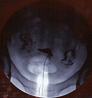
Hysterosalpingography (HSG), also known as uterosalpingography, is a radiologic procedure to investigate the shape of the uterine cavity and the shape and patency of the Fallopian tubes. It is a special x-ray procedure using dye to look at the womb (uterus) and Fallopian tubes. In this procedure a radio-opaque material is injected into the cervical canal, and radiographs are taken. A normal result shows the filling of the uterine cavity and the bilateral filling of the Fallopian tube with the injection material. To demonstrate tubal rupture, spillage of the material into the peritoneal cavity needs to be observed. Hysterosalpingography has vital role in treatment of infertility, especially in the case of fallopian tube blockage.
