Mesonephric duct

Last updated
Mesonephric duct
Gray1109.png
Urogenital sinus of female human embryo of eight and a half to nine weeks old
Details
Carnegie stage 11
Days28
Precursor intermediate mesoderm
Gives rise to vasa deferentia, seminal vesicles, epididymides, Gartner's duct
Identifiers
Latin ductus mesonephricus; ductus Wolffi
MeSH D014928
TE duct_by_E5.6.2.0.0.0.4 E5.6.2.0.0.0.4
Anatomical terminology

The mesonephric duct, also known as the Wolffian duct, archinephric duct, Leydig's duct or nephric duct, is a paired organ that develops in the early stages of embryonic development in humans and other mammals. It is an important structure that plays a critical role in the formation of male reproductive organs. The duct is named after Caspar Friedrich Wolff, a German physiologist and embryologist who first described it in 1759. [1]

Contents

During embryonic development, the mesonephric duct forms as a part of the urogenital system. [2]

Structure

The mesonephric duct connects the primitive kidney, the mesonephros , to the cloaca. It also serves as the primordium for male urogenital structures including the epididymides, vasa deferentia, and seminal vesicles.

Development

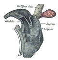
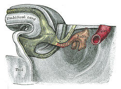
In both males and females, the mesonephric duct develops into the trigone of urinary bladder, a part of the bladder wall, but the sexes differentiate in other ways during development of the urinary and reproductive organs.

Male

In a male, it develops into a system of connected organs between the efferent ducts of the testis and the prostate, namely the epididymis, the vas deferens, and the seminal vesicle. The prostate forms from the urogenital sinus and the efferent ducts form from the mesonephric tubules.

For this it is critical that the ducts are exposed to testosterone during embryogenesis. Testosterone binds to and activates androgen receptor, affecting intracellular signals and modifying the expression of numerous genes. [3]

In the mature male, the function of this system is to store and mature sperm, and provide accessory semen fluid.

Female

In the female, with the absence of anti-Müllerian hormone secretion by the Sertoli cells and subsequent Müllerian apoptosis, the mesonephric duct regresses, although inclusions may persist. The epoophoron and Skene's glands may be present. Also, lateral to the wall of the vagina a Gartner's duct or cyst could develop as a remnant.

Function

Sexual differentiation

History

It is named after Caspar Friedrich Wolff who described the mesonephros and its ducts in his dissertation in 1759. [1]

Additional images

Diagrams to illustrate the changes in the cloaca in mammals during development. A, early embryonic stage, showing the cloaca receiving the urinary bladder, the rectum, and the Wolffian duct, as in the lower vertebrates. B, later stage, showing the beginning of the fold which divides the cloaca into a ventral urogenital sinus which receives the urinary bladder, Wolffian ducts, and ureters, and into a dorsal part which receives the rectum. C, further progress of the fold, dividing the cloaca into urogenital sinus and rectum; the ureter has separated from the Wolffian duct and is shifting anteriorly. D, completion of the fold, showing complete separation of the cloaca into ventral urogenital sinus and dorsal rectum. A laboratory manual for comparative vertebrate anatomy (1922) (20754316592).jpg
Diagrams to illustrate the changes in the cloaca in mammals during development. A, early embryonic stage, showing the cloaca receiving the urinary bladder, the rectum, and the Wolffian duct, as in the lower vertebrates. B, later stage, showing the beginning of the fold which divides the cloaca into a ventral urogenital sinus which receives the urinary bladder, Wolffian ducts, and ureters, and into a dorsal part which receives the rectum. C, further progress of the fold, dividing the cloaca into urogenital sinus and rectum; the ureter has separated from the Wolffian duct and is shifting anteriorly. D, completion of the fold, showing complete separation of the cloaca into ventral urogenital sinus and dorsal rectum.

See also

Related Research Articles

<span class="mw-page-title-main">Prostate</span> Gland of the male reproductive system in most mammals

The prostate is both an accessory gland of the male reproductive system and a muscle-driven mechanical switch between urination and ejaculation. It is found in all male mammals. It differs between species anatomically, chemically, and physiologically. Anatomically, the prostate is found below the bladder, with the urethra passing through it. It is described in gross anatomy as consisting of lobes and in microanatomy by zone. It is surrounded by an elastic, fibromuscular capsule and contains glandular tissue, as well as connective tissue.

<span class="mw-page-title-main">Seminal vesicles</span> Pair of simple tubular glands posteroinferior to the urinary bladder of male mammals

The seminal vesicles are a pair of convoluted tubular accessory glands that lie behind the urinary bladder of male mammals. They secrete fluid that partly composes the semen.

<span class="mw-page-title-main">Urogenital sinus</span> A part of the human body only present in the development of the urinary and reproductive organs

The urogenital sinus is a part of the human body only present in the development of the urinary and reproductive organs. It is the ventral part of the cloaca, formed after the cloaca separates from the anal canal during the fourth to seventh weeks of development.

<span class="mw-page-title-main">Genitourinary system</span> Organ system of the reproductive organs and the urinary system

The genitourinary system, or urogenital system, are the organs of the reproductive system and the urinary system. These are grouped together because of their proximity to each other, their common embryological origin and the use of common pathways, like the male urethra. Also, because of their proximity, the systems are sometimes imaged together.

Gartner's duct, also known as Gartner's canal or the ductus longitudinalis epoophori, is a potential embryological remnant in human female development of the mesonephric duct in the development of the urinary and reproductive organs. It was discovered and described in 1822 by Hermann Treschow Gartner.

<span class="mw-page-title-main">Paramesonephric duct</span> Paired ducts in the embryo in the primitive urogenital structures

The paramesonephric ducts are paired ducts of the embryo in the female reproductive system that run down the lateral sides of the genital ridge and terminate at the sinus tubercle in the primitive urogenital sinus. In the female, they will develop to form the fallopian tubes, uterus, cervix, and the upper one-third of the vagina.

<span class="mw-page-title-main">Persistent Müllerian duct syndrome</span> Medical condition

Persistent Müllerian duct syndrome (PMDS) is the presence of Müllerian duct derivatives in what would be considered a genetically and otherwise physically normal male animal by typical human based standards. In humans, PMDS typically is due to an autosomal recessive congenital disorder and is considered by some to be a form of pseudohermaphroditism due to the presence of Müllerian derivatives.

The development of the urinary system begins during prenatal development, and relates to the development of the urogenital system – both the organs of the urinary system and the sex organs of the reproductive system. The development continues as a part of sexual differentiation.

<span class="mw-page-title-main">Male reproductive system</span> Reproductive system of the human male

The male reproductive system consists of a number of sex organs that play a role in the process of human reproduction. These organs are located on the outside of the body, and within the pelvis.

<span class="mw-page-title-main">Human reproductive system</span> Organs involved in reproduction

The human reproductive system includes the male reproductive system which functions to produce and deposit sperm; and the female reproductive system which functions to produce egg cells, and to protect and nourish the fetus until birth. Humans have a high level of sexual differentiation. In addition to differences in nearly every reproductive organ, there are numerous differences in typical secondary sex characteristics.

<span class="mw-page-title-main">Mesonephros</span> Principal excretory organ during early human embryonic life

The mesonephros is one of three excretory organs that develop in vertebrates. It serves as the main excretory organ of aquatic vertebrates and as a temporary kidney in reptiles, birds, and mammals. The mesonephros is included in the Wolffian body after Caspar Friedrich Wolff who described it in 1759.

Kidney development, or nephrogenesis, describes the embryologic origins of the kidney, a major organ in the urinary system. This article covers a 3 part developmental process that is observed in most reptiles, birds and mammals, including humans. Nephrogenesis is often considered in the broader context of the development of the urinary and reproductive organs.

Pronephros is the most basic of the three excretory organs that develop in vertebrates, corresponding to the first stage of kidney development. It is succeeded by the mesonephros, which in fish and amphibians remains as the adult kidney. In amniotes, the mesonephros is the embryonic kidney and a more complex metanephros acts as the adult kidney. Once a more advanced kidney forms, the previous version typically degenerates by apoptosis or becomes part of the male reproductive system.

<span class="mw-page-title-main">Intermediate mesoderm</span> Layer of cells in mammalian embryos

Intermediate mesoderm or intermediate mesenchyme is a narrow section of the mesoderm located between the paraxial mesoderm and the lateral plate of the developing embryo. The intermediate mesoderm develops into vital parts of the urogenital system.

<span class="mw-page-title-main">Sex cords</span> Structures that develop from the genital ridges that further differentiate based on an embryos sex

Sex cords are embryonic structures which eventually will give rise (differentiate) to the adult gonads. They are formed from the genital ridges - which will develop into the gonads - in the first 2 months of gestation which depending on the sex of the embryo will give rise to male or female sex cords. These epithelial cells penetrate and invade the underlying mesenchyme to form the primitive sex cords. This occurs shortly before and during the arrival of the primordial germ cells (PGCs) to the paired genital ridges. If there is a Y chromosome present, testicular cords will develop via the Sry gene : repressing the female sex cord genes and activating the male. If there is no Y chromosome present the opposite will occur, developing ovarian cords. Prior to giving rise to sex cords, both XX and XY embryos have Müllerian ducts and Wolffian ducts. One of these structures will be repressed to induce the other to further differentiate into the external genitalia.

<span class="mw-page-title-main">Sexual differentiation in humans</span> Process of development of sex differences in humans

Sexual differentiation in humans is the process of development of sex differences in humans. It is defined as the development of phenotypic structures consequent to the action of hormones produced following gonadal determination. Sexual differentiation includes development of different genitalia and the internal genital tracts and body hair plays a role in sex identification.

<span class="mw-page-title-main">Cloaca (embryology)</span> Structure in the embryo

The cloaca is a structure in the development of the urinary and reproductive organs.

<span class="mw-page-title-main">Human embryonic development</span> Development and formation of the human embryo

Human embryonic development or human embryogenesis is the development and formation of the human embryo. It is characterised by the processes of cell division and cellular differentiation of the embryo that occurs during the early stages of development. In biological terms, the development of the human body entails growth from a one-celled zygote to an adult human being. Fertilization occurs when the sperm cell successfully enters and fuses with an egg cell (ovum). The genetic material of the sperm and egg then combine to form the single cell zygote and the germinal stage of development commences. Embryonic development in the human, covers the first eight weeks of development; at the beginning of the ninth week the embryo is termed a fetus. The eight weeks has 23 stages.

The development of the reproductive system is the part of embryonic growth that results in the sex organs and contributes to sexual differentiation. Due to its large overlap with development of the urinary system, the two systems are typically described together as the genitourinary system.

The epididymis, which is a tube that connects a testicle to a vas deferens in the male reproductive system, evolved by retention of the mesonephric duct during regression and replacement of the mesonephros with the metanephric kidney. Similarly, during embryological involution of the paired mesonephric kidneys, each mesonephric duct is retained to become the epididymis, vas deferens, seminal vesicle and ejaculatory duct. In reptiles and birds both the testes and excurrent ducts occur in an intra-abdominal location (testicond). Primitive mammals, such as the monotremes (prototheria), also are testicond. Marsupial (metatheria) and placental (eutheria) mammals exhibit differing degrees of testicular descent into an extra-abdominal scrotum. In scrotal mammals the epididymis is attached to the testes in an extra-abdominal position where the cauda epididymis extends beyond the lowest extremity of the testis. Hence, the cauda epididymis is exposed to the coolest of temperatures compared to all other reproductive structures.

References

  1. 1 2 synd/2845 at Who Named It?
  2. Du, Hongling; Taylor, Hugh S. (January 1, 2015). "Chapter 27 - Development of the Genital System". In Moody, Sally A. (ed.). Principles of Developmental Genetics (Second ed.). Academic Press. pp. 487–504. doi:10.1016/b978-0-12-405945-0.00027-2. ISBN   9780124059450 via ScienceDirect.
  3. Hannema SE, Print CG, Charnock-Jones DS, Coleman N, Hughes IA (2006). "Changes in gene expression during Wolffian duct development". Horm. Res. 65 (4): 200–9. doi:10.1159/000092408. PMID   16567946. S2CID   2444520.