This article needs additional citations for verification .(June 2017) |
This list of related male and female reproductive organs shows how the male and female reproductive organs and the development of the reproductive system are related, sharing a common developmental path. This makes them biological homologues. These organs differentiate into the respective sex organs in males and females.
Embryological precursor | Female | Male |
---|---|---|
3. Ureter | Ureter | Ureter |
4. Urinary bladder | Urinary bladder | Urinary bladder |
5. Urachus | Urachus | Urachus |
i. Lower part of the intestine | i. Lower part of the intestine | i. Lower part of the intestine |
cl. Cloaca | ||
cp. Elevation which becomes clitoris or penis (genital tubercle) | cc. Corpus cavernosum clitoridis | cp. Corpus cavernosum penis cut short |
ug. Sinus urogenitalis | C. Greater vestibular gland, and immediately above it the urethra | C. Bulbourethral gland of one side |
f. Abdominal opening of the left uterine tube | ||
g. Round ligament, corresponding to gubernaculum | g. Gubernaculum | |
h. Situation of the hymen | ||
m, m. Right and left Müllerian ducts uniting together and running with the Wolffian ducts in gc, the genital cord | m. Müllerian duct, the upper part of which remains as the hydatid of Morgagni; the lower part, represented by a dotted line descending to the prostatic utricle, constitutes the occasionally existing cornu and tube of the uterus masculinus | |
ot. The genital ridge from which either the ovary or testis is formed. | o. The left ovary | t. Testis in the place of its original formation; t', together with the dotted lines above, indicates the direction in which the testis and epididymis descend from the abdomen into the scrotum. |
Skene's glands a.k.a. paraurethral gland | pr. Prostate | |
u. Uterus. The uterine tube of the right side is marked m. | ||
va. Vagina | ||
vh. Ductus aberrans | ||
vs. Vesicula seminalis | ||
W. Left Wolffian body | W. Scattered remains of the Wolffian body, constituting the organ of Giraldès, or the paradidymis of Waldeyer. | |
w, w. Right and left Wolffian ducts | W. Scattered remains of Wolffian tubes near it (paroöphoron of Waldeyer); dG. Remains of the left Wolffian duct, such as give rise to the duct of Gartner, represented by dotted lines; that of the right side is marked w. | |
po. Epoophoron |
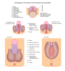
The external genitalia of both males and females have similar origins. They arise from the genital tubercle that forms anterior to the cloacal folds (proliferating mesenchymal cells around the cloacal membrane). The caudal aspect of the cloacal folds further subdivides into the posterior anal folds and the anterior urethral folds. Bilateral to the urethral fold, genital swellings become prominent. These structures are the future scrotum and labia majora in males and females, respectively.
The genital tubercles of an eight-week-old embryo of either sex are identical. They both have a glans area, which will go on to form the clitoral glans (females) or penile glans (males), a urogenital fold and groove, and an anal tubercle. At around ten weeks, the external genitalia are still similar. At the base of the glans, there is a groove known as the coronal sulcus or corona glandis. It is the site of attachment of the future prepuce. Just anterior to the anal tubercle, the caudal end of the left and right urethral folds fuse to form the urethral raphe. The lateral part of the genital tubercle (called the lateral tubercle) grows longitudinally and is about the same length in either sex.
The male external genitalia include the penis and the scrotum. The female external genitalia include the clitoris, the labia, and the vaginal opening, which are collectively called the vulva. External genitalia vary widely in external appearance among different people.
One difference between the glans penis and the glans clitoridis is that the glans clitoridis packs nerve endings into a volume only about one-tenth the size of the glans penis. Therefore, the glans clitoridis has greater variability in cutaneous corpuscular receptor density (1-14 per 100× high-powered field) compared with the glans penis (1-3 per 100× high-power field). Touch for touch, this concentration of nerves makes the glans clitoridis more sensitive than the glans penis. As a result, many women can feel discomfort or pain with anything more than a gentle touch. [3]
In amniotes, the clitoris is a female sex organ. In humans, it is the vulva's most erogenous area and generally the primary anatomical source of female sexual pleasure. The clitoris is a complex structure, and its size and sensitivity can vary. The visible portion, the glans, of the clitoris is roughly the size and shape of a pea and is estimated to have at least 8,000 sensory nerve endings.
The labia minora, also known as the inner labia, inner lips, or nymphae, are two flaps of skin that are part of the primate vulva, extending outwards from the vaginal and urethral openings to encompass the vestibule. The labia minora are situated between the labia majora and together form the labia. They vary widely in size, color and shape from individual to individual.
A sex organ, also known as a reproductive organ, is a part of an organism that is involved in sexual reproduction. Sex organs constitute the primary sex characteristics of an organism. Sex organs are responsible for producing and transporting gametes, as well as facilitating fertilization and supporting the development and birth of offspring. Sex organs are found in many species of animals and plants, with their features varying depending on the species.
In female humans and other mammals, the clitoral hood is a fold of skin that surrounds and protects the glans of the clitoris; it also covers the external clitoral shaft, develops as part of the labia minora and is homologous with the foreskin in the male reproductive system. The clitoral hood is composed of mucocutaneous tissues; these tissues are between the mucous membrane and the skin, and they may have immunological importance because they may be a point of entry of mucosal vaccines.
A frenulum or frenum is a small fold of tissue that secures the motion of a mobile organ in the body.
The glans is a vascular structure located at the tip of the penis in male mammals or a homologous genital structure of the clitoris in female mammals.
The female reproductive system is made up of the internal and external sex organs that function in the reproduction of new offspring. The human female reproductive system is immature at birth and develops to maturity at puberty to be able to produce gametes, and to carry a fetus to full term. The internal sex organs are the vagina, uterus, fallopian tubes, and ovaries. The female reproductive tract includes the vagina, uterus, and fallopian tubes and is prone to infections. The vagina allows for sexual intercourse and childbirth, and is connected to the uterus at the cervix. The uterus or womb accommodates the embryo, which develops into the fetus. The uterus also produces secretions, which help the transit of sperm to the fallopian tubes, where sperm fertilize ova produced by the ovaries. The external sex organs are also known as the genitals and these are the organs of the vulva including the labia, clitoris, and vaginal opening.
A genital tubercle, phallic tubercle, or clitorophallic structure is a body of tissue present in the development of the reproductive system of amniotes. It forms in the ventral, caudal region of mammalian embryos of both sexes, and eventually develops into a primordial phallus. In the human fetus, the genital tubercle develops around week four of gestation, and by week nine, becomes recognizably either a clitoris or penis. This should not be confused with the sinus tubercle which is a proliferation of endoderm induced by paramesonephric ducts. Even after the phallus is developed, the term genital tubercle remains, but only as the terminal end of it, which develops into either the glans penis or the glans clitoridis.
The development of the urinary system begins during prenatal development, and relates to the development of the urogenital system – both the organs of the urinary system and the sex organs of the reproductive system. The development continues as a part of sexual differentiation.
The male reproductive system consists of a number of sex organs that play a role in the process of human reproduction. These organs are located on the outside of the body, and within the pelvis.
The labia are the major externally visible portions of the vulva. In humans and other primates, there are two pairs of labia: the labia majora are large and thick folds of skin that cover the vulva's other parts while the labia minora are the inner folds of skin between the outer labia that surround and protect the urethral and vaginal openings.
The vulval vestibule is the part of the vulva between the labia minora. On the inside, the urinary meatus and the vaginal opening open to the vestibule, while the outer edge is marked by Hart's line, named after David Berry Hart.
The corpus cavernosum of clitoris is one of a pair of sponge-like regions of erectile tissue of the clitoris. It is made of a sponge-like tissue that fills with blood during erection. This is homologous to the corpus cavernosum penis. The term corpora cavernosa literally means "cave-like bodies".
The urinary meatus, also known as the external urethral orifice, is the opening of the urethra. It is the point where urine exits the urethra in both sexes and where semen exits the urethra in males. The meatus has varying degrees of sensitivity to touch.
The development of the reproductive system is the part of embryonic growth that results in the sex organs and contributes to sexual differentiation. Due to its large overlap with development of the urinary system, the two systems are typically described together as the genitourinary system.
The reproductive system of an organism, also known as the genital system, is the biological system made up of all the anatomical organs involved in sexual reproduction. Many non-living substances such as fluids, hormones, and pheromones are also important accessories to the reproductive system. Unlike most organ systems, the sexes of differentiated species often have significant differences. These differences allow for a combination of genetic material between two individuals, which allows for the possibility of greater genetic fitness of the offspring.
Diphallia, penile duplication (PD), diphallic terata, or diphallasparatus is an extremely rare developmental abnormality in which a male is born with two penises. The first reported case was by Johannes Jacob Wecker in 1609. Its occurrence is 1 in 5.5 million boys in the United States.
In mammals, the vulva consists of the external female genitalia. The human vulva includes the mons pubis, labia majora, labia minora, clitoris, vulval vestibule, urinary meatus, the vaginal opening, hymen, and Bartholin's and Skene's vestibular glands. The urinary meatus is also included as it opens into the vulval vestibule. The vulva includes the entrance to the vagina, which leads to the uterus, and provides a double layer of protection for this by the folds of the outer and inner labia. Pelvic floor muscles support the structures of the vulva. Other muscles of the urogenital triangle also give support.
The Prader scale or Prader staging, named after Andrea Prader, is a coarse rating system for the measurement of the degree of virilization of the genitalia of the human body and is similar to the Quigley scale. It primarily relates to virilization of the female genitalia in cases of congenital adrenal hyperplasia (CAH) and identifies five distinct stages, but in recent times has been used to describe the range of differentiation of genitalia, with normal infant presentation being shown on either end of the scale, female on the left (0) and male on the right (6).
The clitoris is a female sex organ present in mammals, ostriches and other amniotes.
This article incorporates text in the public domain from the 20th edition of Gray's Anatomy (1918)