Urachus

Last updated
Urachus
Gray1156.png
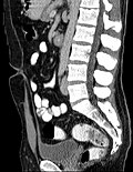
Vertical section of bladder, penis, and urethra. Urachus is seen at top
Illu quiz bladder 01.jpg
Urachus is #1
Identifiers
MeSH D014497
Anatomical terminology

The urachus forms from the distal end of the allantois in the embryo, and develops into a closed cord between the base of the bladder, and the navel. [1] It drains the bladder of the fetus that joins and runs within the umbilical cord. [2] The fibrous remnant lies in the space of Retzius, between the transverse fascia anteriorly and the peritoneum posteriorly. At birth, the urachus develops into the median umbilical ligament. [3] [4]

Contents

Development

The part of the urogenital sinus related to the bladder and urethra absorbs the ends of the Wolffian ducts and the associated ends of the renal diverticula. This gives rise to the trigone of the bladder and part of the prostatic urethra.

The remainder of this part of the urogenital sinus forms the body of the bladder and part of the prostatic urethra. The apex of the bladder stretches and is connected to the umbilicus as a narrow canal. This canal is initially open, but later closes as the urachus goes on to definitively form the median umbilical ligament.

Clinical significance

Failure of the inside of the urachus to be filled in leaves the urachus open. The telltale sign is leakage of urine through the umbilicus. This is often managed surgically. There are four anatomical causes:

The urachus is also subject to neoplasia. Urachal adenocarcinoma is histologically similar to adenocarcinoma of the bowel. Rarely, urachus carcinomas can metastasise to other regions of the body, including pelvic bones and the lung. [7]

One urachal mass has been reported that was found to be a manifestation of IgG4-related disease. [8]

Additional images

References

PD-icon.svgThis article incorporates text in the public domain from page 1213 of the 20th edition of Gray's Anatomy (1918)

  1. "Urachus". www.cancer.gov. 2 February 2011. Retrieved 21 December 2024.
  2. Larsen, "Human Embryology," 3rd ed., pg. 258
  3. Tan C, Simon MA, Dolin N, Gesner L (August 2020). "Incidental vesicourachal diverticulum in a young female". Radiology Case Reports. 15 (8): 1305–1308. doi:10.1016/j.radcr.2020.05.046. PMC   7322240 . PMID   32612730.
  4. Mrad Daly K, Ben Rhouma S, Zaghbib S, Oueslati A, Gharbi M, Nouira Y (September 2019). "Infected urachal cyst in an adult: A case report". Urology Case Reports. 26 100976. doi:10.1016/j.eucr.2019.100976. PMC   6661533 . PMID   31380223.
  5. Le, Tao; Bhushan, Vikas; Vasan, Neil (2010). First Aid for the USMLE Step 1: 2010 (20th Anniversary ed.). US: The McGraw-Hill Companies, Inc. pp.  122. ISBN   978-0-07-163340-6.
  6. Guray, Sogut, et al. (2000) Urachal Cyst. Eastern Journal of Medicine 5(2):76-78.
  7. Elser, C; Sweet, J; Cheran, S K; Haider, M A; Jewett, M; Sridhar, S S (February 2012). "A case of metastatic urachal adenocarcinoma treated with several different chemotherapeutic regimens". Can Urol Assoc J. 6 (1): E27 –E31. doi:10.5489/cuaj.11109 (inactive 12 July 2025). PMC   3289708 . PMID   22396380.{{cite journal}}: CS1 maint: DOI inactive as of July 2025 (link)
  8. Travis W. Dum; Da Zhang; Eugene K. Lee (2015). "IgG4-Related Disease in a Urachal Tumor". Case Reports in Urology. 2015: 275850. doi: 10.1155/2014/275850 . PMC   4151357 . PMID   25202466. 275850.