Related Research Articles

A Meckel's diverticulum, a true congenital diverticulum, is a slight bulge in the small intestine present at birth and a vestigial remnant of the vitelline duct. It is the most common malformation of the gastrointestinal tract and is present in approximately 2% of the population, with males more frequently experiencing symptoms.

The allantois is a hollow sac-like structure filled with clear fluid that forms part of a developing amniote's conceptus. It helps the embryo exchange gases and handle liquid waste.

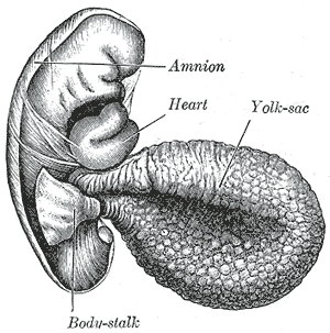
The yolk sac is a membranous sac attached to an embryo, formed by cells of the hypoblast layer of the bilaminar embryonic disc. This is alternatively called the umbilical vesicle by the Terminologia Embryologica (TE), though yolk sac is far more widely used. In humans, the yolk sac is important in early embryonic blood supply, and much of it is incorporated into the primordial gut during the fourth week of embryonic development.

In medicine or biology, a diverticulum is an outpouching of a hollow structure in the body. Depending upon which layers of the structure are involved, diverticula are described as being either true or false.
The development of the urinary system begins during prenatal development, and relates to the development of the urogenital system – both the organs of the urinary system and the sex organs of the reproductive system. The development continues as a part of sexual differentiation.

The urachus is a fibrous remnant of the allantois, a canal that drains the urinary bladder of the fetus that joins and runs within the umbilical cord. The fibrous remnant lies in the space of Retzius, between the transverse fascia anteriorly and the peritoneum posteriorly.

In human anatomy, the median umbilical ligament is an unpaired midline ligamentous structure upon the lower inner surface of the anterior abdominal wall. It is covered by the median umbilical fold.
A urachal cyst is a sinus remaining from the allantois during embryogenesis. It is a cyst which occurs in the remnants between the umbilicus and bladder. This is a type of cyst occurring in a persistent portion of the urachus, presenting as an extraperitoneal mass in the umbilical region. It is characterized by abdominal pain, and fever if infected. It may rupture, leading to peritonitis, or it may drain through the umbilicus. Urachal cysts are usually silent clinically until infection, calculi or adenocarcinoma develop.

Related to the urinary bladder, anteriorly there are the following folds:

The cloaca is a structure in the development of the urinary and reproductive organs.
The development of the reproductive system is the part of embryonic growth that results in the sex organs and contributes to sexual differentiation. Due to its large overlap with development of the urinary system, the two systems are typically described together as the urogenital or genitourinary system.

The urorectal septum is an invagination of the cloaca. It divides it into a dorsal part and a ventral part. It invaginates from cranial to caudal, formed from the endodermal cloaca, and fuses with the cloacal membrane. Malformations can cause fistulas.

The hepatic diverticulum is a primordial cellular extension of the embryonic foregut endoderm that gives rise to the parenchyma of the liver and the bile duct. It typically differentiates from the endoderm in the third or fourth week of gestation and is reabsorbed in tubular structures of the septum transversum by the eighth week.

The fetal membranes are the four extraembryonic membranes, associated with the developing embryo, and fetus in humans and other mammals. They are the amnion, chorion, allantois, and yolk sac. The amnion and the chorion are the chorioamniotic membranes that make up the amniotic sac which surrounds and protects the embryo. The fetal membranes are four of six accessory organs developed by the conceptus that are not part of the embryo itself, the other two are the placenta, and the umbilical cord.

The connecting stalk, or body stalk is an embryonic structure that is formed by the third week of development and connects the embryo to its shell of trophoblasts. The connecting stalk is derived from the extraembryonic mesoderm. Initially it lies caudally to the trilaminar germ disc, but, with subsequent embryonic folding, the body stalk assume a more ventral position. Progressive expansion of the amnion from the umbilical ring creates a tube with a covering of amniotic membrane with allantois and umbilical vessels as its content and mesoderm of the connecting stalk as the ground substance. This extraembryonic mesodermal ground substance forms the future wharton's jelly. The amniotic membrane and its contents form the umbilical cord that connects the embryo and the placenta.

Invasive urothelial carcinoma is a type of transitional cell carcinoma. It is a type of cancer that develops in the urinary system: the kidney, urinary bladder, and accessory organs. Transitional cell carcinoma is the most common type of bladder cancer and cancer of the ureter, urethra, renal pelvis, the ureters, the bladder, and parts of the urethra and urachus. It originates from tissue lining the inner surface of these hollow organs - transitional epithelium. The invading tumors can extend from the kidney collecting system to the bladder.
A urethral diverticulum is a condition where the urethra or the periurethral glands push into the connective tissue layers (fascia) that surround it.
A urachal fistula is a congenital disorder caused by the persistence of the allantois, the structure that connects an embryo's bladder to the yolk sac. Normally, the urachus closes off to become the median umbilical ligament; however, if it remains open, urine can drain from the bladder to an opening by the umbilicus.
Umbilical-urachal sinus is a congenital disorder of the urinary bladder caused by failure of obliteration of proximal or distal part of the allantois, and the presentation of this anomaly is more common in children and rarer in adults.
References
- ↑ Sadler, Thomas W. (2011-12-15). Langman's Medical Embryology. Lippincott Williams & Wilkins. ISBN 9781451113426.