Related Research Articles
A testicle or testis is the male gonad in all bilaterians, including humans. It is homologous to the female ovary. The functions of the testicles are to produce both sperm and androgens, primarily testosterone. Testosterone release is controlled by the anterior pituitary luteinizing hormone, whereas sperm production is controlled both by the anterior pituitary follicle-stimulating hormone and gonadal testosterone.
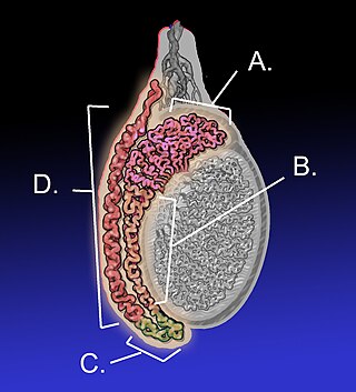
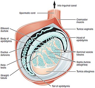
The epididymis is an elongated tubular structure attached to the posterior side of each one of the two male reproductive glands, the testicles. It is a single, narrow, tightly coiled tube in adult humans, 6 to 7 centimetres in length; uncoiled the tube would be approximately 6 m long. It connects the testicle to the vas deferens in the male reproductive system. The epididymis serves as an interconnection between the multiple efferent ducts at the rear of a testicle (proximally), and the vas deferens (distally). Its primary function is the storage, maturation and transport of sperm cells.

The mesonephric duct, also known as the Wolffian duct, archinephric duct, Leydig's duct or nephric duct, is a paired organ that develops in the early stages of embryonic development in humans and other mammals. It is an important structure that plays a critical role in the formation of male reproductive organs. The duct is named after Caspar Friedrich Wolff, a German physiologist and embryologist who first described it in 1759.

Spermatogenesis is the process by which haploid spermatozoa develop from germ cells in the seminiferous tubules of the testicle. This process starts with the mitotic division of the stem cells located close to the basement membrane of the tubules. These cells are called spermatogonial stem cells. The mitotic division of these produces two types of cells. Type A cells replenish the stem cells, and type B cells differentiate into primary spermatocytes. The primary spermatocyte divides meiotically into two secondary spermatocytes; each secondary spermatocyte divides into two equal haploid spermatids by Meiosis II. The spermatids are transformed into spermatozoa (sperm) by the process of spermiogenesis. These develop into mature spermatozoa, also known as sperm cells. Thus, the primary spermatocyte gives rise to two cells, the secondary spermatocytes, and the two secondary spermatocytes by their subdivision produce four spermatozoa and four haploid cells.

Seminiferous tubules are located within the testicles, and are the specific location of meiosis, and the subsequent creation of male gametes, namely spermatozoa.

The rete testis is an anastomosing network of delicate tubules located in the hilum of the testicle that carries sperm from the seminiferous tubules to the efferent ducts. It is the homologue of the rete ovarii in females. Its function is to provide a site for fluid reabsorption.

Virility refers to any of a wide range of masculine characteristics viewed positively. Virile means "marked by strength or force". Virility is commonly associated with vigour, health, sturdiness, and constitution, especially in the fathering of children. In this last sense, virility is to men as fertility is to women. Virile has become obsolete in referring to a "nubile" young woman, or "a maid that is Marriageable or ripe for a Husband, or Virill".

The male reproductive system consists of a number of sex organs that play a role in the process of human reproduction. These organs are located on the outside of the body, and within the pelvis.

Fish reproductive organs include testes and ovaries. In most species, gonads are paired organs of similar size, which can be partially or totally fused. There may also be a range of secondary organs that increase reproductive fitness. The genital papilla is a small, fleshy tube behind the anus in some fishes, from which the sperm or eggs are released; the sex of a fish can often be determined by the shape of its papilla.

Spermiogenesis is the final stage of spermatogenesis, during which the spermatids develop into mature spermatozoa. At the beginning of the stage, the spermatid is a more or less circular cell containing a nucleus, Golgi apparatus, centriole and mitochondria; by the end of the process, it has radically transformed into an elongated spermatozoon, complete with a head, midpiece, and tail.

The blood–testis barrier is a physical barrier between the blood vessels and the seminiferous tubules of the animal testes. The name "blood-testis barrier" is misleading as it is not a blood-organ barrier in a strict sense, but is formed between Sertoli cells of the seminiferous tubule and isolates the further developed stages of germ cells from the blood. A more correct term is the Sertoli cell barrier (SCB).

The mesonephros is one of three excretory organs that develop in vertebrates. It serves as the main excretory organ of aquatic vertebrates and as a temporary kidney in reptiles, birds, and mammals. The mesonephros is included in the Wolffian body after Caspar Friedrich Wolff who described it in 1759.
Testicular sperm extraction (TESE) is a surgical procedure in which a small portion of tissue is removed from the testicle and any viable sperm cells from that tissue are extracted for use in further procedures, most commonly intracytoplasmic sperm injection (ICSI) as part of in vitro fertilisation (IVF). TESE is often recommended to patients who cannot produce sperm by ejaculation due to azoospermia.
The term paradidymis is applied to a small collection of convoluted tubules, situated in front of the lower part of the spermatic cord, above the head of the epididymis.
The germinal epithelium is the epithelial layer of the seminiferous tubules of the testicles. It is also known as the wall of the seminiferous tubules. The cells in the epithelium are connected via tight junctions.
The development of the reproductive system is the part of embryonic growth that results in the sex organs and contributes to sexual differentiation. Due to its large overlap with development of the urinary system, the two systems are typically described together as the genitourinary system.
The development of the gonads is part of the prenatal development of the reproductive system and ultimately forms the testicles in males and the ovaries in females. The immature ova originate from cells from the dorsal endoderm of the yolk sac. Once they have reached the gonadal ridge they are called oogonia. Development proceeds and the oogonia become fully surrounded by a layer of connective tissue cells. In this way, the rudiments of the ovarian follicles are formed.

Sertoli cell-only syndrome (SCOS), also known as germ cell aplasia, is defined by azoospermia where the testicular seminiferous tubules are lined solely with sertoli cells. Sertoli cells contribute to the formation of the blood-testis barrier and aid in sperm generation. These cells respond to follicle-stimulating hormone, which is secreted by the hypothalamus and aids in spermatogenesis.
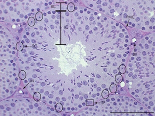
A peritubular myoid (PTM) cell is one of the smooth muscle cells which surround the seminiferous tubules in the testis. These cells are present in all mammals but their organization and abundance varies between species. The exact role of PTM cells is still somewhat uncertain and further work into this is needed. However, a number of functions of these cells have been established. They are contractile cells which contain actin filaments and are primarily involved in transport of spermatozoa through the tubules. They provide structural integrity to the tubules through their involvement in laying down the basement membrane. This has also been shown to affect Sertoli cell function and PTM cells also communicate with Sertoli cells through the secretion of growth factors and ECM components. Studies have shown PTM cells to be critical in achieving normal spermatogenesis. Overall, PTM cells have a role in both maintaining the structure of the tubules and regulating spermatogenesis through cellular interaction.
The monotremes represent the order of extant mammals most distantly related to humans. The platypus is indigenous to eastern Australia; the short-beaked echidna is indigenous to Australia and Papua New Guinea; whereas the long-beaked echidna is restricted to Papua New Guinea and Irian Jaya. Since monotremes exhibit characteristics common with both reptiles and therian mammals, they are of great interest for the study of mammalian evolution.
References
- 1 2 Fix, James D.; Dudek, Ronald W. (1998). Embryology . Baltimore: Williams & Wilkins. ISBN 0-683-30272-8.