
In primates, and specifically in humans, the labia majora, also known as the outer lips or outer labia, are two prominent longitudinal skin folds that extend downward and backward from the mons pubis to the perineum. Together with the labia minora, they form the labia of the vulva.
A sex organ, also known as a reproductive organ, is a part of an organism that is involved in sexual reproduction. Sex organs constitute the primary sex characteristics of an organism. Sex organs are responsible for producing and transporting gametes, as well as facilitating fertilization and supporting the development and birth of offspring. Sex organs are found in many species of animals and plants, with their features varying depending on the species.

The glans is a vascular structure located at the tip of the penis in male mammals or a homologous genital structure of the clitoris in female mammals.

The inguinal canal is a passage in the anterior abdominal wall on each side of the body, which in males, convey the spermatic cords and in females, the round ligament of the uterus. The inguinal canals are larger and more prominent in males.

The paramesonephric ducts are paired ducts of the embryo in the female reproductive system that run down the lateral sides of the genital ridge and terminate at the sinus tubercle in the primitive urogenital sinus. In the female, they will develop to form the fallopian tubes, uterus, cervix, and the upper one-third of the vagina.

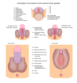
A genital tubercle, phallic tubercle, or clitorophallic structure is a body of tissue present in the development of the reproductive system of amniotes. It forms in the ventral, caudal region of mammalian embryos of both sexes, and eventually develops into a primordial phallus. In the human fetus, the genital tubercle develops around week four of gestation, and by week nine, becomes recognizably either a clitoris or penis. This should not be confused with the sinus tubercle which is a proliferation of endoderm induced by paramesonephric ducts. Even after the phallus is developed, the term genital tubercle remains, but only as the terminal end of it, which develops into either the glans penis or the glans clitoridis.
The development of the urinary system begins during prenatal development, and relates to the development of the urogenital system – both the organs of the urinary system and the sex organs of the reproductive system. The development continues as a part of sexual differentiation.

The male reproductive system consists of a number of sex organs that play a role in the process of human reproduction. These organs are located on the outside of the body, and within the pelvis.

The labia are the major externally visible portions of the vulva. In humans and other primates, there are two pairs of labia: the labia majora are large and thick folds of skin that cover the vulva's other parts while the labia minora are the inner folds of skin between the outer labia that surround and protect the urethral and vaginal openings.

XX male syndrome, also known as de la Chapelle syndrome, is a rare congenital intersex condition in which an individual with a 46,XX karyotype develops a male phenotype. Synonyms include 46,XX testicular difference of sex development, 46,XX sex reversal, nonsyndromic 46,XX testicular DSD, and XX sex reversal.

The mesovarium is the portion of the broad ligament of the uterus that suspends the ovaries. The ovary is not covered by the mesovarium; rather, it is covered by germinal epithelium.

Sexual differentiation in humans is the process of development of sex differences in humans. It is defined as the development of phenotypic structures consequent to the action of hormones produced following gonadal determination. Sexual differentiation includes development of different genitalia and the internal genital tracts and body hair plays a role in sex identification.
The development of the reproductive system is the part of embryonic growth that results in the sex organs and contributes to sexual differentiation. Due to its large overlap with development of the urinary system, the two systems are typically described together as the genitourinary system.
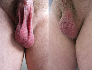
In most terrestrial mammals, the scrotum or scrotal sac is a part of the external male genitalia located at the base of the penis. It consists of a sac of skin containing the external spermatic fascia, testicles, epididymides, and vasa deferentia. The scrotum will usually tighten during penile erection and when exposed to cold temperatures.
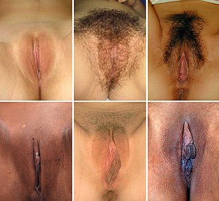
In mammals, the vulva consists of the external female genitalia. The human vulva includes the mons pubis, labia majora, labia minora, clitoris, vulval vestibule, urinary meatus, vaginal opening, hymen, and Bartholin's and Skene's vestibular glands. The vulva includes the entrance to the vagina, which leads to the uterus, and provides a double layer of protection for this by the folds of the outer and inner labia. Pelvic floor muscles support the structures of the vulva. Other muscles of the urogenital triangle also give support.
An intromittent organ is any external organ of a male organism that is specialized to deliver sperm during copulation. Intromittent organs are found most often in terrestrial species, as most non-mammalian aquatic species fertilize their eggs externally, although there are exceptions. For many species in the animal kingdom, the male intromittent organ is a hallmark characteristic of internal fertilization.

The study of the genitalia of Lepidoptera is important for Lepidoptera taxonomy in addition to development, anatomy and natural history. The genitalia are complex and provide the basis for species discrimination in most families and also in family identification. The genitalia are attached onto the tenth or most distal segment of the abdomen. Lepidoptera have some of the most complex genital structures in the insect groups with a wide variety of complex spines, setae, scales and tufts in males, claspers of different shapes and different modifications of the ductus bursae in females.
Male genital examination is a physical examination of the genital in males to detect ailments and to assess sexual development, and is normally a component of an annual physical examination. The examination includes checking the penis, scrotum, and urethral meatus. A comprehensive assessment of the male genitals assesses the pubic hair based on Sexual Maturity Rating and the size of the testicles and penis. The exam can also be conducted to verify a person's age and biological sex. The genitourinary system can also be assessed as part of the male genital examination. During a genital examination, the doctor can detect any of the following: structural abnormalities, urethral opening abnormalities, problems related to not being circumcised, lumps, tumors, redness, excoriation, edema, lesions, swelling, cancer, hair-related issues, and many others. In some instances where a physical examination of the male genitals is not sufficient to diagnose an individual, then an internal genital examination using imaging or ultrasounds will be needed for further evaluation.
Sexual anomalies, also known as sexual abnormalities, are a set of clinical conditions due to chromosomal, gonadal and/or genitalia variation. Individuals with congenital (inborn) discrepancy between sex chromosome, gonadal, and their internal and external genitalia are categorised as individuals with a disorder of sex development (DSD). Afterwards, if the family or individual wishes, they can partake in different management and treatment options for their conditions.