Intermediate mesoderm

Last updated
Intermediate mesoderm
Intermediate mesoderm.png
Intermediate mesoderm location during early gastrulation and outline of organ derivatives in relation to mammals.
* neural tube: grey.
* Paraxial mesoderm: pink, at.
* Intermediate mesoderm: green.
* Lateral plate mesoderm: purple.
* notochord: blue.
* yolk sac: yellow.
Details
Gives rise to kidneys, gonads
Identifiers
Latin mesenchyma intermedium
TE mesoderm_by_E5.6.0.0.0.0.2 E5.6.0.0.0.0.2
Anatomical terminology

Intermediate mesoderm or intermediate mesenchyme is a narrow section of the mesoderm (one of the three primary germ layers) located between the paraxial mesoderm and the lateral plate of the developing embryo. [1] The intermediate mesoderm develops into vital parts of the urogenital system (kidneys, gonads and respective tracts).

Contents

Early formation

Factors regulating the formation of the intermediate mesoderm are not fully understood. It is believed that bone morphogenic proteins, or BMPs, specify regions of growth along the dorsal-ventral axis of the mesoderm and plays a central role in formation of the intermediate mesoderm. [2] Vg1/Nodal signalling is an identified regulator of intermediate mesoderm formation acting through BMP signalling. [3] Excess Vg1/Nodal signalling during early gastrulation stages results in expansion of the intermediate mesoderm at the expense of the adjacent paraxial mesoderm, whereas inhibition of Vg1/Nodal signalling represses intermediate mesoderm formation. [4] A link has been established between Vg1/Nodal signalling and BMP signalling, whereby Vg1/Nodal signalling regulates intermediate mesoderm formation by modulating the growth-inducing effects of BMP signalling. [4]

Other necessary markers of intermediate mesoderm induction include the odd-skipped related gene ( Osr1 ) and paired-box-2 gene ( Pax2 ) which require intermediate levels of BMP signalling to activate [3] Markers of early intermediate mesoderm formation are often not exclusive to the intermediate mesoderm. This can be seen in early stages of intermediate mesoderm differentiation where higher levels of BMP stimulate growth of lateral plate tissue, whilst lower concentrations lead to paraxial mesoderm and somite formation. [5] Osr1, which encodes a zinc-finger DNA-binding protein, and LIM-type homeobox gene ( Lhx1 ) expression overlaps the intermediate mesoderm as well as the lateral plate. Osr1 has expression domains encompassing the entire length of the anterior-posterior (AP) axis from the first somites. It is not until the 4th-8th somite stage that markers with greater specificity to the intermediate mesoderm are identified including Pax2/8 genes activated from the 6th somite (Bouchard, 2002). Lhx1 expression also becomes more restricted to the intermediate mesoderm. [1] Genetic analyses in animal studies show that Lhx1, Osr1 and Pax2/8 signalling are all critical in specification of the intermediate mesoderm into its early derivatives. [5]

Derived organs and tissues

As development proceeds, the intermediate mesoderm differentiates sequentially along the anterior-posterior axis into three successive stages of the early mammalian and avian urogenital system, named pronephros, mesonephros and metanephros respectively (anamniote embryos form only a pronephros and mesonephros). [2] The intermediate mesoderm will eventually develop into the kidney and parts of both male and female reproductive systems.

Kidneys

Early kidney structures include the pronephros and mesonephros, whose complexity, size and duration can vary greatly between vertebrate species. [1] The adult kidney, also referred to as the metanephric kidney, forms at the posterior end of the intermediate mesoderm after the degeneration of previous, less complex kidney structures. [1]

Pronephros

During early development (approximately day 22 in humans), the pronephric duct forms from the intermediate mesoderm, ventral to the anterior somites. The cells of the pronephric duct migrate caudally whilst inducing adjacent mesenchyme to form the tubules of the initial kidney-like structure called the pronephros. [6] This process is regulated by Pax2/8 markers. [7] The pronephros is active in adult forms of some primitive fish and acts as the primary excretory system in amphibious larvae and embryonic forms of more advanced fish. [8] In mammals however, the pronephric tubules and the anterior portion of the pronephric duct degenerates in 3.5 weeks to be succeeded by the mesonephros, the embryonic kidney. [6]

Mesonephros

The mesonephros is constituted of a set of new tubules formed from the lateral and ventral sides of the gonadal ridge joining the cloaca. [5] The mesonephros functions between the 6th and 10th weeks of embryological life of mammals as a temporary kidney, but serves as the permanent excretory organ of aquatic vertebrates. By 8 weeks post-conception, the human mesonephros reaches maximum size and begins to regress, with complete regression occurring by week 16. [6] Despite its transiency, the mesonephros is crucial for the development of structures such as the Wolffian duct (or mesonephric duct), which in turn gives rise to the ureteric bud of the metanephric kidney. [9]

Metanephros

The permanent kidney of amniotes, the metanephros, develops during the 10th week in human embryos and is formed by the reciprocal interactions of the metanephrogenic blastema (or metaneophrogenic mesenchyme) and the ureteric bud. [6] Gonadal derived neurotrophic factor (GDNF) secreted by the metanephrogenic blastema activates the receptor tyrosine kinase RET, via the co-receptor GFRα1 and triggers outgrowth of Ret positive cells from the nephric duct towards the GDNF signal, promoting ureteric bud outgrowth and invasion. [1] Once the bud invades the metanephrogenic blastema, a permissive signal in the form of Wnt proteins is activated and stimulates the condensation of metanephric mesenchymal cells around the ureteric bud tips, beginning the polarisation of the blastema to generate the epithelial cells of parts of the nephron: the proximal tubules, loops of Henle and the distal convoluted tubules. [1] The ureteric bud secretes FGF2 (fibroblast growth factor 2) and BMP7 (bone morphogenic protein 7) to prevent apoptosis in the kidney mesenchyme. [2] Condensing mesenchyme then secretes paracrine factors that mediate branching of the ureteric bud to give rise to the ureter and collecting duct of the adult kidney. [10]

Wilm's tumor

Wilms' tumor (WT), also known as nephroblastoma, is an embryonic tumor originating from metanephric blastemal cells that are incapable of completing the mesenchymal-epithelial transition (MET), a crucial process during kidney differentiation involving the transition from a multipolar, spindle-shaped mesenchymal cell to a planar assembly of polarized epithelial cells. [11] As a consequence, WTs have a triphasic histology composed of three morphogenically distinct cell types: undifferentiated blastemal cells, epithelial cells, and stromal cells. [11] The Wnt/βcatenin signalling pathway is crucial for initiating MET, where specifically the WNT4 protein is required for induction of epithelial renal vesicles and the transition from mesenchymal to epithelial cells. [12] WTs are often a result of a genetic deletions or inactivating mutations in WT1 (Wilms tumor 1), which subsequently inhibits Wnt/βcatenin signalling and prevents MET progression. [11] [12]

Persistent Müllerian duct syndrome

Persistent Müllerian duct syndrome (PMDS) is a congenital disorder of male sexual development and is a form of pseudohermaphroditism. Males with PMDS retain normal male reproductive organs and external genitalia, but also possess internal female reproductive organs such as the uterus and fallopian tubes. [13] PMDS is primarily caused by a mutation in the anti-Müllerian hormone (AMH) gene (PMDS Type 1) or AMHR2 gene (PMDS Type 2). In PMDS Type 1, AMH is either not produced, produced in deficient quantities, defective, or secreted at the wrong critical time for male differentiation. PMDS Type 2 is a result of AMH receptor insensitivity to AMH molecules. [14] In a smaller percentage of cases, the cause of PMDS is not fully understood but is related to complex malformations of the urogenital region and paramesonephric ducts during male gonadal development. [13]

Related Research Articles

<span class="mw-page-title-main">Mesoderm</span> Middle germ layer of embryonic development

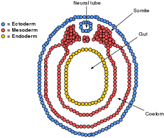
The mesoderm is the middle layer of the three germ layers that develops during gastrulation in the very early development of the embryo of most animals. The outer layer is the ectoderm, and the inner layer is the endoderm.

<span class="mw-page-title-main">Genitourinary system</span> Organ system of the reproductive organs and the urinary system

The genitourinary system, or urogenital system, are the organs of the reproductive system and the urinary system. These are grouped together because of their proximity to each other, their common embryological origin and the use of common pathways, like the male urethra. Also, because of their proximity, the systems are sometimes imaged together.

<span class="mw-page-title-main">Somitogenesis</span>

Somitogenesis is the process by which somites form. Somites are bilaterally paired blocks of paraxial mesoderm that form along the anterior-posterior axis of the developing embryo in segmented animals. In vertebrates, somites give rise to skeletal muscle, cartilage, tendons, endothelium, and dermis.

The development of the urinary system begins during prenatal development, and relates to the development of the urogenital system – both the organs of the urinary system and the sex organs of the reproductive system. The development continues as a part of sexual differentiation.

<span class="mw-page-title-main">Mesonephros</span> Principal excretory organ during early human embryonic life

The mesonephros is one of three excretory organs that develop in vertebrates. It serves as the main excretory organ of aquatic vertebrates and as a temporary kidney in reptiles, birds, and mammals. The mesonephros is included in the Wolffian body after Caspar Friedrich Wolff who described it in 1759.

<span class="mw-page-title-main">Primitive streak</span> Structure in early amniote embryogenesis

The primitive streak is a structure that forms in the early embryo in amniotes. In amphibians, the equivalent structure is the blastopore. During early embryonic development, the embryonic disc becomes oval shaped, and then pear-shaped with the broad end towards the anterior, and the narrower region projected to the posterior. The primitive streak forms a longitudinal midline structure in the narrower posterior (caudal) region of the developing embryo on its dorsal side. At first formation, the primitive streak extends for half the length of the embryo. In the human embryo, this appears by stage 6, about 17 days.

Kidney development, or nephrogenesis, describes the embryologic origins of the kidney, a major organ in the urinary system. This article covers a 3 part developmental process that is observed in most reptiles, birds and mammals, including humans. Nephrogenesis is often considered in the broader context of the development of the urinary and reproductive organs.

Pronephros is the most basic of the three excretory organs that develop in vertebrates, corresponding to the first stage of kidney development. It is succeeded by the mesonephros, which in fish and amphibians remains as the adult kidney. In amniotes, the mesonephros is the embryonic kidney and a more complex metanephros acts as the adult kidney. Once a more advanced kidney forms, the previous version typically degenerates by apoptosis or becomes part of the male reproductive system.

The metanephrogenic blastema or metanephric blastema is one of the two embryological structures that give rise to the kidney, the other being the ureteric bud.

<span class="mw-page-title-main">Lateral plate mesoderm</span>

The lateral plate mesoderm is the mesoderm that is found at the periphery of the embryo. It is to the side of the paraxial mesoderm, and further to the axial mesoderm. The lateral plate mesoderm is separated from the paraxial mesoderm by a narrow region of intermediate mesoderm. The mesoderm is the middle layer of the three germ layers, between the outer ectoderm and inner endoderm.

<span class="mw-page-title-main">Paraxial mesoderm</span>

Paraxial mesoderm, also known as presomitic or somitic mesoderm, is the area of mesoderm in the neurulating embryo that flanks and forms simultaneously with the neural tube. The cells of this region give rise to somites, blocks of tissue running along both sides of the neural tube, which form muscle and the tissues of the back, including connective tissue and the dermis.

<span class="mw-page-title-main">Bone morphogenetic protein 4</span> Human protein and coding gene

Bone morphogenetic protein 4 is a protein that in humans is encoded by BMP4 gene. BMP4 is found on chromosome 14q22-q23.

<span class="mw-page-title-main">Mesenchyme</span> Type of animal embryonic connective tissue

Mesenchyme is a type of loosely organized animal embryonic connective tissue of undifferentiated cells that give rise to most tissues, such as skin, blood or bone. The interactions between mesenchyme and epithelium help to form nearly every organ in the developing embryo.

The limb bud is a structure formed early in vertebrate limb development. As a result of interactions between the ectoderm and underlying mesoderm, formation occurs roughly around the fourth week of development. In the development of the human embryo the upper limb bud appears in the third week and the lower limb bud appears four days later.

Gremlin is an inhibitor in the TGF beta signaling pathway. It primarily inhibits bone morphogenesis and is implicated in disorders of increased bone formation and several cancers.

<span class="mw-page-title-main">Human embryonic development</span> Development and formation of the human embryo

Human embryonic development, or human embryogenesis, is the development and formation of the human embryo. It is characterised by the processes of cell division and cellular differentiation of the embryo that occurs during the early stages of development. In biological terms, the development of the human body entails growth from a one-celled zygote to an adult human being. Fertilization occurs when the sperm cell successfully enters and fuses with an egg cell (ovum). The genetic material of the sperm and egg then combine to form the single cell zygote and the germinal stage of development commences. Embryonic development in the human, covers the first eight weeks of development; at the beginning of the ninth week the embryo is termed a fetus. The eight weeks has 23 stages.

<span class="mw-page-title-main">OSR1</span> Protein-coding gene in the species Homo sapiens

Protein odd-skipped-related 1 is a transcription factor that in humans is encoded by the OSR1 gene. The OSR1 and OSR2 transcription factors participate in the normal development of body parts such as the kidney.

A mesenchymal–epithelial transition (MET) is a reversible biological process that involves the transition from motile, multipolar or spindle-shaped mesenchymal cells to planar arrays of polarized cells called epithelia. MET is the reverse process of epithelial–mesenchymal transition (EMT) and it has been shown to occur in normal development, induced pluripotent stem cell reprogramming, cancer metastasis and wound healing.

Lim-1 is a homeobox transcription factor. This transcription factor is found in adults in the cerebellum, kidneys, and cerebrum, but plays a larger role in development of the fetal head and the female reproductive tract during gestation. During development it is found in the anterior visceral endoderm, is in tissues formed by the primitive streak, and is required in both tissues for head formation. Lim1 is a member of the LIM homeobox gene and encodes a 406 amino acid protein.

The kidneys are a pair of organs of the excretory system in vertebrates, which maintains the balance of water and electrolytes in the body (osmoregulation), filters the blood, removes metabolic waste products, and in many vertebrates also produces hormones and maintains blood pressure. In healthy vertebrates, the kidneys maintain homeostasis of extracellular fluid in the body. When the blood is being filtered, the kidneys form urine, which consists of water and excess or unnecessary substances, the urine is then excreted from the body through other organs, which in vertebrates, depending on the species, may include the ureter, urinary bladder, cloaca, and urethra.

References

  1. 1 2 3 4 5 6 Dressler, GR (Dec 2009). "Advances in early kidney specification, development and patterning". Development. 136 (23): 3863–3874. doi:10.1242/dev.034876. PMC   2778737 . PMID   19906853.
  2. 1 2 3 Barak, Hila; Rosenfelder, Lea; Scultheiss, Thomas.M; Reshef, Ram (9 March 2005). "Cell fate specification along the anterior–posterior axis of the intermediate mesoderm". Developmental Dynamics. 232 (4): 901–904. doi: 10.1002/dvdy.20263 . PMID   15759277. S2CID   35055809.
  3. 1 2 Katsu, K; Tatsumi, N; Niki, D; Yamamura, K; Yokouchi, Y (1 Feb 2013). "Multi-modal effects of BMP signaling on Nodal expression in the lateral plate mesoderm during left-right axis formation in the chick embryo". Developmental Biology. 374 (1): 71–84. doi: 10.1016/j.ydbio.2012.11.027 . PMID   23206893.
  4. 1 2 Fleming, BM; Yelin, R; James, RG; Scultheiss, TM (April 2013). "A role for Vg1/Nodal signaling in specification of the intermediate mesoderm". Development. 140 (8): 1819–1829. doi:10.1242/dev.093740. PMC   3621495 . PMID   23533180.
  5. 1 2 3 Patel, SR; Dressler, GR (July 2013). "The genetics and epigenetics of kidney development". Seminars in Nephrology. 33 (4): 314–326. doi:10.1016/j.semnephrol.2013.05.004. PMC   3767926 . PMID   24011574.
  6. 1 2 3 4 Gilbert, Scott F (2000). Developmental Biology (6 ed.). Sunderland (MA): Sinauer Associates. ISBN   0-87893-243-7.
  7. Bouchard, M; Souabni, A; Neubuser, A; Busslinger, M (15 Nov 2002). "Nephric lineage specification by Pax2 and Pax8". Genes & Development. 16 (22): 2958–2970. doi:10.1101/gad.240102. PMC   187478 . PMID   12435636.
  8. Wingert, RA; Davidson, AJ (2 May 2008). "The zebrafish pronephros: A model to study nephron segmentation". Kidney International. 73 (10): 1120–1127. doi: 10.1038/ki.2008.37 . PMID   18322540.
  9. Hannema, SE; Hughes, IA (2007). "Regulation of Wolffian duct development". Hormone Research. 67 (3): 142–151. doi: 10.1159/000096644 . PMID   17077643.
  10. Moritz, KM; Wintour, EM (Feb 1999). "Functional development of the meso- and metanephros". Pediatric Nephrology (Berlin, Germany). 13 (2): 171–178. doi:10.1007/s004670050587. PMID   10229008. S2CID   10356536.
  11. 1 2 3 Carraro, DM; Ramalho, RF; Maschietto, M (23 March 2016). Wilms Tumor. Utrecht, The Netherlands: Codon Publications. pp. 149–162. ISBN   978-0-9944381-1-9.
  12. 1 2 Hastie, ND (15 Aug 2017). "Wilms' tumour 1 (WT1) in development, homeostasis and disease". Development. 144 (16): 2862–2872. doi: 10.1242/dev.153163 . hdl: 20.500.11820/264c369d-27a6-4a25-9837-b4a9d2c861b8 . PMID   28811308.
  13. 1 2 Josso, N; Belville, C; Clemente, N; Picard, JY (5 May 2005). "AMH and AMH receptor defects in persistent Müllerian duct syndrome". Human Reproduction Update. 11 (4): 351–356. doi: 10.1093/humupd/dmi014 . PMID   15878900.
  14. Rey, R; Josso, N; Racine, C (12 June 2016). Sexual Differentiation. South Dartmouth (MA): MDText.com, Inc.