Related Research Articles

The collecting duct system of the kidney consists of a series of tubules and ducts that physically connect nephrons to a minor calyx or directly to the renal pelvis. The collecting duct system participates in electrolyte and fluid balance through reabsorption and excretion, processes regulated by the hormones aldosterone and vasopressin.

A hematoma, also spelled haematoma, is a localized bleeding outside of blood vessels, due to either disease or trauma including injury or surgery and may involve blood continuing to seep from broken capillaries. A hematoma is benign and is initially in liquid form spread among the tissues including in sacs between tissues where it may coagulate and solidify before blood is reabsorbed into blood vessels. An ecchymosis is a hematoma of the skin larger than 10mm.
A tubule is:
Kidney development, or nephrogenesis, describes the embryologic origins of the kidney, a major organ in the urinary system. This article covers a 3 part developmental process that is observed in most reptiles, birds and mammals, including humans. Nephrogenesis is often considered in the broader context of the development of the urinary and reproductive organs.

Intermediate mesoderm or intermediate mesenchyme is a narrow section of the mesoderm located between the paraxial mesoderm and the lateral plate of the developing embryo. The intermediate mesoderm develops into vital parts of the urogenital system, as well as the reproductive system.
The metanephrogenic blastema or metanephric blastema is one of the two embryological structures that give rise to the kidney, the other being the ureteric bud.
A supernumerary kidney is an additional kidney to the number usually present in an organism. This often develops as the result of splitting of the nephrogenic blastema, or from separate metanephric blastemas into which partially or completely reduplicated ureteral stalks enter to form separate capsulated kidneys; in some cases the separation of the reduplicated organ is incomplete.
Gremlin is an inhibitor in the TGF beta signaling pathway. It primarily inhibits bone morphogenesis and is implicated in disorders of increased bone formation and several cancers.

Medullary sponge kidney is a congenital disorder of the kidneys characterized by cystic dilatation of the collecting tubules in one or both kidneys. Individuals with medullary sponge kidney are at increased risk for kidney stones and urinary tract infection (UTI). Patients with MSK typically pass twice as many stones per year as do other stone formers without MSK. While having a low morbidity rate, as many as 10% of patients with MSK have an increased risk of morbidity associated with frequent stones and UTIs. While many patients report increased chronic kidney pain, the source of the pain, when a UTI or blockage is not present, is unclear at this time. Renal colic is present in 55% of patients. Women with MSK experience more stones, UTIs, and complications than men. MSK was previously believed not to be hereditary but there is more evidence coming forth that may indicate otherwise.
A pelvic kidney is a normal kidney located in the pelvis, instead of the abdomen. This occurs when a kidney does not ascend from its original location in the pelvis to its final location during fetal development. Typically, the kidney functions normally despite being in the wrong location. Often a person with a pelvic kidney will go through their whole life not even knowing they have this condition, unless it is discovered on newborn kidney ultrasound screening or if complications arise later in life for this or a completely different reason, and during investigations the condition is diagnosed. Pelvic kidneys occur in 1 in every 500 people in the U.S. It is not a harmful condition generally, but can develop complications.

Multicystic dysplastic kidney (MCDK) is a condition that results from the malformation of the kidney during fetal development. The kidney consists of irregular cysts of varying sizes. Multicystic dysplastic kidney is a common type of renal cystic disease, and it is a cause of an abdominal mass in infants.

Homeobox protein SIX1 is a protein that in humans is encoded by the SIX1 gene.

Protein odd-skipped-related 1 is a transcription factor that in humans is encoded by the OSR1 gene. The OSR1 and OSR2 transcription factors participate in the normal development of body parts such as the kidney.
A ureteral neoplasm is a type of tumor that can be primary, or associated with a metastasis from another site.
Juxtaglomerular cell tumor is an extremely rare kidney tumour of the juxtaglomerular cells, with less than 100 cases reported in literature. This tumor typically secretes renin, hence the former name of reninoma. It often causes severe hypertension that is difficult to control, in adults and children, although among causes of secondary hypertension it is rare. It develops most commonly in young adults, but can be diagnosed much later in life. It is generally considered benign, but its malignant potential is uncertain.

Kidney tumours are tumours, or growths, on or in the kidney. These growths can be benign or malignant.

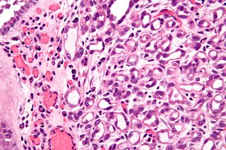
Metanephric adenoma (MA)is a rare, benign tumour of the kidney, that can have a microscopic appearance similar to a nephroblastoma, or a papillary renal cell carcinoma.
Nephrogenic adenoma is a benign growth typically found in the urinary bladder.
Metanephric dysplastic hematoma of the sacral region (MDHSR) has been described by Cozzutto and Lazzaroni-Fossati in 1980, by Posalaki et al. in 1981 and by Cozzutto et al. in 1982. Three additional cases were seen by Finegold.
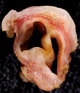
A Gartner's duct cyst is a benign vaginal cyst that originates from the Gartner's duct, which is a vestigial remnant of the mesonephric duct in females. They are typically small asymptomatic cysts that occur along the lateral walls of the vagina, following the course of the duct. They can present in adolescence with painful menstruation (dysmenorrhea) or difficulty inserting a tampon. They can also enlarge to substantial proportions and be mistaken for urethral diverticulum or cystocele. In some rare instances, they can be congenital.
References
- ↑ Mitchell, Barry; Sharma, Ram (2009). Embriology, 2nd edition. Churchill Livingstone Elsevier.
- ↑ genital-017 —Embryo Images at University of North Carolina
- ↑ Diagram at Nature.com