Renal pelvis

Last updated
Renal pelvis
Illu kidney2.jpg
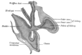
Cross-section of the kidney, with major structures labelled. The renal pelvis, located in the middle of the image, collects urine from the urinary calices.
Renal pelves and calices.png
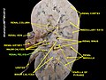
An image showing just the pelvis and calices of the kidneys, with the rest of the kidney removed, from a dissected cow and seal specimen. These vary greatly in size and number depending on species.[ citation needed ]
Details
Precursor Ureteric bud
System Urinary system
Identifiers
Latin pelvis renalis
MeSH D007682
TA98 A08.1.05.001
TA2 3384
FMA 15575
Anatomical terminology

The renal pelvis or pelvis of the kidney is the funnel-like dilated part of the ureter in the kidney. It is formed by the convergence of the major calyces, acting as a funnel for urine flowing from the major calyces to the ureter. It has a mucous membrane and is covered with transitional epithelium and an underlying lamina propria of loose-to-dense connective tissue.

Contents

The renal pelvis is situated within the renal sinus alongside the other structures of the renal sinus. [1]

Clinical significance

The renal pelvis is the location of several kinds of kidney cancer and is affected by infection in pyelonephritis.[ citation needed ] A large "staghorn" kidney stone may block all or part of the renal pelvis.

The size of the renal pelvis plays a major role in the grading of hydronephrosis. Normally, the anteroposterior diameter of the renal pelvis is less than 4 mm in fetuses up to 32 weeks of gestational age and 7 mm afterwards. [2] In adults, 13% of the normal population have a transverse pelvic diameter of over 10 mm. [3]

Etymology and pronunciation

Like the bony pelvis, the renal pelvis ( /ˈrnəlˈpɛlvɪs/ ) gets its English name via Neo-Latin from the older Latin word pelvis , "basin", as in "wash basin". [4] In both cases the name reflects the shape of the structure, and in the case of the renal pelvis, it also reflects the function. The name reflects that each renal pelvis collects urine from the calyces and funnels it into the ureter like a wash basin collects water and funnels it into a drain pipe. The renal pelvis is occasionally called the pyelum (from Greek πύελος pýelos, "trough", ‘anything hollow’), and the combining form pyelo- denotes the renal pelvis (pyelo- is not to be confused with pyo- ). The words infundibulum and choana are other words for funnel-shaped cavities (which medical English got from the Latin and Greek words for "funnel", respectively), and the renal pelvis is sometimes called the renal infundibulum. The form *renal choana is logical but is not used.

Additional images

See also

Related Research Articles

An infundibulum is a funnel-shaped cavity or organ.

<span class="mw-page-title-main">Kidney</span> Organ that filters blood and produces urine

In humans, the kidneys are two reddish-brown bean-shaped blood-filtering organs that are a multilobar, multipapillary form of mammalian kidneys, usually without signs of external lobulation. They are located on the left and right in the retroperitoneal space, and in adult humans are about 12 centimetres in length. They receive blood from the paired renal arteries; blood exits into the paired renal veins. Each kidney is attached to a ureter, a tube that carries excreted urine to the bladder.

<span class="mw-page-title-main">Bladder</span> Organ in humans and vertebrates that collects and stores urine from the kidneys before disposal

The bladder is a hollow organ in humans and other vertebrates that stores urine from the kidneys before disposal by urination. In humans, the bladder is a distensible organ that sits on the pelvic floor. Urine enters the bladder via the ureters and exits via the urethra. The typical adult human bladder will hold between 300 and 500 ml before the urge to empty occurs, but can hold considerably more.

<span class="mw-page-title-main">Urinary system</span> Human anatomical system consisting of the kidneys, ureters, urinary bladder, and the urethra

The urinary system, also known as the urinary tract or renal system, consists of the kidneys, ureters, bladder, and the urethra. The purpose of the urinary system is to eliminate waste from the body, regulate blood volume and blood pressure, control levels of electrolytes and metabolites, and regulate blood pH. The urinary tract is the body's drainage system for the eventual removal of urine. The kidneys have an extensive blood supply via the renal arteries which leave the kidneys via the renal vein. Each kidney consists of functional units called nephrons. Following filtration of blood and further processing, wastes exit the kidney via the ureters, tubes made of smooth muscle fibres that propel urine towards the urinary bladder, where it is stored and subsequently expelled from the body by urination. The female and male urinary system are very similar, differing only in the length of the urethra.

The excretory system is a passive biological system that removes excess, unnecessary materials from the body fluids of an organism, so as to help maintain internal chemical homeostasis and prevent damage to the body. The dual function of excretory systems is the elimination of the waste products of metabolism and to drain the body of used up and broken down components in a liquid and gaseous state. In humans and other amniotes, most of these substances leave the body as urine and to some degree exhalation, mammals also expel them through sweating.

<span class="mw-page-title-main">Ureter</span> Tubes used in the urinary system in most animals

The ureters are tubes made of smooth muscle that propel urine from the kidneys to the urinary bladder. In a human adult, the ureters are usually 20–30 cm (8–12 in) long and around 3–4 mm (0.12–0.16 in) in diameter. The ureter is lined by urothelial cells, a type of transitional epithelium, and has an additional smooth muscle layer that assists with peristalsis in its lowest third.

<span class="mw-page-title-main">Renal calyx</span> Anatomical structure in the kidneys

The renal calyces are conduits in the kidney through which urine passes. The minor calyces form a cup-shaped drain around the apex of the renal pyramids. Urine formed in the kidney passes through a renal papilla at the apex into the minor calyx; 4-5 minor calyces converge to form a major calyx through which urine passes into the renal pelvis.

<span class="mw-page-title-main">Renal medulla</span> Innermost part of the kidney

The renal medulla is the innermost part of the kidney. The renal medulla is split up into a number of sections, known as the renal pyramids. Blood enters into the kidney via the renal artery, which then splits up to form the segmental arteries which then branch to form interlobar arteries. The interlobar arteries each in turn branch into arcuate arteries, which in turn branch to form interlobular arteries, and these finally reach the glomeruli. At the glomerulus the blood reaches a highly disfavourable pressure gradient and a large exchange surface area, which forces the serum portion of the blood out of the vessel and into the renal tubules. Flow continues through the renal tubules, including the proximal tubule, the loop of Henle, through the distal tubule and finally leaves the kidney by means of the collecting duct, leading to the renal pelvis, the dilated portion of the ureter.

<span class="mw-page-title-main">Pyelonephritis</span> Medical condition

Pyelonephritis is inflammation of the kidney, typically due to a bacterial infection. Symptoms most often include fever and flank tenderness. Other symptoms may include nausea, burning with urination, and frequent urination. Complications may include pus around the kidney, sepsis, or kidney failure.

<span class="mw-page-title-main">Renal artery</span> Vessel supplying blood to kidney

The renal arteries are paired arteries that supply the kidneys with blood. Each is directed across the crus of the diaphragm, so as to form nearly a right angle.

The development of the urinary system begins during prenatal development, and relates to the development of the urogenital system – both the organs of the urinary system and the sex organs of the reproductive system. The development continues as a part of sexual differentiation.

<span class="mw-page-title-main">Hydronephrosis</span> Medical condition

Hydronephrosis describes hydrostatic dilation of the renal pelvis and calyces as a result of obstruction to urine flow downstream. Alternatively, hydroureter describes the dilation of the ureter, and hydronephroureter describes the dilation of the entire upper urinary tract.

Horseshoe kidney, also known as ren arcuatus, renal fusion or super kidney, is a congenital disorder affecting about 1 in 500 people that is more common in men, often asymptomatic, and usually diagnosed incidentally. In this disorder, the patient's kidneys fuse to form a horseshoe-shape during development in the womb. The fused part is the isthmus of the horseshoe kidney. The abnormal anatomy can affect kidney drainage resulting in increased frequency of kidney stones and urinary tract infections as well as increase risk of certain renal cancers.

<span class="mw-page-title-main">Nephrostomy</span> Surgical procedure that creates a long-term opening between the kidney and the skin

A nephrostomy or percutaneous nephrostomy is an artificial opening created between the kidney and the skin which allows for the urinary diversion directly from the upper part of the urinary system. It is an interventional radiology/surgical procedure in which the renal pelvis is punctured whilst using imaging as guidance. Images are obtained once an antegrade pyelogram, with a fine needle, has been performed. A nephrostomy tube may then be placed to allow drainage.

<span class="mw-page-title-main">Vesicoureteral reflux</span> Medical condition

Vesicoureteral reflux (VUR), also known as vesicoureteric reflux, is a condition in which urine flows retrograde, or backward, from the bladder into one or both ureters and then to the renal calyx or kidneys. Urine normally travels in one direction from the kidneys to the bladder via the ureters, with a one-way valve at the vesicoureteral (ureteral-bladder) junction preventing backflow. The valve is formed by oblique tunneling of the distal ureter through the wall of the bladder, creating a short length of ureter (1–2 cm) that can be compressed as the bladder fills. Reflux occurs if the ureter enters the bladder without sufficient tunneling, i.e., too "end-on".

Kidney development, or nephrogenesis, describes the embryologic origins of the kidney, a major organ in the urinary system. This article covers a 3 part developmental process that is observed in most reptiles, birds and mammals, including humans. Nephrogenesis is often considered in the broader context of the development of the urinary and reproductive organs.

Pyelogram is a form of imaging of the renal pelvis and ureter.

Bladder outlet obstruction occurs when urine is unable to flow from the kidneys through the ureters and out of the bladder through the urethra. Decreased flow of urine leads to swelling of the urinary tract, called hydronephrosis. This process of decreased flow of urine through the urinary tract can begin as early as during intrauterine life and it prevents normal development of fetal kidneys and fetal urine. Low levels of fetal urine leads to low amniotic fluid levels and incomplete lung maturation. Older children and adults can also experience bladder outlet obstruction; however, this process is usually reversible and isn't associated with as many poor outcomes as in infants with congenital bladder outlet obstruction.

<span class="mw-page-title-main">Fraley syndrome</span> Medical condition

Fraley syndrome is a condition where the superior infundibulum of the upper calyx of the kidney is obstructed by the crossing renal artery branch, causing distension and dilatation of the calyx and presenting clinically as haematuria and nephralgia. Furthermore, when the renal artery obstructs the proximal collecting system, filling defects can occur anywhere in the calyces, pelvis, or ureter.

<span class="mw-page-title-main">Renal ultrasonography</span> Examination of one or both kidneys using medical ultrasound

Renal ultrasonography is the examination of one or both kidneys using medical ultrasound.

References

  1. Sobotta anatomy textbook. Friedrich Paulsen, Tobias M. Böckers, J. Waschke, Stephan Winkler, Katja Dalkowski, Jörg Mair, Sonja Klebe, Elsevier ClinicalKey. Munich. 2019. p. 354. ISBN   978-0-7020-6760-0. OCLC   1132300315.{{cite book}}: CS1 maint: location missing publisher (link) CS1 maint: others (link)
  2. Page 189 in: V. D'Addario (2014). Donald School Basic Textbook of Ultrasound in Obstetrics & Gynecology. JP Medical Ltd. ISBN   9789351523376.
  3. Emamian SA, Nielsen MB, Pedersen JF, Ytte L (1993). "Sonographic evaluation of renal appearance in 665 adult volunteers. Correlation with age and obesity". Acta Radiol. 34 (5): 482–5. doi:10.3109/02841859309175388. PMID   8369185.
  4. Merriam-Webster, Merriam-Webster's Unabridged Dictionary, Merriam-Webster, archived from the original on 2020-05-25, retrieved 2016-02-08.