Lamina propria

Last updated
Lamina propria
Oral mucosa.png
The lamina propria, a thin layer of connective tissue, is part of the mucosa. Here is an example of the mucosa of the mouth.
Details
Part of Mucosa
Identifiers
Latin lamina propria mucosæ
FMA 62517
Anatomical terminology

The lamina propria is a thin layer of connective tissue that forms part of the moist linings known as mucous membranes or mucosae, which line various tubes in the body, such as the respiratory tract, the gastrointestinal tract, and the urogenital tract.

Contents

The lamina propria is a thin layer of loose (areolar) connective tissue, which lies beneath the epithelium, and together with the epithelium and basement membrane constitutes the mucosa. As its Latin name indicates, it is a characteristic component of the mucosa, or the mucosa's "own special layer." Thus, the term mucosa or mucous membrane refers to the combination of the epithelium and the lamina propria. [1]

The connective tissue of the lamina propria is loose and rich in cells. The cells of the lamina propria are variable and can include fibroblasts, lymphocytes, plasma cells, macrophages, eosinophilic leukocytes, and mast cells. [2] It provides support and nutrition to the epithelium, as well as the means to bind to the underlying tissue. Irregularities in the connective tissue surface, such as papillae found in the tongue, increase the area of contact of the lamina propria and the epithelium. [3]

Structure

The lamina propria is a loose connective tissue, hence it is not as fibrous as the underlying connective tissue of the submucosa. [4] [ self-published source? ] The connective tissue and architecture of the lamina propria is very compressible and elastic, this can be seen in organs that require expansion such as the bladder. [5] The collagen in the lamina propria of elastic organs has been shown to play a major role in mechanical function. In the bladder the collagen composition of its lamina propria allows for structure, tensile strength, and compliance, through complex coiling. [6] It has been suggested that myofibroblasts also reside in the lamina propria of several organs. These cells have characteristics of both smooth muscle and fibroblasts. [7]

The lamina propria may also be rich in vascular networks, lymphatic vessels, elastic fibers, and smooth muscle fascicles from the muscularis mucosae. Afferent and efferent nerve endings can be found in the lamina propria as well. [6] Immune cells as well as lymphoid tissue, including lymphoid nodules and capillaries, may be present. Smooth muscle fibers may be in the lamina propria, such as in the intestinal villi. It is practically void of fat cells. [4] Lymphatics penetrate the mucosa and lie below the basement membrane of the epithelium, from there they drain the lamina propria. [8] The fast rate of cell death and regeneration of the epithelium leaves behind many apoptotic cell bodies. These have been found to go into the lamina propria, most of which are inside its macrophages. [9]

Function

Role in the immune system

Because the epithelium is often under external stress and is somewhat delicate, the lamina propria hosts many immune cells. [4] In the intestinal tract the immune system must have tolerance to the normal intestinal flora, yet respond to pathogenic microorganisms. Imbalance of this causes inflammation diseases such as inflammatory bowel disease. [10] The lamina propria’s richness in macrophages and lymphoid cells makes it a key place for immune responses to occur. It forms part of the barrier that protects internal tissues from external pathogenic microorganisms, especially from the gastrointestinal tract. [11]

Clinical significance

Progression of epithelial cancer often relies on deep and regional lymph node invasion. [12] The lamina propria, being one of the barriers to the submucosa, is an area where epithelial cancer invasion is of significance since lymphatic invasion is an independent predictor of lymph node metastasis, especially in gastric cancer. [13] As soon as the tumors breach the basement membrane and reach the lamina propria, they are exposed to lymphatics which may increase the rate of metastasis and cancer progression. Deeper invasion into the submucosa will increase the exposure to lymphatics. [8]

Long-standing inflammation is a risk factor for the development of cancer. The lamina propria macrophages when under much stress release pro-inflammatory signals that may lead to increased probability of developing cancer. An example of this is the over activation of the IL-6/STAT3 pathway, which has been linked to colitis-associated cancer. [14]

See also

Related Research Articles

<span class="mw-page-title-main">Mucous membrane</span> Protective layer which lines the interior of hollow organs

A mucous membrane or mucosa is a membrane that lines various cavities in the body of an organism and covers the surface of internal organs. It consists of one or more layers of epithelial cells overlying a layer of loose connective tissue. It is mostly of endodermal origin and is continuous with the skin at body openings such as the eyes, eyelids, ears, inside the nose, inside the mouth, lips, the genital areas, the urethral opening and the anus. Some mucous membranes secrete mucus, a thick protective fluid. The function of the membrane is to stop pathogens and dirt from entering the body and to prevent bodily tissues from becoming dehydrated.

<span class="mw-page-title-main">Lymphatic system</span> Organ system in vertebrates

The lymphatic system, or lymphoid system, is an organ system in vertebrates that is part of the immune system, and complementary to the circulatory system. It consists of a large network of lymphatic vessels, lymph nodes, lymphoid organs, lymphoid tissues and lymph. Lymph is a clear fluid carried by the lymphatic vessels back to the heart for re-circulation..

<span class="mw-page-title-main">Lymph node</span> Organ of the lymphatic system

A lymph node, or lymph gland, is a kidney-shaped organ of the lymphatic system and the adaptive immune system. A large number of lymph nodes are linked throughout the body by the lymphatic vessels. They are major sites of lymphocytes that include B and T cells. Lymph nodes are important for the proper functioning of the immune system, acting as filters for foreign particles including cancer cells, but have no detoxification function.

<span class="mw-page-title-main">Ileum</span> Final section of the small intestine

The ileum is the final section of the small intestine in most higher vertebrates, including mammals, reptiles, and birds. In fish, the divisions of the small intestine are not as clear and the terms posterior intestine or distal intestine may be used instead of ileum. Its main function is to absorb vitamin B12, bile salts, and whatever products of digestion that were not absorbed by the jejunum.

<span class="mw-page-title-main">Epithelium</span> Tissue lining the surfaces of organs in animals

Epithelium or epithelial tissue is a thin, continuous, protective layer of compactly packed cells with a little intercellular matrix. Epithelial tissues line the outer surfaces of organs and blood vessels throughout the body, as well as the inner surfaces of cavities in many internal organs. An example is the epidermis, the outermost layer of the skin. Epithelial tissue is one of the four basic types of animal tissue, along with connective tissue, muscle tissue and nervous tissue. These tissues also lack blood or lymph supply. The tissue is supplied by nerves.

<span class="mw-page-title-main">Peyer's patch</span> Lymphatic tissue in the lower small intestine

Peyer's patches are organized lymphoid follicles, named after the 17th-century Swiss anatomist Johann Conrad Peyer. They are an important part of gut associated lymphoid tissue usually found in humans in the lowest portion of the small intestine, mainly in the distal jejunum and the ileum, but also could be detected in the duodenum.

<span class="mw-page-title-main">Loose connective tissue</span> Type of connective tissue in animals

Loose connective tissue, also known as areolar tissue, is a cellular connective tissue with thin and relatively sparse collagen fibers. They have a semi-fluid matrix with lesser proportions of fibers. Its ground substance occupies more volume than the fibers do. It has a viscous to gel-like consistency and plays an important role in the diffusion of oxygen and nutrients from the capillaries that course through this connective tissue as well as in the diffusion of carbon dioxide and metabolic wastes back to the vessels. Moreover, loose connective tissue is primarily located beneath the epithelia that cover the body surfaces and line the internal surfaces of the body. It is also associated with the epithelium of glands and surrounds the smallest blood vessels. This tissue is thus the initial site where pathogenic agents, such as bacteria that have breached an epithelial surface, are challenged and destroyed by cells of the immune system.

The oral mucosa is the mucous membrane lining the inside of the mouth. It comprises stratified squamous epithelium, termed "oral epithelium", and an underlying connective tissue termed lamina propria. The oral cavity has sometimes been described as a mirror that reflects the health of the individual. Changes indicative of disease are seen as alterations in the oral mucosa lining the mouth, which can reveal systemic conditions, such as diabetes or vitamin deficiency, or the local effects of chronic tobacco or alcohol use. The oral mucosa tends to heal faster and with less scar formation compared to the skin. The underlying mechanism remains unknown, but research suggests that extracellular vesicles might be involved.

Gut-associated lymphoid tissue (GALT) is a component of the mucosa-associated lymphoid tissue (MALT) which works in the immune system to protect the body from invasion in the gut.

The mucosa-associated lymphoid tissue (MALT), also called mucosa-associated lymphatic tissue, is a diffuse system of small concentrations of lymphoid tissue found in various submucosal membrane sites of the body, such as the gastrointestinal tract, nasopharynx, thyroid, breast, lung, salivary glands, eye, and skin. MALT is populated by lymphocytes such as T cells and B cells, as well as plasma cells, dendritic cells and macrophages, each of which is well situated to encounter antigens passing through the mucosal epithelium. In the case of intestinal MALT, M cells are also present, which sample antigen from the lumen and deliver it to the lymphoid tissue. MALT constitute about 50% of the lymphoid tissue in human body. Immune responses that occur at mucous membranes are studied by mucosal immunology.

<span class="mw-page-title-main">Muscularis mucosae</span> Thin layer of muscle of the gastrointestinal tract

The lamina muscularis mucosae is a thin layer of muscle of the gastrointestinal tract, located outside the lamina propria, and separating it from the submucosa. It is present in a continuous fashion from the esophagus to the upper rectum. A discontinuous muscularis mucosae–like muscle layer is present in the urinary tract, from the renal pelvis to the bladder; as it is discontinuous, it should not be regarded as a true muscularis mucosae.

Microfold cells are found in the gut-associated lymphoid tissue (GALT) of the Peyer's patches in the small intestine, and in the mucosa-associated lymphoid tissue (MALT) of other parts of the gastrointestinal tract. These cells are known to initiate mucosal immunity responses on the apical membrane of the M cells and allow for transport of microbes and particles across the epithelial cell layer from the gut lumen to the lamina propria where interactions with immune cells can take place.

<span class="mw-page-title-main">Submucosa</span> Thin layer of tissue in various organs

The submucosa is a thin layer of tissue in various organs of the gastrointestinal, respiratory, and genitourinary tracts. It is the layer of dense irregular connective tissue that supports the mucosa and joins it to the muscular layer, the bulk of overlying smooth muscle.

In anatomy and histology, the term wandering cell is used to describe cells that are found in connective tissue, but are not fixed in place. This term is used occasionally and usually refers to blood leukocytes in particular mononuclear phagocytes. Frequently, the term refers to circulating macrophages and has been used also for stationary macrophages fixed in tissues (histiocytes), which are sometimes referred to as "resting wandering cells".
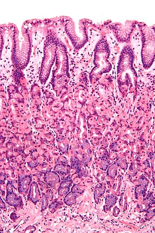
Histology is the study of the minute structure, composition, and function of tissues. Mature human vocal cords are composed of layered structures which are quite different at the histological level.

<span class="mw-page-title-main">Mucosal immunology</span> Field of study

Mucosal immunology is the study of immune system responses that occur at mucosal membranes of the intestines, the urogenital tract, and the respiratory system. The mucous membranes are in constant contact with microorganisms, food, and inhaled antigens. In healthy states, the mucosal immune system protects the organism against infectious pathogens and maintains a tolerance towards non-harmful commensal microbes and benign environmental substances. Disruption of this balance between tolerance and deprivation of pathogens can lead to pathological conditions such as food allergies, irritable bowel syndrome, susceptibility to infections, and more.

<span class="mw-page-title-main">Gastrointestinal wall</span> Digestive system structure

The gastrointestinal wall of the gastrointestinal tract is made up of four layers of specialised tissue. From the inner cavity of the gut outwards, these are:

  1. Mucosa
  2. Submucosa
  3. Muscular layer
  4. Serosa or adventitia
<span class="mw-page-title-main">Tuft cell</span>

Tuft cells are chemosensory cells in the epithelial lining of the intestines. Similar tufted cells are found in the respiratory epithelium where they are known as brush cells. The name "tuft" refers to the brush-like microvilli projecting from the cells. Ordinarily there are very few tuft cells present but they have been shown to greatly increase at times of a parasitic infection. Several studies have proposed a role for tuft cells in defense against parasitic infection. In the intestine, tuft cells are the sole source of secreted interleukin 25 (IL-25).

<span class="mw-page-title-main">Intestinal mucosal barrier</span>

The intestinal mucosal barrier, also referred to as intestinal barrier, refers to the property of the intestinal mucosa that ensures adequate containment of undesirable luminal contents within the intestine while preserving the ability to absorb nutrients. The separation it provides between the body and the gut prevents the uncontrolled translocation of luminal contents into the body proper. Its role in protecting the mucosal tissues and circulatory system from exposure to pro-inflammatory molecules, such as microorganisms, toxins, and antigens is vital for the maintenance of health and well-being. Intestinal mucosal barrier dysfunction has been implicated in numerous health conditions such as: food allergies, microbial infections, irritable bowel syndrome, inflammatory bowel disease, celiac disease, metabolic syndrome, non-alcoholic fatty liver disease, diabetes, and septic shock.

<span class="mw-page-title-main">Anatomical terms of microanatomy</span> Anatomical terminology is used to describe microanatomical (or histological) structures

Anatomical terminology is used to describe microanatomical structures. This helps describe precisely the structure, layout and position of an object, and minimises ambiguity. An internationally accepted lexicon is Terminologia Histologica.

References

  1. Burkitt, H. George; Young, Barbara; Heath, John W., eds. (1993). Wheater's Functional Histology (3rd ed.). Churchill Livingstone. ISBN   978-0-443-04691-9.[ page needed ]
  2. Slomianka, Lutz (2009). "Blue Histology-Gastrointestinal Tract". The University of Western Australia.
  3. Mescher, Anthony (2009). Junqueira's Basic Histology: Text & Atlas (12th ed.). McGraw Hill Professional. ISBN   978-0-07-171475-4.[ page needed ]
  4. 1 2 3 King, David (2009). "Study Guide: Histology of the Gastrointestinal System".
  5. Chang, S. L.; Chung, J. S.; Yeung, M. K.; Howard, P. S.; MacArak, E. J. (1999). "Roles of the Lamina Propria and the Detrusor in Tension Transfer during Bladder Filling". Scandinavian Journal of Urology and Nephrology. 33 (201): 38–45. doi:10.1080/003655999750042132. PMID   10573775.
  6. 1 2 Andersson, Karl-Erik; McCloskey, Karen D. (2014). "Lamina propria: The functional center of the bladder?". Neurourology and Urodynamics. 33 (1): 9–16. doi: 10.1002/nau.22465 . PMID   23847015.
  7. Wiseman, O.J.; Fowler, C.J.; Landon, D.N. (2003). "The role of the human bladder lamina propria myofibroblast". BJU International. 91 (1): 89–93. doi:10.1046/j.1464-410X.2003.03802.x. PMID   12614258. S2CID   7628206.
  8. 1 2 Rice, Thomas W; Zuccaro, Gregory; Adelstein, David J; Rybicki, Lisa A; Blackstone, Eugene H; Goldblum, John R (1998). "Esophageal Carcinoma: Depth of Tumor Invasion is Predictive of Regional Lymph Node Status". The Annals of Thoracic Surgery. 65 (3): 787–92. doi:10.1016/S0003-4975(97)01387-8. PMID   9527214.
  9. Hall, Peter A.; Coates, Philip J.; Ansari, Bijan; Hopwood, David (1994). "Regulation of cell number in the mammalian gastrointestinal tract: The importance of apoptosis". Journal of Cell Science. 107 (12): 3569–77. doi:10.1242/jcs.107.12.3569. PMID   7706406.
  10. Varol, Chen; Vallon-Eberhard, Alexandra; Elinav, Eran; Aychek, Tegest; Shapira, Yami; Luche, Hervé; Fehling, Hans Jörg; Hardt, Wolf-Dietrich; Shakhar, Guy; Jung, Steffen (2009). "Intestinal Lamina Propria Dendritic Cell Subsets Have Different Origin and Functions". Immunity. 31 (3): 502–12. doi: 10.1016/j.immuni.2009.06.025 . PMID   19733097.
  11. Bischoff, Stephan C; Barbara, Giovanni; Buurman, Wim; Ockhuizen, Theo; Schulzke, Jörg-Dieter; Serino, Matteo; Tilg, Herbert; Watson, Alastair; Wells, Jerry M (2014-11-18). "Intestinal permeability – a new target for disease prevention and therapy". BMC Gastroenterology. 14: 189. doi: 10.1186/s12876-014-0189-7 . ISSN   1471-230X. PMC   4253991 . PMID   25407511.
  12. Lee, Ji Yong; Joo, Hee Jae; Cho, Dae Sung; Kim, Sun Il; Ahn, Hyun Soo; Kim, Se Joong (2012). "Prognostic Significance of Substaging according to the Depth of Lamina Propria Invasion in Primary T1 Transitional Cell Carcinoma of the Bladder". Korean Journal of Urology. 53 (5): 317–23. doi:10.4111/kju.2012.53.5.317. PMC   3364470 . PMID   22670190.
  13. Yonemura, Yutaka; Endou, Yoshio; Tabachi, Kayoko; Kawamura, Taiichi; Yun, Hyo-Yung; Kameya, Toru; Hayashi, Isamu; Bandou, Etsurou; Sasaki, Takuma; Miura, Masahiro (2006). "Evaluation of lymphatic invasion in primary gastric cancer by a new monoclonal antibody, D2-40". Human Pathology. 37 (9): 1193–9. doi:10.1016/j.humpath.2006.04.014. PMID   16938525.
  14. Matsumoto, Satoshi; Hara, Taeko; Mitsuyama, Keiichi; Yamamoto, Mayuko; Tsuruta, Osamu; Sata, Michio; Scheller, Jürgen; Rose-John, Stefan; Kado, Sho-ichi; Takada, Toshihiko (2009). "Essential Roles of IL-6 Trans-Signaling in Colonic Epithelial Cells, Induced by the IL-6/Soluble-IL-6 Receptor Derived from Lamina Propria Macrophages, on the Development of Colitis-Associated Premalignant Cancer in a Murine Model". The Journal of Immunology. 184 (3): 1543–51. doi: 10.4049/jimmunol.0801217 . PMID   20042582.