Respiratory tract

Last updated
Respiratory tract
Illu conducting passages.svg
Conducting passages
Details
System Respiratory system
Identifiers
FMA 265130
Anatomical terminology

The respiratory tract is the subdivision of the respiratory system involved with the process of conducting air to the alveoli for the purposes of gas exchange in mammals. [1] The respiratory tract is lined with respiratory epithelium as respiratory mucosa. [2]

Contents

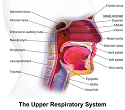
Air is breathed in through the nose to the nasal cavity, where a layer of nasal mucosa acts as a filter and traps pollutants and other harmful substances found in the air. Next, air moves into the pharynx, a passage that contains the intersection between the oesophagus and the larynx. The opening of the larynx has a special flap of cartilage, the epiglottis, that opens to allow air to pass through but closes to prevent food from moving into the airway.

From the larynx, air moves into the trachea and down to the intersection known as the carina that branches to form the right and left primary (main) bronchi. Each of these bronchi branches into a secondary (lobar) bronchus that branches into tertiary (segmental) bronchi, that branch into smaller airways called bronchioles that eventually connect with tiny specialized structures called alveoli that function in gas exchange.

The lungs which are located in the thoracic cavity, are protected from physical damage by the rib cage. At the base of the lungs is a sheet of skeletal muscle called the diaphragm. The diaphragm separates the lungs from the stomach and intestines. The diaphragm is also the main muscle of respiration involved in breathing, and is controlled by the sympathetic nervous system.

The lungs are encased in a serous membrane that folds in on itself to form the pleurae – a two-layered protective barrier. The inner visceral pleura covers the surface of the lungs, and the outer parietal pleura is attached to the inner surface of the thoracic cavity. The pleurae enclose a cavity called the pleural cavity that contains pleural fluid. This fluid is used to decrease the amount of friction that lungs experience during breathing.

Structure

Complete respiratory system Respiratory system complete en.svg
Complete respiratory system

The respiratory tract is divided into the upper airways and lower airways. The upper airways or upper respiratory tract includes the nose and nasal passages, paranasal sinuses, the pharynx, and the portion of the larynx above the vocal folds (cords). The lower airways or lower respiratory tract includes the portion of the larynx below the vocal folds, trachea, bronchi and bronchioles. The lungs can be included in the lower respiratory tract or as separate entity and include the respiratory bronchioles, alveolar ducts, alveolar sacs, and alveoli. [3]

The respiratory tract can also be divided into a conducting zone and a respiratory zone, based on the distinction of transporting gases or exchanging them.

The conducting zone includes structures outside of the lungs – the nose, pharynx, larynx, and trachea, and structures inside the lungs – the bronchi, bronchioles, and terminal bronchioles. The conduction zone conducts air breathed in that is filtered, warmed, and moistened, into the lungs. It represents the 1st through the 16th division of the respiratory tract. The conducting zone is most of the respiratory tract that conducts gases into and out of the lungs but excludes the respiratory zone that exchanges gases. The conducting zone also functions to offer a low resistance pathway for airflow. It provides a major defense role in its filtering abilities.

The respiratory zone includes the respiratory bronchioles, alveolar ducts, and alveoli, and is the site of oxygen and carbon dioxide exchange with the blood. The respiratory bronchioles and the alveolar ducts are responsible for 10% of the gas exchange. The alveoli are responsible for the other 90%. The respiratory zone represents the 16th through the 23rd division of the respiratory tract.

From the bronchi, the dividing tubes become progressively smaller with an estimated 20 to 23 divisions before ending at an alveolus. [1]

Upper respiratory tract

Details of upper respiratory tract. Blausen 0872 UpperRespiratorySystem.png
Details of upper respiratory tract.

The upper respiratory tract can refer to the parts of the respiratory system lying above the sternal angle (outside of the thorax), [4] above the vocal folds, or above the cricoid cartilage. [5] [6] The larynx is sometimes included in both the upper and lower airways. [7] The larynx is also called the voice box and has the associated cartilage that produces sound. The tract consists of the nasal cavity and paranasal sinuses, the pharynx (nasopharynx, oropharynx and laryngopharynx) and sometimes includes the larynx.

Lower respiratory tract

Parts of the lower respiratory tract. Diagram showing the parts of the respiratory system CRUK 335.svg
Parts of the lower respiratory tract.
Trachea
Mainstem bronchus
Lobar bronchus
Segmental bronchus
Bronchiole
Alveolar duct
Alveolus Illu quiz lung05.jpg

The lower respiratory tract or lower airway is derived from the developing foregut and consists of the trachea, bronchi (primary, secondary and tertiary), bronchioles (including terminal and respiratory), and lungs (including alveoli). [8] It also sometimes includes the larynx.

The lower respiratory tract is also called the respiratory tree or tracheobronchial tree, to describe the branching structure of airways supplying air to the lungs, and includes the trachea, bronchi and bronchioles. [9]

At each division point or generation, one airway branches into two smaller airways. The human respiratory tree may consist on average of 23 generations, while the respiratory tree of the mouse has up to 13 generations. Proximal divisions (those closest to the top of the tree, such as the bronchi) mainly function to transmit air to the lower airways. Later divisions including the respiratory bronchiole, alveolar ducts, and alveoli, are specialized for gas exchange.

The trachea is the largest tube in the respiratory tract and consists of tracheal rings of hyaline cartilage. It branches off into two bronchial tubes, a left and a right main bronchus. The bronchi branch off into smaller sections inside the lungs, called bronchioles. These bronchioles give rise to the air sacs in the lungs called the alveoli. [11]

The lungs are the largest organs in the lower respiratory tract. The lungs are suspended within the pleural cavity of the thorax. The pleurae are two thin membranes, one cell layer thick, which surround the lungs. The inner (visceral pleura) covers the lungs and the outer (parietal pleura) lines the inner surface of the chest wall. This membrane secretes a small amount of fluid, allowing the lungs to move freely within the pleural cavity while expanding and contracting during breathing. The lungs are divided into different lobes. The right lung is larger in size than the left, because of the heart's being situated to the left of the midline. The right lung has three lobes – upper, middle, and lower (or superior, middle, and inferior), and the left lung has two – upper and lower (or superior and inferior), plus a small tongue-shaped portion of the upper lobe known as the lingula. Each lobe is further divided up into segments called bronchopulmonary segments. Each lung has a costal surface, which is adjacent to the ribcage; a diaphragmatic surface, which faces downward toward the diaphragm; and a mediastinal surface, which faces toward the center of the chest, and lies against the heart, great vessels, and the carina where the two mainstem bronchi branch off from the base of the trachea.

The alveoli are tiny air sacs in the lungs where gas exchange takes place. The mean number of alveoli in a human lung is 480 million. [12] When the diaphragm contracts, a negative pressure is generated in the thorax and air rushes in to fill the cavity. When that happens, these sacs fill with air, making the lung expand. The alveoli are rich with capillaries, called alveolar capillaries. Here the red blood cells absorb oxygen from the air and then carry it back in the form of oxyhaemaglobin, to nourish the cells. The red blood cells also carry carbon dioxide (CO2) away from the cells in the form of carbaminohemoglobin and release it into the alveoli through the alveolar capillaries. When the diaphragm relaxes, a positive pressure is generated in the thorax and air rushes out of the alveoli expelling the carbon dioxide.

Microanatomy

Respiratory epithelium Blausen 0750 PseudostratifiedCiliatedColumnar.png
Respiratory epithelium

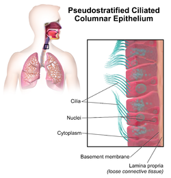
The respiratory tract is covered in epithelium, which varies down the tract. There are glands and mucus produced by goblet cells in parts, as well as smooth muscle, elastin or cartilage. The epithelium from the nose to the bronchioles is covered in ciliated pseudostratified columnar epithelium, commonly called respiratory epithelium. [13] The cilia beat in one direction, moving mucus towards the throat where it is swallowed. Moving down the bronchioles, the cells get more cuboidal in shape but are still ciliated.

Glands are abundant in the upper respiratory tract, but there are fewer lower down and they are absent starting at the bronchioles. The same goes for goblet cells, although there are scattered ones in the first bronchioles.

Cartilage is present until the small bronchi. In the trachea, they are C-shaped rings of hyaline cartilage, whereas in the bronchi the cartilage takes the form of interspersed plates. Smooth muscle starts in the trachea, where it joins the C-shaped rings of cartilage. It continues down the bronchi and bronchioles, which it completely encircles. Instead of hard cartilage, the bronchi and bronchioles are composed of elastic tissue.

The lungs are made up of thirteen different kinds of cells, eleven types of epithelial cell and two types of mesenchymal cell. [14] The epithelial cells form the lining of the tracheal, and bronchial tubes, while the mesenchymal cells line the lungs.

Function

Most of the respiratory tract exists merely as a piping system for air to travel in the lungs, and alveoli are the only part of the lung that exchanges oxygen and carbon dioxide with the blood.

Respiration

Respiration is the rhythmical process of breathing, in which air is drawn into the alveoli of the lungs via inhalation and subsequently expelled via exhalation. When a human being inhales, air travels down the trachea, through the bronchial tubes, and into the lungs. The entire tract is protected by the rib cage, spine, and sternum. In the lungs, oxygen from the inhaled air is transferred into the blood and circulated throughout the body. Carbon dioxide (CO2) is transferred from returning blood back into gaseous form in the lungs and exhaled through the lower respiratory tract and then the upper, to complete the process of breathing.

Unlike the trachea and bronchi, the upper airway is a collapsible, compliant tube. As such, it has to be able to withstand suction pressures generated by the rhythmic expansion of the thoracic cavity that sucks air into the lungs. This is accomplished by the contraction of upper airway muscles during inhalation, such as the genioglossus (tongue) and the hyoid muscles. In addition to rhythmic innervation from the respiratory center in the medulla oblongata, the motor neurons controlling the muscles also receive tonic innervation that sets a baseline level of stiffness and size.

The diaphragm is the primary muscle that allows for lung expansion and contraction. Smaller muscles between the ribs, the external intercostals, assist with this process.

Defences against infection

The epithelial lining of the upper respiratory tract is interspersed with goblet cells that secrete a protective mucus. This helps to filter waste, which is eventually either swallowed into the highly acidic stomach environment or expelled via spitting. The epithelium lining the respiratory tract is covered in small hairs called cilia. These beat rhythmically out from the lungs, moving secreted mucus foreign particles toward the laryngopharynx upwards and outwards, in a process called mucociliary clearance, they prevent mucus accumulation in the lungs. Macrophages in the alveoli are part of the immune system which engulf and digest any inhaled harmful agents.

Hair in the nostrils plays a protective role, trapping particulate matter such as dust. [15] The cough reflex expels all irritants within the mucous membrane to the outside. The airways of the lungs contain rings of muscle. When the passageways are irritated by some allergen, these muscles can constrict.

Clinical significance

The respiratory tract is a common site for infections.

Infection

Upper respiratory infection

Upper respiratory tract infections are probably the most common infections in the world.

The respiratory system is very prone to developing infections in the lungs. Infants and older adults are more likely to develop infections in their lungs because their lungs are not as strong in fighting off these infections. Most of these infections used to be fatal, but with new research and medicine, they are now treatable. With bacterial infections, antibiotics are prescribed, while viral infections are harder to treat but still curable.

The common cold, and flu are the most common causes of an upper respiratory tract infection, which can cause more serious illness that can develop in the lower respiratory tract.

Lower respiratory tract infections

Pneumonia is the most common, and frequent lower respiratory tract infection. This can be either viral, bacterial, or fungal. This infection is very common because pneumonia can be airborne, and when you inhale this infection in the air, the particles enter the lungs and move into the air sacs. This infection quickly develops in the lower part of the lung and fills the lung with fluid, and excess mucus. This causes difficulty in breathing and coughing as the lower respiratory tract tries to get rid of the fluid in the lungs. You can be more prone to developing this infection if you have asthma, flu, heart disease, or cancer [16]

Bronchitis is another common infection that takes place in the lower respiratory tract. It is an inflammation of the bronchial tubes. There are two forms of this infection: acute bronchitis, which is treatable and can go away without treatment, or chronic bronchitis, which comes and goes, but will always affect one's lungs. Bronchitis increases the amount of mucus that is natural in your respiratory tract. Chronic bronchitis is common in smokers, because the tar from smoking accumulates over time, causing the lungs to work harder to repair themselves. [17]

Tuberculosis is one of many other infections that occurs in the lower respiratory tract. You can contract this infection from airborne droplets, and if inhaled you are at risk of this disease. This is a bacterial infection that deteriorates the lung tissue resulting in coughing up blood. [18] This infection is deadly if not treated.

Cancer

3D still showing increased mucus. Respiratory Tract.jpg
3D still showing increased mucus.

Some of these cancers have environmental causes such as smoking. When a tobacco product is inhaled, the smoke paralyzes the cilia, causing mucus to enter the lungs. Frequent smoking, over time, causes the cilia hairs to die and can no longer filter mucus. Tar from the smoke inhaled enters the lungs, turning the pink-coloured lungs black. The accumulation of this tar could eventually lead to lung cancer, or chronic obstructive pulmonary disease. [8]

COPD

Chronic obstructive pulmonary disease (COPD) is a common lower respiratory disease that can be caused by exposure to harmful chemicals, or prolonged use of tobacco. This disease is chronic and progressive, the damage to the lungs is irreversible and eventually fatal. COPD destroys the alveoli, and lung tissue which makes breathing very difficult, causing shortness of breath, hyperventilation, and raised chest. The decreased number of alveoli causes loss of oxygen supply to the lungs and an increased accumulation of carbon dioxide. There are two types of COPD: primary and secondary.[ citation needed ] Primary COPD can be found in younger adults. This type of COPD deteriorates the air sacs, and lung mass. Secondary COPD can be found in older adults who smoke or have smoked and have a history of bronchitis. [ citation needed ] COPD includes symptoms of emphysema and chronic bronchitis. [19]

Asthma

3D still showing constricted airways. Respiratory tract.jpg
3D still showing constricted airways.

The bronchi are the main passages to the right and left lungs. These airways carry oxygen to the bronchioles inside the lungs. Inflammation of the bronchii and bronchioles can cause them to swell up, which could lead to an asthma attack. This results in wheezing, tightness of the chest, and severe difficulty in breathing. There are different types of asthma that affect the functions of the bronchial tubes. Allergies can also set off an allergic reaction, causing swelling of the bronchial tubes; as a result, the air passage will swell up, or close up completely. [20]

Mouth breathing

In general, air is inhaled through the nose. It can be inhaled through the mouth if it is not possible to breathe through the nose. However, chronic mouth breathing can cause a dry mouth and lead to infections. [21]

See also

Related Research Articles

<span class="mw-page-title-main">Lung</span> Primary organ of the respiratory system

The lungs are the central organs of the respiratory system in humans and most other animals, including some snails and a small number of fish. In mammals and most other vertebrates, two lungs are located near the backbone on either side of the heart. Their function in the respiratory system is to extract oxygen from the air and transfer it into the bloodstream, and to release carbon dioxide from the bloodstream into the atmosphere, in a process of gas exchange. The pleurae, which are thin, smooth, and moist, serve to reduce friction between the lungs and chest wall during breathing, allowing for easy and effortless movements of the lungs.

<span class="mw-page-title-main">Respiratory system</span> Biological system in animals and plants for gas exchange

The respiratory system is a biological system consisting of specific organs and structures used for gas exchange in animals and plants. The anatomy and physiology that make this happen varies greatly, depending on the size of the organism, the environment in which it lives and its evolutionary history. In land animals, the respiratory surface is internalized as linings of the lungs. Gas exchange in the lungs occurs in millions of small air sacs; in mammals and reptiles, these are called alveoli, and in birds, they are known as atria. These microscopic air sacs have a very rich blood supply, thus bringing the air into close contact with the blood. These air sacs communicate with the external environment via a system of airways, or hollow tubes, of which the largest is the trachea, which branches in the middle of the chest into the two main bronchi. These enter the lungs where they branch into progressively narrower secondary and tertiary bronchi that branch into numerous smaller tubes, the bronchioles. In birds, the bronchioles are termed parabronchi. It is the bronchioles, or parabronchi that generally open into the microscopic alveoli in mammals and atria in birds. Air has to be pumped from the environment into the alveoli or atria by the process of breathing which involves the muscles of respiration.

<span class="mw-page-title-main">Trachea</span> Cartilaginous tube that connects the pharynx and larynx to the lungs

The trachea, also known as the windpipe, is a cartilaginous tube that connects the larynx to the bronchi of the lungs, allowing the passage of air, and so is present in almost all animals with lungs. The trachea extends from the larynx and branches into the two primary bronchi. At the top of the trachea the cricoid cartilage attaches it to the larynx. The trachea is formed by a number of horseshoe-shaped rings, joined together vertically by overlying ligaments, and by the trachealis muscle at their ends. The epiglottis closes the opening to the larynx during swallowing.

<span class="mw-page-title-main">Pulmonary alveolus</span> Hollow cavity found in the lungs

A pulmonary alveolus, also known as an air sac or air space, is one of millions of hollow, distensible cup-shaped cavities in the lungs where pulmonary gas exchange takes place. Oxygen is exchanged for carbon dioxide at the blood–air barrier between the alveolar air and the pulmonary capillary. Alveoli make up the functional tissue of the mammalian lungs known as the lung parenchyma, which takes up 90 percent of the total lung volume.

<span class="mw-page-title-main">Sputum</span> Mucus that is coughed up from the lower airways

Sputum is mucus that is coughed up from the lower airways. In medicine, sputum samples are usually used for a naked eye examination, microbiological investigation of respiratory infections and cytological investigations of respiratory systems. It is crucial that the specimen does not include any mucoid material from the nose or oral cavity.

<span class="mw-page-title-main">Bronchus</span> Airway in the respiratory tract

A bronchus is a passage or airway in the lower respiratory tract that conducts air into the lungs. The first or primary bronchi to branch from the trachea at the carina are the right main bronchus and the left main bronchus. These are the widest bronchi, and enter the right lung, and the left lung at each hilum. The main bronchi branch into narrower secondary bronchi or lobar bronchi, and these branch into narrower tertiary bronchi or segmental bronchi. Further divisions of the segmental bronchi are known as 4th order, 5th order, and 6th order segmental bronchi, or grouped together as subsegmental bronchi. The bronchi, when too narrow to be supported by cartilage, are known as bronchioles. No gas exchange takes place in the bronchi.

<span class="mw-page-title-main">Bronchiole</span> Passageways by which air passes through the nose or mouth to the alveoli of the lungs

The bronchioles or bronchioli are the smaller branches of the bronchial airways in the lower respiratory tract. They include the terminal bronchioles, and finally the respiratory bronchioles that mark the start of the respiratory zone delivering air to the gas exchanging units of the alveoli. The bronchioles no longer contain the cartilage that is found in the bronchi, or glands in their submucosa.

<span class="mw-page-title-main">Inhalation</span> Flow of the respiratory current into an organism

Inhalation is the process of drawing air or other gases into the respiratory tract, primarily for the purpose of bringing oxygen into the body. It is a fundamental physiological function in humans and many other organisms, essential for sustaining life. Inhalation is the first phase of breathing, allowing the exchange of oxygen and carbon dioxide between the body and the environment, vital for the body's metabolic processes. This article delves into the mechanics of inhalation, its significance in various contexts, and its potential impact on health.

<span class="mw-page-title-main">Mucus</span> Secretion produced by mucous membranes

Mucus is a slippery aqueous secretion produced by, and covering, mucous membranes. It is typically produced from cells found in mucous glands, although it may also originate from mixed glands, which contain both serous and mucous cells. It is a viscous colloid containing inorganic salts, antimicrobial enzymes, immunoglobulins, and glycoproteins such as lactoferrin and mucins, which are produced by goblet cells in the mucous membranes and submucosal glands. Mucus serves to protect epithelial cells in the linings of the respiratory, digestive, and urogenital systems, and structures in the visual and auditory systems from pathogenic fungi, bacteria and viruses. Most of the mucus in the body is produced in the gastrointestinal tract.

<span class="mw-page-title-main">Atelectasis</span> Partial collapse of a lung causing reduced gas exchange

Atelectasis is the partial collapse or closure of a lung resulting in reduced or absent gas exchange. It is usually unilateral, affecting part or all of one lung. It is a condition where the alveoli are deflated down to little or no volume, as distinct from pulmonary consolidation, in which they are filled with liquid. It is often referred to informally as a collapsed lung, although more accurately it usually involves only a partial collapse, and that ambiguous term is also informally used for a fully collapsed lung caused by a pneumothorax.

<span class="mw-page-title-main">Bronchoconstriction</span> Constriction of the terminal airways in the lungs

Bronchoconstriction is the constriction of the airways in the lungs due to the tightening of surrounding smooth muscle, with consequent coughing, wheezing, and shortness of breath.

<span class="mw-page-title-main">Respiratory disease</span> Disease of the respiratory system

Respiratory diseases, or lung diseases, are pathological conditions affecting the organs and tissues that make gas exchange difficult in air-breathing animals. They include conditions of the respiratory tract including the trachea, bronchi, bronchioles, alveoli, pleurae, pleural cavity, the nerves and muscles of respiration. Respiratory diseases range from mild and self-limiting, such as the common cold, influenza, and pharyngitis to life-threatening diseases such as bacterial pneumonia, pulmonary embolism, tuberculosis, acute asthma, lung cancer, and severe acute respiratory syndromes, such as COVID-19. Respiratory diseases can be classified in many different ways, including by the organ or tissue involved, by the type and pattern of associated signs and symptoms, or by the cause of the disease.

The cough reflex occurs when stimulation of cough receptors in the respiratory tract by dust or other foreign particles produces a cough, which causes rapidly moving air which usually remove the foreign material before it reaches the lungs. This typically clears particles from the bronchi and trachea, the tubes that feed air to lung tissue from the nose and mouth. The larynx and carina are especially sensitive. Cough receptors in the surface cells (epithelium) of the respiratory tract are also sensitive to chemicals. Terminal bronchioles and even the alveoli are sensitive to chemicals such as sulfur dioxide gas or chlorine gas.

<span class="mw-page-title-main">Respiratory epithelium</span> Mucosa that serves to moisten and protect the airways

Respiratory epithelium, or airway epithelium, is a type of ciliated columnar epithelium found lining most of the respiratory tract as respiratory mucosa, where it serves to moisten and protect the airways. It is not present in the vocal cords of the larynx, or the oropharynx and laryngopharynx, where instead the epithelium is stratified squamous. It also functions as a barrier to potential pathogens and foreign particles, preventing infection and tissue injury by the secretion of mucus and the action of mucociliary clearance.

<span class="mw-page-title-main">Respiratory system of the horse</span> Biological system by which a horse circulates air for the purpose of gaseous exchange

The respiratory system of the horse is the biological system by which a horse circulates air for the purpose of gaseous exchange.

<span class="mw-page-title-main">Obstructive lung disease</span> Category of respiratory disease characterized by airway obstruction

Obstructive lung disease is a category of respiratory disease characterized by airway obstruction. Many obstructive diseases of the lung result from narrowing (obstruction) of the smaller bronchi and larger bronchioles, often because of excessive contraction of the smooth muscle itself. It is generally characterized by inflamed and easily collapsible airways, obstruction to airflow, problems exhaling, and frequent medical clinic visits and hospitalizations. Types of obstructive lung disease include asthma, bronchiectasis, bronchitis and chronic obstructive pulmonary disease (COPD). Although COPD shares similar characteristics with all other obstructive lung diseases, such as the signs of coughing and wheezing, they are distinct conditions in terms of disease onset, frequency of symptoms, and reversibility of airway obstruction. Cystic fibrosis is also sometimes included in obstructive pulmonary disease.

<span class="mw-page-title-main">Mucociliary clearance</span>

Mucociliary clearance (MCC), mucociliary transport, or the mucociliary escalator describes the self-clearing mechanism of the airways in the respiratory system. It is one of the two protective processes for the lungs in removing inhaled particles including pathogens before they can reach the delicate tissue of the lungs. The other clearance mechanism is provided by the cough reflex. Mucociliary clearance has a major role in pulmonary hygiene.

<span class="mw-page-title-main">Tracheobronchial injury</span> Damage to the tracheobronchial tree

Tracheobronchial injury is damage to the tracheobronchial tree. It can result from blunt or penetrating trauma to the neck or chest, inhalation of harmful fumes or smoke, or aspiration of liquids or objects.

<span class="mw-page-title-main">Ventilation–perfusion coupling</span> Relationship between respiratory and cardiovascular processes

Ventilation-perfusion coupling is the relationship between ventilation and perfusion processes, which take place in the respiratory system and the cardiovascular system. Ventilation is the movement of gas during breathing, and perfusion is the process of pulmonary blood circulation, which delivers oxygen to body tissues. Anatomically, the lung structure, alveolar organization, and alveolar capillaries contribute to the physiological mechanism of ventilation and perfusion. Ventilation-perfusion coupling maintains a constant ventilation/perfusion ratio near 0.8 on average, while the regional variation exists within the lungs due to gravity. When the ratio gets above or below 0.8, it is considered abnormal ventilation-perfusion coupling, also known as a ventilation–perfusion mismatch. Lung diseases, cardiac shunts, and smoking can cause a ventilation-perfusion mismatch that results in significant symptoms and diseases, which can be treated through treatments like bronchodilators and oxygen therapy.

Development of the respiratory system begins early in the fetus. It is a complex process that includes many structures, most of which arise from the endoderm. Towards the end of development, the fetus can be observed making breathing movements. Until birth, however, the mother provides all of the oxygen to the fetus as well as removes all of the fetal carbon dioxide via the placenta.

References

  1. 1 2 Patwa A, Shah A (September 2015). "Anatomy and physiology of respiratory system relevant to anaesthesia". Indian Journal of Anaesthesia. 59 (9): 533–541. doi: 10.4103/0019-5049.165849 . PMC   4613399 . PMID   26556911.
  2. "Respiratory mucosa". mesh..nlm.nih.gov. Retrieved 26 July 2019.
  3. "Respiratory tract". www.cancer.gov. 2 February 2011.
  4. Alcamo IE, Bergdahl J (29 July 2003). Anatomy Coloring Workbook. The Princeton Review. pp. 238–. ISBN   978-0-375-76342-7 . Retrieved 26 April 2010.
  5. Ronald M. Perkin; James D Swift; Dale A Newton (1 September 2007). Pediatric hospital medicine: textbook of inpatient management. Lippincott Williams & Wilkins. pp. 473–. ISBN   978-0-7817-7032-3 . Retrieved 26 April 2010.
  6. Jeremy P. T. Ward; Jane Ward; Charles M. Wiener (2006). The respiratory system at a glance. Wiley-Blackwell. pp. 11–. ISBN   978-1-4051-3448-4 . Retrieved 26 April 2010.
  7. Sabyasachi Sircar (2008). Principles of medical physiology. Thieme. pp. 309–. ISBN   978-3-13-144061-7 . Retrieved 26 April 2010.
  8. 1 2 "Bronchial Anatomy" . Retrieved 5 Mar 2014.
  9. "Tracheobronchial tree | Radiology Reference Article". Radiopaedia . Retrieved 7 September 2017.
  10. 1 2 3 Page 81 in Robert M. Kacmarek, Steven Dimas & Craig W. Mack (2013). Essentials of Respiratory Care. Elsevier Health Sciences. ISBN   978-032327778-5.
  11. "Human Respiratory System". Archived from the original on 2008-10-15. Retrieved 5 Oct 2008.
  12. Ochs M, Nyengaard JR, Jung A, Knudsen L, Voigt M, Wahlers T, et al. (January 2004). "The number of alveoli in the human lung". American Journal of Respiratory and Critical Care Medicine. 169 (1): 120–124. doi:10.1164/rccm.200308-1107OC. PMID   14512270.
  13. Moore EJ, Feliciano DV, Mattox KL (2004). Trauma. New York: McGraw-Hill, Medical Pub. Division. p. 545. ISBN   0-07-137069-2 . Retrieved 2008-06-15.
  14. Breeze R, Turk M (April 1984). "Cellular structure, function and organization in the lower respiratory tract". Environmental Health Perspectives. 55: 3–24. doi:10.1289/ehp.84553. PMC   1568358 . PMID   6376102.
  15. Blaivas AJ (29 June 2012). "Anatomy and function of the respiratory system". Penn State Hershey Medical Center. Retrieved 2017-09-20.
  16. "Pneumonia". The Lecturio Medical Concept Library. Retrieved 2 July 2021.
  17. "Chest Cold (Acute Bronchitis)". Centers For Disease Control And Prevention. Retrieved 2 July 2021.
  18. "Tuberculosis". The Lecturio Medical Concept Library. Retrieved 1 July 2021.
  19. "Emphysema: Background, Pathophysiology, Etiology". 26 March 2020. Retrieved 3 July 2020.
  20. "Asthma". The Lecturio Medical Concept Library. Retrieved 2 July 2021.
  21. "Your Nose, the Guardian of Your Lungs". Boston Medical Center . Retrieved 2020-06-29.