Epiglottis

Last updated

Epiglottis
Gray958.png
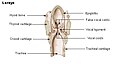
View of the larynx from behind. The epiglottis is the structure at the top of the image.
Details
Precursor Fourth pharyngeal arch [1]
FunctionPrevent food from entering the respiratory tract
Identifiers
Latin epiglottis
MeSH D004825
TA98 A06.2.07.001
TA2 3190
FMA 55130
Anatomical terminology

The epiglottis (pl.: epiglottises or epiglottides) is a leaf-shaped flap in the throat that prevents food and water from entering the trachea and the lungs. It stays open during breathing, allowing air into the larynx. During swallowing, it closes to prevent aspiration of food into the lungs, forcing the swallowed liquids or food to go along the esophagus toward the stomach instead. It is thus the valve that diverts passage to either the trachea or the esophagus.

Contents

The epiglottis is made of elastic cartilage covered with a mucous membrane, attached to the entrance of the larynx. It projects upwards and backwards behind the tongue and the hyoid bone.

The epiglottis may be inflamed in a condition called epiglottitis, which is most commonly due to the vaccine-preventable bacterium Haemophilus influenzae . Dysfunction may cause the inhalation of food, called aspiration, which may lead to pneumonia or airway obstruction. The epiglottis is also an important landmark for intubation.

The epiglottis has been identified as early as Aristotle, and gets its name from being above the glottis ( epi- + glottis).

Structure

Location of the epiglottis Epiglottis (PSF).png
Location of the epiglottis

The epiglottis sits at the entrance of the larynx. It is shaped like a leaf of purslane and has a free upper part that rests behind the tongue, and a lower stalk (Latin : petiolus). [2] The stalk originates from the back surface of the thyroid cartilage, connected by a thyroepiglottic ligament. At the sides, the stalk is connected to the arytenoid cartilages at the walls of the larynx by folds. [2]

The epiglottis originates at the entrance of the larynx, and is attached to the hyoid bone. From there, it projects upwards and backwards behind the tongue. [3] The space between the epiglottis and the tongue is called the vallecula. [3]

Microanatomy

The epiglottis has two surfaces; a forward-facing anterior surface, and a posterior surface facing the larynx. [2] The forward-facing surface is covered with several layers of thin cells (stratified squamous epithelium), and is not covered with keratin, the same surface as the back of the tongue. [2] The back surface is covered in a layer of column-shaped cells with cilia, similar to the rest of the respiratory tract. It also has mucous-secreting goblet cells. [2] There is an intermediate zone between these surfaces that contains cells that transition in shape. [4] The body of the epiglottis consists of elastic cartilage. [2]

Development

The epiglottis arises from the fourth pharyngeal arch. It can be seen as a distinct structure later than the other cartilage of the pharynx, visible around the fifth month of development. [1] The position of the epiglottis also changes with ageing. In infants, it touches the soft palate, whereas in adults, its position is lower. [3]

Variation

A high rising epiglottis (with forward-facing anterior surface being visible) High rising epiglottis.JPG
A high rising epiglottis (with forward-facing anterior surface being visible)

A high-rising epiglottis is a normal anatomical variation, visible during an examination of the mouth. It does not cause any serious problem apart from maybe a mild sensation of a foreign body in the throat. It is seen more often in children than adults and does not need any medical or surgical intervention. [5] The front surface of the epiglottis is occasionally notched. [2]

Function

The epiglottis is normally pointed upward during breathing with its underside functioning as part of the pharynx. [2] There are taste buds on the epiglottis. [6]

Swallowing

During swallowing, the epiglottis bends backwards, folding over the entrance to the trachea, and preventing food from going into it. [2] The folding backwards is a complex movement the causes of which are not completely understood. [2] It is likely that during swallowing the hyoid bone and the larynx move upwards and forwards, which increases passive pressure from the back of the tongue; because the aryepiglottic muscles contract; because of the passive weight of the food pushing down; and because of contraction of laryngeal and because of contraction of thyroarytenoid muscles. [2] The consequence of this is that during swallowing the bent epiglottis blocks off the trachea, preventing food from going into it; food instead travels down the esophagus, which is behind it. [3]

Speech sounds

In many languages, the epiglottis is not essential for producing sounds. [2] In some languages, the epiglottis is used to produce epiglottal consonant speech sounds, though this sound-type is rather rare. [7]

Clinical significance

Inflammation

Inflammation of the epiglottis is known as epiglottitis. Epiglottitis is mainly caused by Haemophilus influenzae . A person with epiglottitis may have a fever, sore throat, difficulty swallowing, and difficulty breathing. For this reason, acute epiglottitis is considered a medical emergency, because of the risk of obstruction of the pharynx. Epiglottitis is often managed with antibiotics, inhaled aerosolised epinephrine to act as a bronchodilator, and may require tracheal intubation or a tracheostomy if breathing is difficult. [8]

The incidence of epiglottitis has decreased significantly in countries where vaccination against Haemophilus influenzae is administered. [9] [10]

Aspiration

When food or other objects travel down the respiratory tract rather than down the esophagus to the stomach, this is called aspiration . This can lead to the obstruction of airways, inflammation of lung tissue, and aspiration pneumonia; and in the long term, atelectasis and bronchiectasis. [3] One reason aspiration can occur is because of failure of the epiglottis to close completely. [2] [3]

If food or liquid enters the airway due to the epiglottis failing to close properly, throat-clearing or a cough reflex may occur to protect the respiratory system and expel material from the airway. [3] [11] Where there is impairment in laryngeal vestibule sensation, silent aspiration (entry of material to the airway that does not result in a cough reflex) may occur. [3] [12]

Other

The epiglottis and vallecula are important anatomical landmarks in intubation. [13] Abnormal positioning of the epiglottis is a rare cause of obstructive sleep apnoea. [14]

Other animals

The epiglottis is present in mammals, [15] including land mammals and cetaceans, [16] also as a cartilaginous structure. [17] Like in humans, it functions to prevent entry of food into the trachea during swallowing. [17] The position of the larynx is flat in mice and other rodents, as well as rabbits. [4] For this reason, because the epiglottis is located behind the soft palate in rabbits, they are obligate nose breathers, [18] [19] as are mice and other rodents. [4] In rodents and mice, there is a unique pouch in front of the epiglottis, and the epiglottis is commonly injured by inhaled substances, particularly at the transition zone between the flattened and cuboidal epithelium. [20] [4] It is also common to see taste buds on the epiglottis in these species. [4]

History

The epiglottis was noted by Aristotle, [15] although the epiglottis' function was first defined by Vesalius in 1543. [21] The word has Greek roots. [22] The epiglottis gets its name from being above (Ancient Greek : ἐπί, romanized: epi-) the glottis (Ancient Greek : γλωττίς, romanized: glottis, lit. 'tongue'). [23]

Additional images

See also

Related Research Articles

<span class="mw-page-title-main">Place of articulation</span> Place in the mouth consonants are articulated

In articulatory phonetics, the place of articulation of a consonant is a location along the vocal tract where its production occurs. It is a point where a constriction is made between an active and a passive articulator. Active articulators are organs capable of voluntary movement which create the constriction, while passive articulators are so called because they are normally fixed and are the parts with which an active articulator makes contact. Along with the manner of articulation and phonation, the place of articulation gives the consonant its distinctive sound.

<span class="mw-page-title-main">Larynx</span> Voice box, an organ in the neck of amphibians, reptiles, and mammals

The larynx, commonly called the voice box, is an organ in the top of the neck involved in breathing, producing sound and protecting the trachea against food aspiration. The opening of larynx into pharynx known as the laryngeal inlet is about 4–5 centimeters in diameter. The larynx houses the vocal cords, and manipulates pitch and volume, which is essential for phonation. It is situated just below where the tract of the pharynx splits into the trachea and the esophagus. The word 'larynx' comes from the Ancient Greek word lárunx ʻlarynx, gullet, throat.ʼ

<span class="mw-page-title-main">Trachea</span> Cartilaginous tube that connects the pharynx and larynx to the lungs

The trachea, also known as the windpipe, is a cartilaginous tube that connects the larynx to the bronchi of the lungs, allowing the passage of air, and so is present in almost all animals with lungs. The trachea extends from the larynx and branches into the two primary bronchi. At the top of the trachea the cricoid cartilage attaches it to the larynx. The trachea is formed by a number of horseshoe-shaped rings, joined together vertically by overlying ligaments, and by the trachealis muscle at their ends. The epiglottis closes the opening to the larynx during swallowing.

<span class="mw-page-title-main">Throat</span> Anterior part of the neck, in front of the vertebra

In vertebrate anatomy, the throat is the front part of the neck, internally positioned in front of the vertebrae. It contains the pharynx and larynx. An important section of it is the epiglottis, separating the esophagus from the trachea (windpipe), preventing food and drinks being inhaled into the lungs. The throat contains various blood vessels, pharyngeal muscles, the nasopharyngeal tonsil, the tonsils, the palatine uvula, the trachea, the esophagus, and the vocal cords. Mammal throats consist of two bones, the hyoid bone and the clavicle. The "throat" is sometimes thought to be synonymous for the fauces.

<span class="mw-page-title-main">Tracheal intubation</span> Placement of a tube into the trachea

Tracheal intubation, usually simply referred to as intubation, is the placement of a flexible plastic tube into the trachea (windpipe) to maintain an open airway or to serve as a conduit through which to administer certain drugs. It is frequently performed in critically injured, ill, or anesthetized patients to facilitate ventilation of the lungs, including mechanical ventilation, and to prevent the possibility of asphyxiation or airway obstruction.

Swallowing, also called deglutition or inglutition in scientific contexts, is the process in the body of a human or other animal that allows for a substance to pass from the mouth, to the pharynx, and into the esophagus, while shutting the epiglottis. Swallowing is an important part of eating and drinking. If the process fails and the material goes through the trachea, then choking or pulmonary aspiration can occur. In the human body the automatic temporary closing of the epiglottis is controlled by the swallowing reflex.

<span class="mw-page-title-main">Hyoid bone</span> Bone situated in the neck between the chin and the thyroid cartilage

The hyoid bone is a horseshoe-shaped bone situated in the anterior midline of the neck between the chin and the thyroid cartilage. At rest, it lies between the base of the mandible and the third cervical vertebra.

<span class="mw-page-title-main">Respiratory tract</span> Organs involved in transmission of air to and from the point where gases diffuse into tissue

The respiratory tract is the subdivision of the respiratory system involved with the process of respiration in mammals. The respiratory tract is lined with respiratory epithelium as respiratory mucosa.

The pharyngeal reflex or gag reflex is a reflex muscular contraction of the back of the throat, evoked by touching the roof of the mouth, back of the tongue, area around the tonsils, uvula, and back of the throat. It, along with other aerodigestive reflexes such as reflexive pharyngeal swallowing, prevents objects in the oral cavity from entering the throat except as part of normal swallowing and helps prevent choking, and is a form of coughing. The pharyngeal reflex is different from the laryngeal spasm, which is a reflex muscular contraction of the vocal cords.

<span class="mw-page-title-main">Epiglottitis</span> Inflammation of the epiglottis

Epiglottitis is the inflammation of the epiglottis—the flap at the base of the tongue that prevents food entering the trachea (windpipe). Symptoms are usually rapid in onset and include trouble swallowing which can result in drooling, changes to the voice, fever, and an increased breathing rate. As the epiglottis is in the upper airway, swelling can interfere with breathing. People may lean forward in an effort to open the airway. As the condition worsens, stridor and bluish skin may occur.

Stridor is a high-pitched extra-thoracic breath sound resulting from turbulent air flow in the larynx or lower in the bronchial tree. It is different from a stertor which is a noise originating in the pharynx.

<span class="mw-page-title-main">Cricoid cartilage</span> Complete ring of cartilage around the trachea

The cricoid cartilage, or simply cricoid or cricoid ring, is the only complete ring of cartilage around the trachea. It forms the back part of the voice box and functions as an attachment site for muscles, cartilages, and ligaments involved in opening and closing the airway and in producing speech.

<span class="mw-page-title-main">Middle pharyngeal constrictor muscle</span> Muscle in the neck

The middle pharyngeal constrictor is a fan-shaped muscle located in the neck. It is one of three pharyngeal constrictor muscles. It is smaller than the inferior pharyngeal constrictor muscle.

<span class="mw-page-title-main">Inferior pharyngeal constrictor muscle</span> Skeletal muscle of the pharynx

The inferior pharyngeal constrictor muscle is a skeletal muscle of the neck. It is the thickest of the three outer pharyngeal muscles. It arises from the sides of the cricoid cartilage and the thyroid cartilage. It is supplied by the vagus nerve. It is active during swallowing, and partially during breathing and speech. It may be affected by Zenker's diverticulum.

<span class="mw-page-title-main">Oropharyngeal dysphagia</span> Difficulty controlling the mouth or throat for swallowing

Oropharyngeal dysphagia is the inability to empty material from the oropharynx into the esophagus as a result of malfunction near the esophagus. Oropharyngeal dysphagia manifests differently depending on the underlying pathology and the nature of the symptoms. Patients with dysphagia can experience feelings of food sticking to their throats, coughing and choking, weight loss, recurring chest infections, or regurgitation. Depending on the underlying cause, age, and environment, dysphagia prevalence varies. In research including the general population, the estimated frequency of oropharyngeal dysphagia has ranged from 2 to 16 percent.

<span class="mw-page-title-main">Lung bud</span> Embryological precursor of the respiratory system

The lung bud sometimes referred to as the respiratory bud forms from the respiratory diverticulum, an embryological endodermal structure that develops into the respiratory tract organs such as the larynx, trachea, bronchi and lungs. It arises from part of the laryngotracheal tube.

<span class="mw-page-title-main">Respiratory system of the horse</span> Biological system by which a horse circulates air for the purpose of gaseous exchange

The respiratory system of the horse is the biological system by which a horse circulates air for the purpose of gaseous exchange.

<span class="mw-page-title-main">Pharynx</span> Part of the throat that is behind the mouth and nasal cavity

The pharynx is the part of the throat behind the mouth and nasal cavity, and above the esophagus and trachea. It is found in vertebrates and invertebrates, though its structure varies across species. The pharynx carries food to the esophagus and air to the larynx. The flap of cartilage called the epiglottis stops food from entering the larynx.

<span class="mw-page-title-main">Human digestive system</span> Digestive system in humans

The human digestive system consists of the gastrointestinal tract plus the accessory organs of digestion. Digestion involves the breakdown of food into smaller and smaller components, until they can be absorbed and assimilated into the body. The process of digestion has three stages: the cephalic phase, the gastric phase, and the intestinal phase.

<span class="mw-page-title-main">Advanced airway management</span>

Advanced airway management is the subset of airway management that involves advanced training, skill, and invasiveness. It encompasses various techniques performed to create an open or patent airway – a clear path between a patient's lungs and the outside world.

References

  1. 1 2 Schoenwolf, Gary C.; et al. (2009). ""Development of the Urogenital system"". Larsen's human embryology (4th ed., Thoroughly rev. and updated. ed.). Philadelphia: Churchill Livingstone/Elsevier. p. 362. ISBN   9780443068119.
  2. 1 2 3 4 5 6 7 8 9 10 11 12 13 Standring, Susan, ed. (2016). Gray's anatomy : the anatomical basis of clinical practice (41st ed.). Philadelphia. pp. 586–8. ISBN   9780702052309. OCLC   920806541.{{cite book}}: CS1 maint: location missing publisher (link)
  3. 1 2 3 4 5 6 7 8 Matsuo, Koichiro; Palmer, Jeffrey B. (November 2008). "Anatomy and Physiology of Feeding and Swallowing – Normal and Abnormal". Physical Medicine and Rehabilitation Clinics of North America. 19 (4): 691–707. doi:10.1016/j.pmr.2008.06.001. ISSN   1047-9651. PMC   2597750 . PMID   18940636.
  4. 1 2 3 4 5 Harkema, Jack R.; Carey, Stephan A.; Wagner, James G.; Dintzis, Suzanne M.; Liggitt, Denny (2018), "Nose, Sinus, Pharynx, and Larynx", Comparative Anatomy and Histology, Elsevier, pp. 89–114, doi:10.1016/b978-0-12-802900-8.00006-3, ISBN   9780128029008
  5. Petkar N, Georgalas C, Bhattacharyya A (2007). "High-rising epiglottis in children: should it cause concern?". J Am Board Fam Med. 20 (5): 495–6. doi: 10.3122/jabfm.2007.05.060212 . PMID   17823468.
  6. Jowett, Adrian; Shrestha, Rajani (November 1998). "Mucosa and taste buds of the human epiglottis". Journal of Anatomy. 193 (4): 617–618. doi:10.1046/j.1469-7580.1998.19340617.x. PMC   1467887 . PMID   10029195.
  7. Shahin, Kimary (2011), "Pharyngeals", The Blackwell Companion to Phonology, American Cancer Society, pp. 1–24, doi:10.1002/9781444335262.wbctp0025, ISBN   9781444335262
  8. Nicki R. Colledge; Brian R. Walker; Stuart H. Ralston, eds. (2010). Davidson's principles and practice of medicine. illustrated by Robert Britton (21st ed.). Edinburgh: Churchill Livingstone/Elsevier. p. 681. ISBN   978-0-7020-3084-0.
  9. Reilly BK, Reddy SK, Verghese ST (April 2013). "Acute epiglottitis in the era of post-Haemophilus influenzae type B (HIB) vaccine". J Anesth. 27 (2): 316–7. doi:10.1007/s00540-012-1500-9. PMID   23076559. S2CID   33540359.
  10. Hermansen MN, Schmidt JH, Krug AH, Larsen K, Kristensen S (April 2014). "Low incidence of children with acute epiglottis after introduction of vaccination". Dan Med J. 61 (4): A4788. PMID   24814584.
  11. Widdicombe, J. (1 July 2006). "Cough: what's in a name?". European Respiratory Journal. 28 (1): 10–15. doi: 10.1183/09031936.06.00096905 . PMID   16816346.
  12. Ramsey, Deborah; Smithard, David; Kalra, Lalit (13 December 2005). "Silent Aspiration: What Do We Know?". Dysphagia. 20 (3): 218–225. doi:10.1007/s00455-005-0018-9. PMID   16362510. S2CID   24880995.
  13. Peitzman, Andrew B.; Rhodes, Michael; Schwab, C. William (2008). The Trauma Manual: Trauma and Acute Care Surgery. Lippincott Williams & Wilkins. p. 86. ISBN   9780781762755.
  14. Catalfumo, Frank J.; Golz, Avishay; Westerman, S. Thomas; Gilbert, Liane M.; Joachims, Henry Z.; Goldenberg, David (2018). "The epiglottis and obstructive sleep apnoea syndrome". The Journal of Laryngology & Otology. 112 (10): 940–943. doi:10.1017/S0022215100142136. ISSN   0022-2151. PMID   10211216. S2CID   23615987.
  15. 1 2 Leroi, Armand Marie (2014-08-28). The Lagoon: How Aristotle Invented Science. Bloomsbury Publishing. p. 145. ISBN   9781408836217.
  16. Perrin, William F.; Würsig, Bernd; Thewissen, J. G. M. (2009-02-26). Encyclopedia of Marine Mammals. Academic Press. p. 225. ISBN   9780080919935.
  17. 1 2 Colville, Thomas P.; Bassert, Joanna M. (2008). Clinical Anatomy and Physiology for Veterinary Technicians. Mosby Elsevier. p. 251. ISBN   9780323046855.[ permanent dead link ]
  18. Suckow, Mark A.; Stevens, Karla A.; Wilson, Ronald P. (2012-01-23). The Laboratory Rabbit, Guinea Pig, Hamster, and Other Rodents. Academic Press. p. 209. ISBN   9780123809209.
  19. Johnson-Delaney, Cathy A.; Orosz, Susan E. (2011). "Rabbit Respiratory System: Clinical Anatomy, Physiology and Disease". Veterinary Clinics of North America: Exotic Animal Practice. 14 (2): 257–266. doi:10.1016/j.cvex.2011.03.002. PMID   21601814.
  20. Treuting, Piper M.; Dintzis, Suzanne M.; Montine, Kathleen S. (2017-08-29). Comparative Anatomy and Histology: A Mouse, Rat, and Human Atlas. Academic Press. pp. 109–110. ISBN   9780128029190.
  21. Issues in Anatomy, Physiology, Metabolism, Morphology, and Human Biology: 2011 Edition. ScholarlyEditions. 2012-01-09. p. 202. ISBN   9781464964770.
  22. Lydiatt DD, Bucher GS (March 2010). "The historical Latin and etymology of selected anatomical terms of the larynx". Clin Anat. 23 (2): 131–44. doi:10.1002/ca.20912. PMID   20069644. S2CID   10234119.
  23. Harper, Douglas. "epiglottis | Origin and meaning of epiglottis by Online Etymology Dictionary". www.etymonline.com. Retrieved 26 October 2019.