Stratified squamous epithelium

Last updated
Stratified squamous epithelium
Tincion hematoxilina-eosina.jpg
H&E stain of biopsy of normal esophagus showing the stratified squamous cell epithelium.
Skinlayers.png
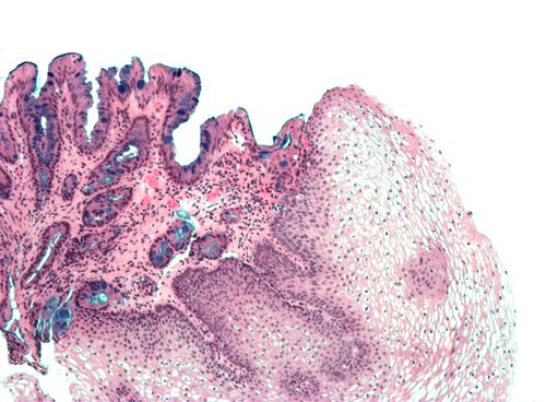
Section of the human skin showing the stratified squamous epithelial surface, referred to as the epidermis. The layer of keratin here is named the stratum corneum.
Details
ShapeMany layers of flat cells
Identifiers
TH H2.00.02.0.02025, H2.00.02.0.02030
FMA 45563
Anatomical terms of microanatomy

A stratified squamous epithelium consists of squamous (flattened) epithelial cells arranged in layers upon a basal membrane. Only one layer is in contact with the basement membrane; the other layers adhere to one another to maintain structural integrity. Although this epithelium is referred to as squamous, many cells within the layers may not be flattened; this is due to the convention of naming epithelia according to the cell type at the surface. In the deeper layers, the cells may be columnar or cuboidal. [1] There are no intercellular spaces. This type of epithelium is well suited to areas in the body subject to constant abrasion, as the thickest layers can be sequentially sloughed off and replaced before the basement membrane is exposed. It forms the outermost layer of the skin and the inner lining of the mouth, esophagus and vagina. [2]

Contents

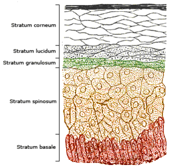
In the epidermis of skin in mammals, reptiles, and birds, the layer of keratin in the outer layer of the stratified squamous epithelial surface is named the stratum corneum. Stratum corneum is made up of squamous cells which are keratinized and dead. These are shed periodically.

Structure

Non-keratinized

Non-keratinized surfaces must be kept moist by bodily secretions to prevent them from drying out. Cells of stratum corneum are sometimes without keratin and living.

Examples of non-keratinized stratified squamous epithelium include some parts of the lining of oral cavity, pharynx, conjunctiva of eye, upper one-third esophagus, rectum, external female genitalia, and vagina.

Even non-keratinized surfaces, consisting as they do of keratinocytes, have a minor superficial keratinized layer of varying thickness, depending on the age of the epithelium and the damage it has experienced.

Keratinized

Keratinized surfaces are protected from absorption by keratin protein. Keratinized epithelium has keratin deposited on the surface which makes it impermeable and dry. Examples of keratinized stratified squamous epithelium include skin, the epidermis of the palm of the hand, and the sole of the foot, [3] and the masticatory mucosa.

Related Research Articles

<span class="mw-page-title-main">Skin</span> Soft outer covering organ of vertebrates

Skin is the layer of usually soft, flexible outer tissue covering the body of a vertebrate animal, with three main functions: protection, regulation, and sensation.

<span class="mw-page-title-main">Integumentary system</span> Skin and other protective organs

The integumentary system is the set of organs forming the outermost layer of an animal's body. It comprises the skin and its appendages, which act as a physical barrier between the external environment and the internal environment that it serves to protect and maintain the body of the animal. Mainly it is the body's outer skin.

<span class="mw-page-title-main">Tissue (biology)</span> Group of cells having similar appearance and performing the same function

In biology, tissue is a historically derived biological organizational level between cells and a complete organ. A tissue is therefore often thought of as assembly of similar cells and their extracellular matrix from the same origin that together carry out a specific function. Organs are then formed by the functional grouping together of multiple tissues.

<span class="mw-page-title-main">Epithelium</span> Tissue lining the surfaces of organs in animals

Epithelium or epithelial tissue is one of the four basic types of animal tissue, along with connective tissue, muscle tissue and nervous tissue. It is a thin, continuous, protective layer of compactly packed cells with a little intercellular matrix. Epithelial tissues line the outer surfaces of organs and blood vessels throughout the body, as well as the inner surfaces of cavities in many internal organs. An example is the epidermis, the outermost layer of the skin.

<span class="mw-page-title-main">Epidermis</span> Outermost of the three layers that make up the skin

The epidermis is the outermost of the three layers that comprise the skin, the inner layers being the dermis and hypodermis. The epidermis layer provides a barrier to infection from environmental pathogens and regulates the amount of water released from the body into the atmosphere through transepidermal water loss.

<span class="mw-page-title-main">Skin condition</span> Any medical condition that affects the integumentary system

A skin condition, also known as cutaneous condition, is any medical condition that affects the integumentary system—the organ system that encloses the body and includes skin, nails, and related muscle and glands. The major function of this system is as a barrier against the external environment.

<span class="mw-page-title-main">Stratum corneum</span> Outermost layer of the epidermis

The stratum corneum is the outermost layer of the epidermis. The human stratum corneum comprises several levels of flattened corneocytes that are divided into two layers: the stratum disjunctum and stratum compactum. The skin's protective acid mantle and lipid barrier sit on top of the stratum disjunctum. The stratum disjunctum is the uppermost and loosest layer of skin. The stratum compactum is the comparatively deeper, more compacted and more cohesive part of the stratum corneum. The corneocytes of the stratum disjunctum are larger, more rigid and more hydrophobic than that of the stratum compactum.

<span class="mw-page-title-main">Squamous metaplasia</span>

Squamous metaplasia is a benign non-cancerous change (metaplasia) of surfacing lining cells (epithelium) to a squamous morphology.

<span class="mw-page-title-main">Transitional epithelium</span> A type of tissue

Transitional epithelium is a type of stratified epithelium. Transitional epithelium is a type of tissue that changes shape in response to stretching. The transitional epithelium usually appears cuboidal when relaxed and squamous when stretched. This tissue consists of multiple layers of epithelial cells which can contract and expand in order to adapt to the degree of distension needed. Transitional epithelium lines the organs of the urinary system and is known here as urothelium. The bladder for example has a need for great distension.

<span class="mw-page-title-main">Hyperkeratosis</span> Medical condition

Hyperkeratosis is thickening of the stratum corneum, often associated with the presence of an abnormal quantity of keratin, and also usually accompanied by an increase in the granular layer. As the corneum layer normally varies greatly in thickness in different sites, some experience is needed to assess minor degrees of hyperkeratosis.

The oral mucosa is the mucous membrane lining the inside of the mouth. It comprises stratified squamous epithelium, termed "oral epithelium", and an underlying connective tissue termed lamina propria. The oral cavity has sometimes been described as a mirror that reflects the health of the individual. Changes indicative of disease are seen as alterations in the oral mucosa lining the mouth, which can reveal systemic conditions, such as diabetes or vitamin deficiency, or the local effects of chronic tobacco or alcohol use. The oral mucosa tends to heal faster and with less scar formation compared to the skin. The underlying mechanism remains unknown, but research suggests that extracellular vesicles might be involved.

<span class="mw-page-title-main">Respiratory epithelium</span> Mucosa that serves to moisten and protect the airways

Respiratory epithelium, or airway epithelium, is a type of ciliated columnar epithelium found lining most of the respiratory tract as respiratory mucosa, where it serves to moisten and protect the airways. It is not present in the vocal cords of the larynx, or the oropharynx and laryngopharynx, where instead the epithelium is stratified squamous. It also functions as a barrier to potential pathogens and foreign particles, preventing infection and tissue injury by the secretion of mucus and the action of mucociliary clearance.

<span class="mw-page-title-main">Corneal epithelium</span>

The corneal epithelium is made up of epithelial tissue and covers the front of the cornea. It acts as a barrier to protect the cornea, resisting the free flow of fluids from the tears, and prevents bacteria from entering the epithelium and corneal stroma.

<span class="mw-page-title-main">Pseudostratified columnar epithelium</span> Tissue type

A pseudostratified epithelium is a type of epithelium that, though comprising only a single layer of cells, has its cell nuclei positioned in a manner suggestive of stratified epithelia. As it rarely occurs as squamous or cuboidal epithelia, it is usually considered synonymous with the term pseudostratified columnar epithelium.

<span class="mw-page-title-main">Human skin</span> Outer covering of the body

The human skin is the outer covering of the body and is the largest organ of the integumentary system. The skin has up to seven layers of ectodermal tissue guarding muscles, bones, ligaments and internal organs. Human skin is similar to most of the other mammals' skin, and it is very similar to pig skin. Though nearly all human skin is covered with hair follicles, it can appear hairless. There are two general types of skin, hairy and glabrous skin (hairless). The adjective cutaneous literally means "of the skin".

Keratohyalin is a protein structure found in cytoplasmic granules of the keratinocytes in the stratum granulosum of the epidermis. Keratohyalin granules (KHG) mainly consist of keratin, profilaggrin, loricrin and trichohyalin proteins which contribute to cornification or keratinization, the process of the formation of epidermal cornified cell envelope. During the keratinocyte differentiation, these granules maturate and expand in size, which leads to the conversion of keratin tonofilaments into a homogenous keratin matrix, an important step in cornification.

<span class="mw-page-title-main">Nasal mucosa</span> Part of the mucus membrane lining the nasal cavity

The nasal mucosa lines the nasal cavity. It is part of the respiratory mucosa, the mucous membrane lining the respiratory tract. The nasal mucosa is intimately adherent to the periosteum or perichondrium of the nasal conchae. It is continuous with the skin through the nostrils, and with the mucous membrane of the nasal part of the pharynx through the choanae. From the nasal cavity its continuity with the conjunctiva may be traced, through the nasolacrimal and lacrimal ducts; and with the frontal, ethmoidal, sphenoidal, and maxillary sinuses, through the several openings in the nasal meatuses. The mucous membrane is thickest, and most vascular, over the nasal conchae. It is also thick over the nasal septum where increased numbers of goblet cells produce a greater amount of nasal mucus. It is very thin in the meatuses on the floor of the nasal cavities, and in the various sinuses. It is one of the most commonly infected tissues in adults and children. Inflammation of this tissue may cause significant impairment of daily activities, with symptoms such as stuffy nose, headache, mouth breathing, etc.

Tissue engineering of oral mucosa combines cells, materials and engineering to produce a three-dimensional reconstruction of oral mucosa. It is meant to simulate the real anatomical structure and function of oral mucosa. Tissue engineered oral mucosa shows promise for clinical use, such as the replacement of soft tissue defects in the oral cavity. These defects can be divided into two major categories: the gingival recessions which are tooth-related defects, and the non tooth-related defects. Non tooth-related defects can be the result of trauma, chronic infection or defects caused by tumor resection or ablation. Common approaches for replacing damaged oral mucosa are the use of autologous grafts and cultured epithelial sheets.

<span class="mw-page-title-main">Vaginal epithelium</span>

The vaginal epithelium is the inner lining of the vagina consisting of multiple layers of (squamous) cells. The basal membrane provides the support for the first layer of the epithelium-the basal layer. The intermediate layers lie upon the basal layer, and the superficial layer is the outermost layer of the epithelium. Anatomists have described the epithelium as consisting of as many as 40 distinct layers. The mucus found on the epithelium is secreted by the cervix and uterus. The rugae of the epithelium create an involuted surface and result in a large surface area that covers 360 cm2. This large surface area allows the trans-epithelial absorption of some medications via the vaginal route.

<span class="mw-page-title-main">Anatomical terms of microanatomy</span> Anatomical terminology is used to describe microanatomical (or histological) structures

Anatomical terminology is used to describe microanatomical structures. This helps describe precisely the structure, layout and position of an object, and minimises ambiguity. An internationally accepted lexicon is Terminologia Histologica.

References

  1. Tortora, Gerard J.; Derrickson, Bryan (22 November 2011). Introduction to the Human Body. The Essentials of Anatomy and Physiology (9th ed.). John Wiley & Sons, Inc. p. 84. ISBN   978-0470-59892-4. [referring to stratified squamous epithelium] Description: Two or more layers of cells; cells in apical layer and several layers deep to it are squamous; those in the deep layers vary in shape from cuboidal to columnar.
  2. Human Anatomy Laboratory Manual with Cat Dissections 7th Edition. Pearson. 18 February 2013. p. 58. ISBN   978-0-321-88418-3.
  3. Pratt, Rebecca. "Stratified Squamous Epithelium (Keratinized)". AnatomyOne. Amirsys, Inc. Archived from the original on 2013-10-29. Retrieved 2012-09-28.