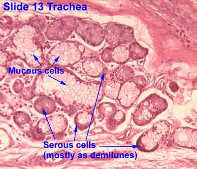
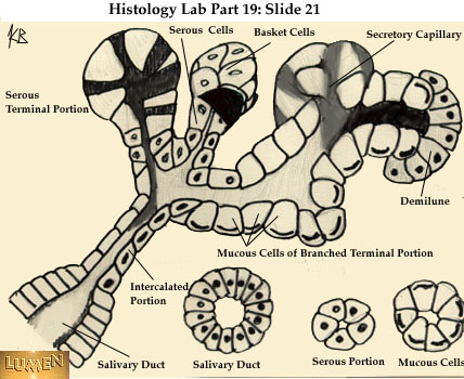
Serous demilunes, also known as Crescents of Giannuzzi or Demilunes of Heidenhain, are cellular formations in the shape of a half-moon (hence the name "demilune") on the mixed submandibular and sublingualsalivary glands.[1]
Serous demilunes are the serous cells at the distal end of mucous acini, the secretory endpieces of certain salivary glands.[1] These demilune cells secrete the proteins that contain the enzyme lysozyme, which degrades the cell walls of bacteria. In this way, lysozyme confers antimicrobial activity to mucus.
The serous demilune is an artifact from traditional methods of preparing samples. Samples are traditionally preserved and fixed in formaldehyde. When samples were preserved by quick-freezing in liquid nitrogen and then fixed with osmium tetraoxide in acetone, no demilunes were found. Examination showed that the serous cells and mucosal cells were aligned in the acinus. The traditional preparation caused mucous cells to swell during fixation which results in the serous cells being popped out of their alignment. After sectioning the serous cells resembled the common demilune shape, and were so named.[2]
Histology, also known as microscopic anatomy or microanatomy, is the branch of biology that studies the microscopic anatomy of biological tissues. Histology is the microscopic counterpart to gross anatomy, which looks at larger structures visible without a microscope. Although one may divide microscopic anatomy into organology, the study of organs, histology, the study of tissues, and cytology, the study of cells, modern usage places all of these topics under the field of histology. In medicine, histopathology is the branch of histology that includes the microscopic identification and study of diseased tissue. In the field of paleontology, the term paleohistology refers to the histology of fossil organisms.
A gland is a cell or an organ in an animal's body that produces and secretes different substances either into the bloodstream or into a body cavity or outer surface that the organism needs. A gland may also function to remove unwanted substances such as urine from the body.
Exocrine glands are glands that secrete substances onto an epithelial surface by way of a duct. Examples of exocrine glands include sweat, salivary, mammary, ceruminous, lacrimal, sebaceous, prostate and mucous. Exocrine glands are one of two types of glands in the human body, the other being endocrine glands, which secrete their products directly into the bloodstream. The liver and pancreas are both exocrine and endocrine glands; they are exocrine glands because they secrete products—bile and pancreatic juice—into the gastrointestinal tract through a series of ducts, and endocrine because they secrete other substances directly into the bloodstream. Exocrine sweat glands are part of the integumentary system; they have eccrine and apocrine types.
The salivary glands in many vertebrates including mammals are exocrine glands that produce saliva through a system of ducts. Humans have three paired major salivary glands, as well as hundreds of minor salivary glands. Salivary glands can be classified as serous, mucous, or seromucous (mixed).
The parotid gland is a major salivary gland in many animals. In humans, the two parotid glands are present on either side of the mouth and in front of both ears. They are the largest of the salivary glands. Each parotid is wrapped around the mandibular ramus, and secretes serous saliva through the parotid duct into the mouth, to facilitate mastication and swallowing and to begin the digestion of starches. There are also two other types of salivary glands; they are submandibular and sublingual glands. Sometimes accessory parotid glands are found close to the main parotid glands.
The paired submandibular glands are major salivary glands located beneath the floor of the mouth. In adult humans, they each weigh about 15 grams and contribute some 60–67% of unstimulated saliva secretion; on stimulation their contribution decreases in proportion as parotid gland secretion rises to 50%. The average length of the normal adult human submandibular salivary gland is approximately 27 mm, while the average width is approximately 14.3 mm.
Brunner's glands are compound tubuloalveolar submucosal glands found in that portion of the duodenum proximal to the hepatopancreatic sphincter.
The sublingual gland is a seromucous polystomatic exocrine gland. Located underneath the oral diaphragm, the sublingual gland is the smallest and most diffuse of the three major salivary glands of the oral cavity, with the other two being the submandibular and parotid. The sublingual gland provides approximately 3-5% of the total salivary volume.
Serous glands secrete serous fluid. They contain serous acini, a grouping of serous cells that secrete serous fluid, isotonic with blood plasma, that contains enzymes such as alpha-amylase.
A ranula is a mucus extravasation cyst involving a sublingual gland and is a type of mucocele found on the floor of the mouth. Ranulae present as a swelling of connective tissue consisting of collected mucin from a ruptured salivary gland caused by local trauma. If small and asymptomatic further treatment may not be needed, otherwise minor oral surgery may be indicated.
Sialadenitis (sialoadenitis) is inflammation of salivary glands, usually the major ones, the most common being the parotid gland, followed by submandibular and sublingual glands. It should not be confused with sialadenosis (sialosis) which is a non-inflammatory enlargement of the major salivary glands.
The esophageal glands are glands that are part of the digestive system of various animals, including humans.
The submucosa is a thin layer of tissue in various organs of the gastrointestinal, respiratory, and genitourinary tracts. It is the layer of dense irregular connective tissue that supports the mucosa and joins it to the muscular layer, the bulk of overlying smooth muscle.
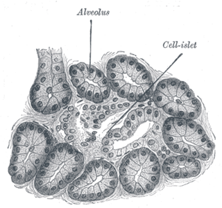
An acinus refers to any cluster of cells that resembles a many-lobed "berry," such as a raspberry. The berry-shaped termination of an exocrine gland, where the secretion is produced, is acinar in form, as is the alveolar sac containing multiple alveoli in the lungs.
Alveolar glands, also called saccular glands, are glands with a saclike secretory portion, in contrast with tubular glands. They typically have an enlarged lumen (cavity), hence the name: they have a shape similar to alveoli, the very small air sacs in the lungs.
In anatomy and physiology, a duct is a circumscribed channel leading from an exocrine gland or organ.
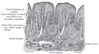
Von Ebner's glands, also called Ebner's glands or gustatory glands, are exocrine glands found in the mouth. More specifically, they are serous salivary glands which reside adjacent to the moats surrounding the circumvallate and foliate papillae just anterior to the posterior third of the tongue in its submucosa, anterior to the terminal sulcus.
Foveolar cells or surface mucous cells are mucus-producing cells which cover the inside of the stomach, protecting it from the corrosive nature of gastric acid. These cells line the gastric mucosa and the gastric pits. Mucous neck cells are found in the necks of the gastric glands. The mucus-secreting cells of the stomach can be distinguished histologically from the intestinal goblet cells, another type of mucus-secreting cell.
Acinic cell carcinoma of the lung is a very uncommon tumor that typically appears close to the right bronchus. As of 2022, only 29 cases have been documented in the English literature since Fechner et al. first described this entity in 1972. Histologically similar to the major and minor salivary glands, pluripotent cells of the submucosal serous and mucous glands of the tracheobronchial tree are thought to be the source of acinic cell carcinoma of the lung. The histologic characteristics of acinic cell carcinoma of the lung are nearly identical to those of the salivary glands.
The human digestive system consists of the gastrointestinal tract plus the accessory organs of digestion. Digestion involves the breakdown of food into smaller and smaller components, until they can be absorbed and assimilated into the body. The process of digestion has three stages: the cephalic phase, the gastric phase, and the intestinal phase.
This page is based on this Wikipedia article Text is available under the CC BY-SA 4.0 license; additional terms may apply. Images, videos and audio are available under their respective licenses.


