133l: ROLE OF ARG 115 IN THE CATALYTIC ACTION OF HUMAN LYSOZYME. X-RAY STRUCTURE OF HIS 115 AND GLU 115 MUTANTS
134l: ROLE OF ARG 115 IN THE CATALYTIC ACTION OF HUMAN LYSOZYME. X-RAY STRUCTURE OF HIS 115 AND GLU 115 MUTANTS
1b5u: CONTRIBUTION OF HYDROGEN BONDS TO THE CONFORMATIONAL STABILITY OF HUMAN LYSOZYME: CALORIMETRY AND X-RAY ANALYSIS OF SIX SER->ALA MUTANT
1b5v: CONTRIBUTION OF HYDROGEN BONDS TO THE CONFORMATIONAL STABILITY OF HUMAN LYSOZYME: CALORIMETRY AND X-RAY ANALYSIS OF SIX SER->ALA MUTANTS
1b5w: CONTRIBUTION OF HYDROGEN BONDS TO THE CONFORMATIONAL STABILITY OF HUMAN LYSOZYME: CALORIMETRY AND X-RAY ANALYSIS OF SIX SER->ALA MUTANTS
1b5x: Contribution of hydrogen bonds to the conformational stability of human lysozyme: calorimetry and x-ray analysis of six ser->ala mutants
1b5y: CONTRIBUTION OF HYDROGEN BONDS TO THE CONFORMATIONAL STABILITY OF HUMAN LYSOZYME: CALORIMETRY AND X-RAY ANALYSIS OF SIX SER->ALA MUTANTS
1b5z: CONTRIBUTION OF HYDROGEN BONDS TO THE CONFORMATIONAL STABILITY OF HUMAN LYSOZYME: CALORIMETRY AND X-RAY ANALYSIS OF SIX SER->ALA MUTANTS
1b7l: VERIFICATION OF SPMP USING MUTANT HUMAN LYSOZYMES
1b7m: VERIFICATION OF SPMP USING MUTANT HUMAN LYSOZYMES
1b7n: VERIFICATION OF SPMP USING MUTANT HUMAN LYSOZYMES
1b7o: VERIFICATION OF SPMP USING MUTANT HUMAN LYSOZYMES
1b7p: VERIFICATION OF SPMP USING MUTANT HUMAN LYSOZYMES
1b7q: VERIFICATION OF SPMP USING MUTANT HUMAN LYSOZYMES
1b7r: VERIFICATION OF SPMP USING MUTANT HUMAN LYSOZYMES
1b7s: VERIFICATION OF SPMP USING MUTANT HUMAN LYSOZYMES
1bb3: HUMAN LYSOZYME MUTANT A96L
1bb4: HUMAN LYSOZYME DOUBLE MUTANT A96L, W109H
1bb5: HUMAN LYSOZYME MUTANT A96L COMPLEXED WITH CHITOTRIOSE
1c43: MUTANT HUMAN LYSOZYME WITH FOREIGN N-TERMINAL RESIDUES
1c45: MUTANT HUMAN LYSOZYME WITH FOREIGN N-TERMINAL RESIDUES
1c46: MUTANT HUMAN LYSOZYME WITH FOREIGN N-TERMINAL RESIDUES
1c7p: CRYSTAL STRUCTURE OF MUTANT HUMAN LYSOZYME WITH FOUR EXTRA RESIDUES (EAEA) AT THE N-TERMINAL
1cj6: T11A MUTANT HUMAN LYSOZYME
1cj7: T11V MUTANT HUMAN LYSOZYME
1cj8: T40A MUTANT HUMAN LYSOZYME
1cj9: T40V MUTANT HUMAN LYSOZYME
1ckc: T43A MUTANT HUMAN LYSOZYME
1ckd: T43V MUTANT HUMAN LYSOZYME
1ckf: T52A MUTANT HUMAN LYSOZYME
1ckg: T52V MUTANT HUMAN LYSOZYME
1ckh: T70V MUTANT HUMAN LYSOZYME
1d6p: HUMAN LYSOZYME L63 MUTANT LABELLED WITH 2',3'-EPOXYPROPYL N,N'-DIACETYLCHITOBIOSE
1d6q: HUMAN LYSOZYME E102 MUTANT LABELLED WITH 2',3'-EPOXYPROPYL GLYCOSIDE OF N-ACETYLLACTOSAMINE
1di3: ROLE OF AMINO ACID RESIDUES AT TURNS IN THE CONFORMATIONAL STABILITY AND FOLDING OF HUMAN LYSOZYME
1di4: ROLE OF AMINO ACID RESIDUES AT TURNS IN THE CONFORMATIONAL STABILITY AND FOLDING OF HUMAN LYSOZYME
1di5: ROLE OF AMINO ACID RESIDUES AT TURNS IN THE CONFORMATIONAL STABILITY AND FOLDING OF HUMAN LYSOZYME
1eq4: CRYSTAL STRUCTURES OF SALT BRIDGE MUTANTS OF HUMAN LYSOZYME
1eq5: CRYSTAL STRUCTURES OF SALT BRIDGE MUTANTS OF HUMAN LYSOZYME
1eqe: CRYSTAL STRUCTURES OF SALT BRIDGE MUTANTS OF HUMAN LYSOZYME
1gay: CRYSTAL STRUCTURE OF MUTANT HUMAN LYSOZYME SUBSTITUTED AT THE SURFACE POSITIONS
1gaz: Crystal Structure of Mutant Human Lysozyme Substituted at the Surface Positions
1gb0: CRYSTAL STRUCTURE OF MUTANT HUMAN LYSOZYME SUBSTITUTED AT THE SURFACE POSITIONS
1gb2: CRYSTAL STRUCTURE OF MUTANT HUMAN LYSOZYME SUBSTITUTED AT THE SURFACE POSITIONS
1gb3: CRYSTAL STRUCTURE OF MUTANT HUMAN LYSOZYME SUBSTITUTED AT THE SURFACE POSITIONS
1gb5: CRYSTAL STRUCTURE OF MUTANT HUMAN LYSOZYME SUBSTITUTED AT THE SURFACE POSITIONS
1gb6: CRYSTAL STRUCTURE OF MUTANT HUMAN LYSOZYME SUBSTITUTED AT THE SURFACE POSITIONS
1gb7: CRYSTAL STRUCTURE OF MUTANT HUMAN LYSOZYME SUBSTITUTED AT THE SURFACE POSITIONS
1gb8: CRYSTAL STRUCTURE OF MUTANT HUMAN LYSOZYME SUBSTITUTED AT THE SURFACE POSITIONS
1gb9: CRYSTAL STRUCTURE OF MUTANT HUMAN LYSOZYME SUBSTITUTED AT THE SURFACE POSITIONS
1gbo: CRYSTAL STRUCTURE OF MUTANT HUMAN LYSOZYME SUBSTITUTED AT THE SURFACE POSITIONS
1gbw: CRYSTAL STRUCTURE OF MUTANT HUMAN LYSOZYME SUBSTITUTED AT THE SURFACE POSITIONS
1gbx: CRYSTAL STRUCTURE OF MUTANT HUMAN LYSOZYME SUBSTITUTED AT THE SURFACE POSITIONS
1gby: CRYSTAL STRUCTURE OF MUTANT HUMAN LYSOZYME SUBSTITUTED AT THE SURFACE POSITIONS
1gbz: CRYSTAL STRUCTURE OF MUTANT HUMAN LYSOZYME SUBSTITUTED AT THE SURFACE POSITIONS
1gdw: CRYSTAL STRUCTURE OF MUTANT HUMAN LYSOZYME SUBSTITUTED AT LEFT-HANDED HELICAL POSITIONS
1gdx: CRYSTAL STRUCTURE OF MUTANT HUMAN LYSOZYME SUBSTITUTED AT LEFT-HANDED HELICAL POSITIONS
1ge0: CRYSTAL STRUCTURE OF MUTANT HUMAN LYSOZYME SUBSTITUTED AT LEFT-HANDED HELICAL POSITIONS
1ge1: CRYSTAL STRUCTURE OF MUTANT HUMAN LYSOZYME SUBSTITUTED AT LEFT-HANDED HELICAL POSITIONS
1ge2: CRYSTAL STRUCTURE OF MUTANT HUMAN LYSOZYME SUBSTITUTED AT LEFT-HANDED HELICAL POSITIONS
1ge3: CRYSTAL STRUCTURE OF MUTANT HUMAN LYSOZYME SUBSTITUTED AT LEFT-HANDED HELICAL POSITIONS
1ge4: CRYSTAL STRUCTURE OF MUTANT HUMAN LYSOZYME SUBSTITUTED AT LEFT-HANDED HELICAL POSITIONS
1gev: BURIED POLAR MUTANT HUMAN LYSOZYME
1gez: BURIED POLAR MUTANT HUMAN LYSOZYME
1gf0: BURIED POLAR MUTANT HUMAN LYSOZYME
1gf3: BURIED POLAR MUTANT HUMAN LYSOZYME
1gf4: BURIED POLAR MUTANT HUMAN LYSOZYME
1gf5: BURIED POLAR MUTANT HUMAN LYSOZYME
1gf6: BURIED POLAR MUTANT HUMAN LYSOZYME
1gf7: BURIED POLAR MUTANT HUMAN LYSOZYME
1gf8: CRYSTAL STRUCTURE OF MUTANT HUMAN LYSOZYME SUBSTITUTED AT THE SURFACE POSITIONS
1gf9: CRYSTAL STRUCTURE OF MUTANT HUMAN LYSOZYME SUBSTITUTED AT THE SURFACE POSITIONS
1gfa: CRYSTAL STRUCTURE OF MUTANT HUMAN LYSOZYME SUBSTITUTED AT THE SURFACE POSITIONS
1gfe: CRYSTAL STRUCTURE OF MUTANT HUMAN LYSOZYME SUBSTITUTED AT THE SURFACE POSITIONS
1gfg: CRYSTAL STRUCTURE OF MUTANT HUMAN LYSOZYME SUBSTITUTED AT THE SURFACE POSITIONS
1gfh: CRYSTAL STRUCTURE OF MUTANT HUMAN LYSOZYME SUBSTITUTED AT THE SURFACE POSITIONS
1gfj: CRYSTAL STRUCTURE OF MUTANT HUMAN LYSOZYME SUBSTITUTED AT THE SURFACE POSITIONS
1gfk: CRYSTAL STRUCTURE OF MUTANT HUMAN LYSOZYME SUBSTITUTED AT THE SURFACE POSITIONS
1gfr: CRYSTAL STRUCTURE OF MUTANT HUMAN LYSOZYME SUBSTITUTED AT THE SURFACE POSITIONS
1gft: CRYSTAL STRUCTURE OF MUTANT HUMAN LYSOZYME SUBSTITUTED AT THE SURFACE POSITIONS
1gfu: CRYSTAL STRUCTURE OF MUTANT HUMAN LYSOZYME SUBSTITUTED AT THE SURFACE POSITIONS
1gfv: CRYSTAL STRUCTURE OF MUTANT HUMAN LYSOZYME SUBSTITUTED AT THE SURFACE POSITIONS
1hnl: CRYSTAL STRUCTURE OF A GLUTATHIONYLATED HUMAN LYSOZYME: A FOLDING INTERMEDIATE MIMIC IN THE FORMATION OF A DISULFIDE BOND
1i1z: MUTANT HUMAN LYSOZYME (Q86D)
1i20: MUTANT HUMAN LYSOZYME (A92D)
1i22: MUTANT HUMAN LYSOZYME (A83K/Q86D/A92D)
1inu: CRYSTAL STRUCTURE OF MUTANT HUMAN LYSOZYME SUBSTITUTED AT THE SURFACE POSITIONS
1ioc: CRYSTAL STRUCTURE OF MUTANT HUMAN LYSOZYME, EAEA-I56T
1ip1: G37A HUMAN LYSOZYME
1ip2: G48A HUMAN LYSOZYME
1ip3: G68A HUMAN LYSOZYME
1ip4: G72A HUMAN LYSOZYME
1ip5: G105A HUMAN LYSOZYME
1ip6: G127A HUMAN LYSOZYME
1ip7: G129A HUMAN LYSOZYME
1iwt: Crystal Structure Analysis of Human lysozyme at 113K.
1iwu: Crystal Structure Analysis of Human lysozyme at 127K.
1iwv: Crystal Structure Analysis of Human lysozyme at 147K.
1iww: Crystal Structure Analysis of Human lysozyme at 152K.
1iwx: Crystal Structure Analysis of Human lysozyme at 161K.
1iwy: Crystal Structure Analysis of Human lysozyme at 170K.
1iwz: Crystal Structure Analysis of Human lysozyme at 178K.
1ix0: I59A-3SS human lysozyme
1iy3: Solution Structure of the Human lysozyme at 4 degree C
1iy4: Solution structure of the human lysozyme at 35 degree C
1jka: HUMAN LYSOZYME MUTANT WITH GLU 35 REPLACED BY ASP
1jkb: HUMAN LYSOZYME MUTANT WITH GLU 35 REPLACED BY ALA
1jkc: HUMAN LYSOZYME MUTANT WITH TRP 109 REPLACED BY PHE
1jkd: HUMAN LYSOZYME MUTANT WITH TRP 109 REPLACED BY ALA
1jsf: FULL-MATRIX LEAST-SQUARES REFINEMENT OF HUMAN LYSOZYME
1jwr: Crystal structure of human lysozyme at 100 K
1laa: X-RAY STRUCTURE OF GLU 53 HUMAN LYSOZYME
1lhh: ROLE OF PROLINE RESIDUES IN HUMAN LYSOZYME STABILITY: A SCANNING CALORIMETRIC STUDY COMBINED WITH X-RAY STRUCTURE ANALYSIS OF PROLINE MUTANTS
1lhi: ROLE OF PROLINE RESIDUES IN HUMAN LYSOZYME STABILITY: A SCANNING CALORIMETRIC STUDY COMBINED WITH X-RAY STRUCTURE ANALYSIS OF PROLINE MUTANTS
1lhj: ROLE OF PROLINE RESIDUES IN HUMAN LYSOZYME STABILITY: A SCANNING CALORIMETRIC STUDY COMBINED WITH X-RAY STRUCTURE ANALYSIS OF PROLINE MUTANTS
1lhk: ROLE OF PROLINE RESIDUES IN HUMAN LYSOZYME STABILITY: A SCANNING CALORIMETRIC STUDY COMBINED WITH X-RAY STRUCTURE ANALYSIS OF PROLINE MUTANTS
1lhl: ROLE OF PROLINE RESIDUES IN HUMAN LYSOZYME STABILITY: A SCANNING CALORIMETRIC STUDY COMBINED WITH X-RAY STRUCTURE ANALYSIS OF PROLINE MUTANTS
1lhm: THE CRYSTAL STRUCTURE OF A MUTANT LYSOZYME C77(SLASH)95A WITH INCREASED SECRETION EFFICIENCY IN YEAST
1lmt: STRUCTURE OF A CONFORMATIONALLY CONSTRAINED ARG-GLY-ASP SEQUENCE INSERTED INTO HUMAN LYSOZYME
1loz: AMYLOIDOGENIC VARIANT (I56T) VARIANT OF HUMAN LYSOZYME
1lyy: AMYLOIDOGENIC VARIANT (ASP67HIS) OF HUMAN LYSOZYME
1lz1: REFINEMENT OF HUMAN LYSOZYME AT 1.5 ANGSTROMS RESOLUTION. ANALYSIS OF NON-BONDED AND HYDROGEN-BOND INTERACTIONS
1lz4: ENTHALPIC DESTABILIZATION OF A MUTANT HUMAN LYSOZYME LACKING A DISULFIDE BRIDGE BETWEEN CYSTEINE-77 AND CYSTEINE-95
1lz5: STRUCTURAL AND FUNCTIONAL ANALYSES OF THE ARG-GLY-ASP SEQUENCE INTRODUCED INTO HUMAN LYSOZYME
1lz6: STRUCTURAL AND FUNCTIONAL ANALYSES OF THE ARG-GLY-ASP SEQUENCE INTRODUCED INTO HUMAN LYSOZYME
1lzr: STRUCTURAL CHANGES OF THE ACTIVE SITE CLEFT AND DIFFERENT SACCHARIDE BINDING MODES IN HUMAN LYSOZYME CO-CRYSTALLIZED WITH HEXA-N-ACETYL-CHITOHEXAOSE AT PH 4.0
1lzs: STRUCTURAL CHANGES OF THE ACTIVE SITE CLEFT AND DIFFERENT SACCHARIDE BINDING MODES IN HUMAN LYSOZYME CO-CRYSTALLIZED WITH HEXA-N-ACETYL-CHITOHEXAOSE AT PH 4.0
1op9: Complex of human lysozyme with camelid VHH HL6 antibody fragment
1oua: CONTRIBUTION OF HYDROPHOBIC RESIDUES TO THE STABILITY OF HUMAN LYSOZYME: X-RAY STRUCTURE OF THE I56T MUTANT
1oub: CONTRIBUTION OF HYDROPHOBIC RESIDUES TO THE STABILITY OF HUMAN LYSOZYME: X-RAY STRUCTURE OF THE V100A MUTANT
1ouc: CONTRIBUTION OF HYDROPHOBIC RESIDUES TO THE STABILITY OF HUMAN LYSOZYME: X-RAY STRUCTURE OF THE V110A MUTANT
1oud: CONTRIBUTION OF HYDROPHOBIC RESIDUES TO THE STABILITY OF HUMAN LYSOZYME: X-RAY STRUCTURE OF THE V121A MUTANT
1oue: CONTRIBUTION OF HYDROPHOBIC RESIDUES TO THE STABILITY OF HUMAN LYSOZYME: X-RAY STRUCTURE OF THE V125A MUTANT
1ouf: CONTRIBUTION OF HYDROPHOBIC RESIDUES TO THE STABILITY OF HUMAN LYSOZYME: X-RAY STRUCTURE OF THE V130A MUTANT
1oug: CONTRIBUTION OF HYDROPHOBIC RESIDUES TO THE STABILITY OF HUMAN LYSOZYME: X-RAY STRUCTURE OF THE V2A MUTANT
1ouh: CONTRIBUTION OF HYDROPHOBIC RESIDUES TO THE STABILITY OF HUMAN LYSOZYME: X-RAY STRUCTURE OF THE V74A MUTANT
1oui: CONTRIBUTION OF HYDROPHOBIC RESIDUES TO THE STABILITY OF HUMAN LYSOZYME: X-RAY STRUCTURE OF THE V93A MUTANT
1ouj: CONTRIBUTION OF HYDROPHOBIC RESIDUES TO THE STABILITY OF HUMAN LYSOZYME: X-RAY STRUCTURE OF THE V99A MUTANT
1qsw: CRYSTAL STRUCTURE ANALYSIS OF A HUMAN LYSOZYME MUTANT W64C C65A
1re2: HUMAN LYSOZYME LABELLED WITH TWO 2',3'-EPOXYPROPYL BETA-GLYCOSIDE OF N-ACETYLLACTOSAMINE
1rem: HUMAN LYSOZYME WITH MAN-B1,4-GLCNAC COVALENTLY ATTACHED TO ASP53
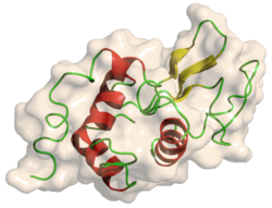
1rex: NATIVE HUMAN LYSOZYME
1rey: HUMAN LYSOZYME-N,N'-DIACETYLCHITOBIOSE COMPLEX
1rez: HUMAN LYSOZYME-N-ACETYLLACTOSAMINE COMPLEX
1tay: DISSECTION OF THE FUNCTIONAL ROLE OF STRUCTURAL ELEMENTS OF TYROSINE-63 IN THE CATALYTIC ACTION OF HUMAN LYSOZYME
1tby: DISSECTION OF THE FUNCTIONAL ROLE OF STRUCTURAL ELEMENTS OF TYROSINE-63 IN THE CATALYTIC ACTION OF HUMAN LYSOZYME
1tcy: DISSECTION OF THE FUNCTIONAL ROLE OF STRUCTURAL ELEMENTS OF TYROSINE-63 IN THE CATALYTIC ACTION OF HUMAN LYSOZYME
1tdy: DISSECTION OF THE FUNCTIONAL ROLE OF STRUCTURAL ELEMENTS OF TYROSINE-63 IN THE CATALYTIC ACTION OF HUMAN LYSOZYME
1ubz: Crystal structure of Glu102-mutant human lysozyme doubly labeled with 2',3'-epoxypropyl beta-glycoside of N-acetyllactosamine
1w08: STRUCTURE OF T70N HUMAN LYSOZYME
1wqm: CONTRIBUTION OF HYDROGEN BONDS TO THE CONFORMATIONAL STABILITY OF HUMAN LYSOZYME
1wqn: CONTRIBUTION OF HYDROGEN BONDS TO THE CONFORMATIONAL STABILITY OF HUMAN LYSOZYME
1wqo: CONTRIBUTION OF HYDROGEN BONDS TO THE CONFORMATIONAL STABILITY OF HUMAN LYSOZYME
1wqp: CONTRIBUTION OF HYDROGEN BONDS TO THE CONFORMATIONAL STABILITY OF HUMAN LYSOZYME
1wqq: CONTRIBUTION OF HYDROGEN BONDS TO THE CONFORMATIONAL STABILITY OF HUMAN LYSOZYME
1wqr: CONTRIBUTION OF HYDROGEN BONDS TO THE CONFORMATIONAL STABILITY OF HUMAN LYSOZYME
1yam: CONTRIBUTION OF HYDROPHOBIC RESIDUES TO THE STABILITY OF HUMAN LYSOZYME: CALORIMETRIC STUDIES AND X-RAY STRUCTURAL ANALYSIS OF THE FIVE ISOLEUCINE TO VALINE MUTANTS
1yan: CONTRIBUTION OF HYDROPHOBIC RESIDUES TO THE STABILITY OF HUMAN LYSOZYME: CALORIMETRIC STUDIES AND X-RAY STRUCTURAL ANALYSIS OF THE FIVE ISOLEUCINE TO VALINE MUTANTS
1yao: CONTRIBUTION OF HYDROPHOBIC RESIDUES TO THE STABILITY OF HUMAN LYSOZYME: CALORIMETRIC STUDIES AND X-RAY STRUCTURAL ANALYSIS OF THE FIVE ISOLEUCINE TO VALINE MUTANTS
1yap: CONTRIBUTION OF HYDROPHOBIC RESIDUES TO THE STABILITY OF HUMAN LYSOZYME: CALORIMETRIC STUDIES AND X-RAY STRUCTURAL ANALYSIS OF THE FIVE ISOLEUCINE TO VALINE MUTANTS
1yaq: CONTRIBUTION OF HYDROPHOBIC RESIDUES TO THE STABILITY OF HUMAN LYSOZYME: CALORIMETRIC STUDIES AND X-RAY STRUCTURAL ANALYSIS OF THE FIVE ISOLEUCINE TO VALINE MUTANTS
207l: MUTANT HUMAN LYSOZYME C77A
208l: MUTANT HUMAN LYSOZYME C77A
2bqa: CONTRIBUTION OF HYDROPHOBIC EFFECT TO THE CONFORMATIONAL STABILITY OF HUMAN LYSOZYME
2bqb: CONTRIBUTION OF HYDROPHOBIC EFFECT TO THE CONFORMATIONAL STABILITY OF HUMAN LYSOZYME
2bqc: CONTRIBUTION OF HYDROPHOBIC EFFECT TO THE CONFORMATIONAL STABILITY OF HUMAN LYSOZYME
2bqd: CONTRIBUTION OF HYDROPHOBIC EFFECT TO THE CONFORMATIONAL STABILITY OF HUMAN LYSOZYME
2bqe: CONTRIBUTION OF HYDROPHOBIC EFFECT TO THE CONFORMATIONAL STABILITY OF HUMAN LYSOZYME
2bqf: CONTRIBUTION OF HYDROPHOBIC EFFECT TO THE CONFORMATIONAL STABILITY OF HUMAN LYSOZYME
2bqg: CONTRIBUTION OF HYDROPHOBIC EFFECT TO THE CONFORMATIONAL STABILITY OF HUMAN LYSOZYME
2bqh: CONTRIBUTION OF HYDROPHOBIC EFFECT TO THE CONFORMATIONAL STABILITY OF HUMAN LYSOZYME
2bqi: CONTRIBUTION OF HYDROPHOBIC EFFECT TO THE CONFORMATIONAL STABILITY OF HUMAN LYSOZYME
2bqj: CONTRIBUTION OF HYDROPHOBIC EFFECT TO THE CONFORMATIONAL STABILITY OF HUMAN LYSOZYME
2bqk: CONTRIBUTION OF HYDROPHOBIC EFFECT TO THE CONFORMATIONAL STABILITY OF HUMAN LYSOZYME
2bql: CONTRIBUTION OF HYDROPHOBIC EFFECT TO THE CONFORMATIONAL STABILITY OF HUMAN LYSOZYME
2bqm: CONTRIBUTION OF HYDROPHOBIC EFFECT TO THE CONFORMATIONAL STABILITY OF HUMAN LYSOZYME
2bqn: CONTRIBUTION OF HYDROPHOBIC EFFECT TO THE CONFORMATIONAL STABILITY OF HUMAN LYSOZYME
2bqo: CONTRIBUTION OF HYDROPHOBIC EFFECT TO THE CONFORMATIONAL STABILITY OF HUMAN LYSOZYME
2hea: CONTRIBUTION OF WATER MOLECULES IN THE INTERIOR OF A PROTEIN TO THE CONFORMATIONAL STABILITY
2heb: CONTRIBUTION OF WATER MOLECULES IN THE INTERIOR OF A PROTEIN TO THE CONFORMATIONAL STABILITY
2hec: CONTRIBUTION OF WATER MOLECULES IN THE INTERIOR OF A PROTEIN TO THE CONFORMATIONAL STABILITY
2hed: CONTRIBUTION OF WATER MOLECULES IN THE INTERIOR OF A PROTEIN TO THE CONFORMATIONAL STABILITY
2hee: CONTRIBUTION OF WATER MOLECULES IN THE INTERIOR OF A PROTEIN TO THE CONFORMATIONAL STABILITY
2hef: CONTRIBUTION OF WATER MOLECULES IN THE INTERIOR OF A PROTEIN TO THE CONFORMATIONAL STABILITY
2lhm: CRYSTAL STRUCTURES OF THE APO-AND HOLOMUTANT HUMAN LYSOZYMES WITH AN INTRODUCED CA2+ BINDING SITE
2mea: CHANGES IN CONFORMATIONAL STABILITY OF A SERIES OF MUTANT HUMAN LYSOZYMES AT CONSTANT POSITIONS
2meb: CHANGES IN CONFORMATIONAL STABILITY OF A SERIES OF MUTANT HUMAN LYSOZYMES AT CONSTANT POSITIONS
2mec: CHANGES IN CONFORMATIONAL STABILITY OF A SERIES OF MUTANT HUMAN LYSOZYMES AT CONSTANT POSITIONS
2med: CONTRIBUTION OF HYDROPHOBIC EFFECT TO THE CONFORMATIONAL STABILITY OF HUMAN LYSOZYME
2mee: CONTRIBUTION OF HYDROPHOBIC EFFECT TO THE CONFORMATIONAL STABILITY OF HUMAN LYSOZYME
2mef: CONTRIBUTION OF HYDROPHOBIC EFFECT TO THE CONFORMATIONAL STABILITY OF HUMAN LYSOZYME
2meg: CHANGES IN CONFORMATIONAL STABILITY OF A SERIES OF MUTANT HUMAN LYSOZYMES AT CONSTANT POSITIONS.
2meh: CONTRIBUTION OF HYDROPHOBIC EFFECT TO THE CONFORMATIONAL STABILITY OF HUMAN LYSOZYME
2mei: CONTRIBUTION OF HYDROPHOBIC EFFECT TO THE CONFORMATIONAL STABILITY OF HUMAN LYSOZYME
2nwd: Structure of chemically synthesized human lysozyme at 1 Angstrom resolution
3lhm: CRYSTAL STRUCTURES OF THE APO-AND HOLOMUTANT HUMAN LYSOZYMES WITH AN INTRODUCED CA2+ BINDING SITE