Identification

Superfamilies of proteins are identified using a number of methods. Closely related members can be identified by different methods to those needed to group the most evolutionarily divergent members.
Sequence similarity
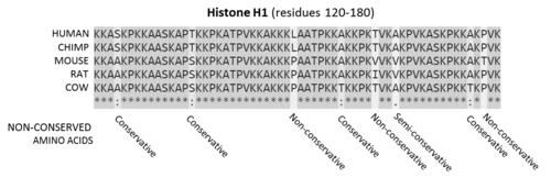
- * conserved sequence,
- : conservative mutations,
- . semi-conservative mutations, and
- ␣ non-conservative mutations.
Historically, the similarity of different amino acid sequences has been the most common method of inferring homology. [6] Sequence similarity is considered a good predictor of relatedness, since similar sequences are more likely the result of gene duplication and divergent evolution, rather than the result of convergent evolution. Amino acid sequence is typically more conserved than DNA sequence (due to the degenerate genetic code), so it is a more sensitive detection method. Since some of the amino acids have similar properties (e.g., charge, hydrophobicity, size), conservative mutations that interchange them are often neutral to function. The most conserved sequence regions of a protein often correspond to functionally important regions like catalytic sites and binding sites, since these regions are less tolerant to sequence changes.
Using sequence similarity to infer homology has several limitations. There is no minimum level of sequence similarity guaranteed to produce identical structures. Over long periods of evolution, related proteins may show no detectable sequence similarity to one another. Sequences with many insertions and deletions can also sometimes be difficult to align and so identify the homologous sequence regions. In the PA clan of proteases, for example, not a single residue is conserved through the superfamily, not even those in the catalytic triad. Conversely, the individual families that make up a superfamily are defined on the basis of their sequence alignment, for example the C04 protease family within the PA clan.
Nevertheless, sequence similarity is the most commonly used form of evidence to infer relatedness, since the number of known sequences vastly outnumbers the number of known tertiary structures. [7] In the absence of structural information, sequence similarity constrains the limits of which proteins can be assigned to a superfamily. [7]
Structural similarity

Structure is much more evolutionarily conserved than sequence, such that proteins with highly similar structures can have entirely different sequences. [8] Over very long evolutionary timescales, very few residues show detectable amino acid sequence conservation, however secondary structural elements and tertiary structural motifs are highly conserved. Some protein dynamics [9] and conformational changes of the protein structure may also be conserved, as is seen in the serpin superfamily. [10] Consequently, protein tertiary structure can be used to detect homology between proteins even when no evidence of relatedness remains in their sequences. Structural alignment programs, such as DALI, use the 3D structure of a protein of interest to find proteins with similar folds. [11] However, on rare occasions, related proteins may evolve to be structurally dissimilar [12] and relatedness can only be inferred by other methods. [13] [14] [15]
Mechanistic similarity
The catalytic mechanism of enzymes within a superfamily is commonly conserved, although substrate specificity may be significantly different. [16] Catalytic residues also tend to occur in the same order in the protein sequence. [17] For the families within the PA clan of proteases, although there has been divergent evolution of the catalytic triad residues used to perform catalysis, all members use a similar mechanism to perform covalent, nucleophilic catalysis on proteins, peptides or amino acids. [18] However, mechanism alone is not sufficient to infer relatedness. Some catalytic mechanisms have been convergently evolved multiple times independently, and so form separate superfamilies, [19] [20] [21] and in some superfamilies display a range of different (though often chemically similar) mechanisms. [16] [22]