Histone

Last updated
Schematic representation of the assembly of the core histones into the nucleosome. Nucleosome structure.png
Schematic representation of the assembly of the core histones into the nucleosome.

In biology, histones are highly basic proteins abundant in lysine and arginine residues that are found in eukaryotic cell nuclei and in most Archaeal phyla. They act as spools around which DNA winds to create structural units called nucleosomes. [1] [2] Nucleosomes in turn are wrapped into 30-nanometer fibers that form tightly packed chromatin. Histones prevent DNA from becoming tangled and protect it from DNA damage. In addition, histones play important roles in gene regulation and DNA replication. Without histones, unwound DNA in chromosomes would be very long. For example, each human cell has about 1.8 meters of DNA if completely stretched out; however, when wound about histones, this length is reduced to about 90 micrometers (0.09 mm) of 30 nm diameter chromatin fibers. [3]

Contents

There are five families of histones which are designated H1/H5 (linker histones), H2, H3, and H4 (core histones). The nucleosome core is formed of two H2A-H2B dimers and a H3-H4 tetramer. The tight wrapping of DNA around histones is to a large degree a result of electrostatic attraction between the positively charged histones and negatively charged phosphate backbone of DNA.

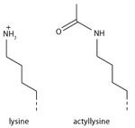
Histones may be chemically modified through the action of enzymes to regulate gene transcription. The most common modification are the methylation of arginine or lysine residues or the acetylation of lysine. Methylation can affect how other protein such as transcription factors interact with the nucleosomes. Lysine acetylation eliminates a positive charge on lysine thereby weakening the electrostatic attraction between histone and DNA resulting in partial unwinding of the DNA making it more accessible for gene expression.

Classes and variants

Histone heterooctamer (H3,H4,H2A,H2B) + DNA fragment, Frog 1aoi.jpg
Histone heterooctamer (H3,H4,H2A,H2B) + DNA fragment, Frog

Five major families of histone proteins exist: H1/H5, H2A, H2B, H3, and H4. [2] [4] [5] [6] Histones H2A, H2B, H3 and H4 are known as the core or nucleosomal histones, while histones H1/H5 are known as the linker histones.

The core histones all exist as dimers, which are similar in that they all possess the histone fold domain: three alpha helices linked by two loops. It is this helical structure that allows for interaction between distinct dimers, particularly in a head-tail fashion (also called the handshake motif). [7] The resulting four distinct dimers then come together to form one octameric nucleosome core, approximately 63 Angstroms in diameter (a solenoid (DNA)-like particle). Around 146 base pairs (bp) of DNA wrap around this core particle 1.65 times in a left-handed super-helical turn to give a particle of around 100 Angstroms across. [8] The linker histone H1 binds the nucleosome at the entry and exit sites of the DNA, thus locking the DNA into place [9] and allowing the formation of higher order structure. The most basic such formation is the 10 nm fiber or beads on a string conformation. This involves the wrapping of DNA around nucleosomes with approximately 50 base pairs of DNA separating each pair of nucleosomes (also referred to as linker DNA). Higher-order structures include the 30 nm fiber (forming an irregular zigzag) and 100 nm fiber, these being the structures found in normal cells. During mitosis and meiosis, the condensed chromosomes are assembled through interactions between nucleosomes and other regulatory proteins.

Histones are subdivided into canonical replication-dependent histones, whose genes are expressed during the S-phase of the cell cycle and replication-independent histone variants, expressed during the whole cell cycle. In mammals, genes encoding canonical histones are typically clustered along chromosomes in 4 different highly-conserved loci, lack introns and use a stem loop structure at the 3' end instead of a polyA tail. Genes encoding histone variants are usually not clustered, have introns and their mRNAs are regulated with polyA tails. [10] Complex multicellular organisms typically have a higher number of histone variants providing a variety of different functions. Recent data are accumulating about the roles of diverse histone variants highlighting the functional links between variants and the delicate regulation of organism development. [11] Histone variants proteins from different organisms, their classification and variant specific features can be found in "HistoneDB 2.0 - Variants" database. [12] [13] Several pseudogenes have also been discovered and identified in very close sequences of their respective functional ortholog genes. [14] [15]

The following is a list of human histone proteins, genes and pseudogenes: [10]

Super familyFamilyReplication-dependent genesReplication-independent genesPseudogenes
Linker H1 H1-1, H1-2, H1-3, H1-4, H1-5, H1-6 H1-0, H1-7, H1-8, H1-10 H1-9P, H1-12P
Core H2A H2AC1, H2AC4, H2AC6, H2AC7, H2AC8, H2AC11, H2AC12, H2AC13, H2AC14, H2AC15, H2AC16, H2AC17, H2AC18, H2AC19, H2AC20, H2AC21, H2AC25 H2AZ1, H2AZ2, MACROH2A1, MACROH2A2, H2AX, H2AJ, H2AB1, H2AB2, H2AB3, H2AP, H2AL1Q, H2AL3 H2AC2P, H2AC3P, H2AC5P, H2AC9P, H2AC10P, H2AQ1P, H2AL1MP
H2B H2BC1, H2BC3, H2BC4, H2BC5, H2BC6, H2BC7, H2BC8, H2BC9, H2BC10, H2BC11, H2BC12, H2BC13, H2BC14, H2BC15, H2BC17, H2BC18, H2BC21, H2BC26, H2BC12L H2BK1, H2BW1, H2BW2, H2BW3P, H2BN1 H2BC2P, H2BC16P, H2BC19P, H2BC20P, H2BC27P, H2BL1P, H2BW3P, H2BW4P
H3 H3C1, H3C2, H3C3, H3C4, H3C6, H3C7, H3C8, H3C10, H3C11, H3C12, H3C13, H3C14, H3C15, H3-4 H3-3A, H3-3B, H3-5, H3-7, H3Y1, H3Y2, CENPA H3C5P, H3C9P, H3P16, H3P44
H4 H4C1, H4C2, H4C3, H4C4, H4C5, H4C6, H4C7, H4C8, H4C9, H4C11, H4C12, H4C13, H4C14, H4C15 H4C16 H4C10P

Structure

Steps in nucleosome assembly Steps in nucleosome assembly.svg
Steps in nucleosome assembly

The nucleosome core is formed of two H2A-H2B dimers and a H3-H4 tetramer, forming two nearly symmetrical halves by tertiary structure (C2 symmetry; one macromolecule is the mirror image of the other). [8] The H2A-H2B dimers and H3-H4 tetramer also show pseudodyad symmetry. The 4 'core' histones (H2A, H2B, H3 and H4) are relatively similar in structure and are highly conserved through evolution, all featuring a 'helix turn helix turn helix' motif (DNA-binding protein motif that recognize specific DNA sequence). They also share the feature of long 'tails' on one end of the amino acid structure - this being the location of post-translational modification (see below). [16]

Archaeal histone only contains a H3-H4 like dimeric structure made out of a single type of unit. Such dimeric structures can stack into a tall superhelix ("hypernucleosome") onto which DNA coils in a manner similar to nucleosome spools. [17] Only some archaeal histones have tails. [18]

The distance between the spools around which eukaryotic cells wind their DNA has been determined to range from 59 to 70 Å. [19]

In all, histones make five types of interactions with DNA:

The highly basic nature of histones, aside from facilitating DNA-histone interactions, contributes to their water solubility.

Histones are subject to post translational modification by enzymes primarily on their N-terminal tails, but also in their globular domains. [20] [21] Such modifications include methylation, citrullination, acetylation, phosphorylation, SUMOylation, ubiquitination, and ADP-ribosylation. This affects their function of gene regulation.

In general, genes that are active have less bound histone, while inactive genes are highly associated with histones during interphase. [22] It also appears that the structure of histones has been evolutionarily conserved, as any deleterious mutations would be severely maladaptive. All histones have a highly positively charged N-terminus with many lysine and arginine residues.

Evolution and species distribution

Core histones are found in the nuclei of eukaryotic cells and in most Archaeal phyla, but not in bacteria. [18] The unicellular algae known as dinoflagellates were previously thought to be the only eukaryotes that completely lack histones, [23] but later studies showed that their DNA still encodes histone genes. [24] Unlike the core histones, homologs of the lysine-rich linker histone (H1) proteins are found in bacteria, otherwise known as nucleoprotein HC1/HC2. [25]

It has been proposed that core histone proteins are evolutionarily related to the helical part of the extended AAA+ ATPase domain, the C-domain, and to the N-terminal substrate recognition domain of Clp/Hsp100 proteins. Despite the differences in their topology, these three folds share a homologous helix-strand-helix (HSH) motif. [16] It's also proposed that they may have evolved from ribosomal proteins (RPS6/RPS15), both being short and basic proteins. [26]

Archaeal histones may well resemble the evolutionary precursors to eukaryotic histones. [18] Histone proteins are among the most highly conserved proteins in eukaryotes, emphasizing their important role in the biology of the nucleus. [2] :939 In contrast mature sperm cells largely use protamines to package their genomic DNA, most likely because this allows them to achieve an even higher packaging ratio. [27]

There are some variant forms in some of the major classes. They share amino acid sequence homology and core structural similarity to a specific class of major histones but also have their own feature that is distinct from the major histones. These minor histones usually carry out specific functions of the chromatin metabolism. For example, histone H3-like CENPA is associated with only the centromere region of the chromosome. Histone H2A variant H2A.Z is associated with the promoters of actively transcribed genes and also involved in the prevention of the spread of silent heterochromatin. [28] Furthermore, H2A.Z has roles in chromatin for genome stability. [29] Another H2A variant H2A.X is phosphorylated at S139 in regions around double-strand breaks and marks the region undergoing DNA repair. [30] Histone H3.3 is associated with the body of actively transcribed genes. [31]

Function

Basic units of chromatin structure Basic units of chromatin structure.svg
Basic units of chromatin structure

Compacting DNA strands

Histones act as spools around which DNA winds. This enables the compaction necessary to fit the large genomes of eukaryotes inside cell nuclei: the compacted molecule is 40,000 times shorter than an unpacked molecule.

Chromatin regulation

Histone tails and their function in chromatin formation Histone tails and their function in chromatin formation.svg
Histone tails and their function in chromatin formation

Histones undergo posttranslational modifications that alter their interaction with DNA and nuclear proteins. The H3 and H4 histones have long tails protruding from the nucleosome, which can be covalently modified at several places. Modifications of the tail include methylation, acetylation, phosphorylation, ubiquitination, SUMOylation, citrullination, and ADP-ribosylation. The core of the histones H2A and H2B can also be modified. Combinations of modifications, known as histone marks, are thought to constitute a code, the so-called "histone code". [32] [33] Histone modifications act in diverse biological processes such as gene regulation, DNA repair, chromosome condensation (mitosis) and spermatogenesis (meiosis). [34]

The common nomenclature of histone modifications is:

So H3K4me1 denotes the monomethylation of the 4th residue (a lysine) from the start (i.e., the N-terminal) of the H3 protein.

Examples of histone modifications in transcriptional regulation
Type of
modification
Histone
H3K4H3K9H3K14H3K27H3K79H3K36H4K20H2BK5H2BK20
mono-methylation activation [35] activation [36] activation [36] activation [36] [37] activation [36] activation [36]
di-methylation repression [38] repression [38] activation [37]
tri-methylation activation [39] repression [36] repression [36] activation, [37]
repression [36]
activation repression [38]
acetylation activation [40] activation [39] activation [39] activation [41] activation

Modification

Schematic representation of histone modifications. Based on Rodriguez-Paredes and Esteller, Nature, 2011 Histone modifications.png
Schematic representation of histone modifications. Based on Rodriguez-Paredes and Esteller, Nature, 2011

A huge catalogue of histone modifications have been described, but a functional understanding of most is still lacking. Collectively, it is thought that histone modifications may underlie a histone code, whereby combinations of histone modifications have specific meanings. However, most functional data concerns individual prominent histone modifications that are biochemically amenable to detailed study.

Chemistry

Lysine methylation

Methyl lysine.svg

The addition of one, two, or many methyl groups to lysine has little effect on the chemistry of the histone; methylation leaves the charge of the lysine intact and adds a minimal number of atoms so steric interactions are mostly unaffected. However, proteins containing Tudor, chromo or PHD domains, amongst others, can recognise lysine methylation with exquisite sensitivity and differentiate mono, di and tri-methyl lysine, to the extent that, for some lysines (e.g.: H4K20) mono, di and tri-methylation appear to have different meanings. Because of this, lysine methylation tends to be a very informative mark and dominates the known histone modification functions.

Glutamine serotonylation

Recently it has been shown, that the addition of a serotonin group to the position 5 glutamine of H3, happens in serotonergic cells such as neurons. This is part of the differentiation of the serotonergic cells. This post-translational modification happens in conjunction with the H3K4me3 modification. The serotonylation potentiates the binding of the general transcription factor TFIID to the TATA box. [42]

Arginine methylation

Methyl arginine.svg

What was said above of the chemistry of lysine methylation also applies to arginine methylation, and some protein domains—e.g., Tudor domains—can be specific for methyl arginine instead of methyl lysine. Arginine is known to be mono- or di-methylated, and methylation can be symmetric or asymmetric, potentially with different meanings.

Arginine citrullination

Enzymes called peptidylarginine deiminases (PADs) hydrolyze the imine group of arginines and attach a keto group, so that there is one less positive charge on the amino acid residue. This process has been involved in the activation of gene expression by making the modified histones less tightly bound to DNA and thus making the chromatin more accessible. [43] PADs can also produce the opposite effect by removing or inhibiting mono-methylation of arginine residues on histones and thus antagonizing the positive effect arginine methylation has on transcriptional activity. [44]

Lysine acetylation

Acetyl lysine.tif

Addition of an acetyl group has a major chemical effect on lysine as it neutralises the positive charge. This reduces electrostatic attraction between the histone and the negatively charged DNA backbone, loosening the chromatin structure; highly acetylated histones form more accessible chromatin and tend to be associated with active transcription. Lysine acetylation appears to be less precise in meaning than methylation, in that histone acetyltransferases tend to act on more than one lysine; presumably this reflects the need to alter multiple lysines to have a significant effect on chromatin structure. The modification includes H3K27ac.

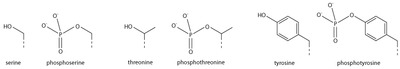
Serine/threonine/tyrosine phosphorylation

Amino acid phosphorylations.tif

Addition of a negatively charged phosphate group can lead to major changes in protein structure, leading to the well-characterised role of phosphorylation in controlling protein function. It is not clear what structural implications histone phosphorylation has, but histone phosphorylation has clear functions as a post-translational modification, and binding domains such as BRCT have been characterised.

Effects on transcription

Most well-studied histone modifications are involved in control of transcription.

Actively transcribed genes

Two histone modifications are particularly associated with active transcription:

Trimethylation of H3 lysine 4 (H3K4me3)
This trimethylation occurs at the promoter of active genes [45] [46] [47] and is performed by the COMPASS complex. [48] [49] [50] Despite the conservation of this complex and histone modification from yeast to mammals, it is not entirely clear what role this modification plays. However, it is an excellent mark of active promoters and the level of this histone modification at a gene's promoter is broadly correlated with transcriptional activity of the gene. The formation of this mark is tied to transcription in a rather convoluted manner: early in transcription of a gene, RNA polymerase II undergoes a switch from initiating' to 'elongating', marked by a change in the phosphorylation states of the RNA polymerase II C terminal domain (CTD). The same enzyme that phosphorylates the CTD also phosphorylates the Rad6 complex, [51] [52] which in turn adds a ubiquitin mark to H2B K123 (K120 in mammals). [53] H2BK123Ub occurs throughout transcribed regions, but this mark is required for COMPASS to trimethylate H3K4 at promoters. [54] [55]
Trimethylation of H3 lysine 36 (H3K36me3)
This trimethylation occurs in the body of active genes and is deposited by the methyltransferase Set2. [56] This protein associates with elongating RNA polymerase II, and H3K36Me3 is indicative of actively transcribed genes. [57] H3K36Me3 is recognised by the Rpd3 histone deacetylase complex, which removes acetyl modifications from surrounding histones, increasing chromatin compaction and repressing spurious transcription. [58] [59] [60] Increased chromatin compaction prevents transcription factors from accessing DNA, and reduces the likelihood of new transcription events being initiated within the body of the gene. This process therefore helps ensure that transcription is not interrupted.

Repressed genes

Three histone modifications are particularly associated with repressed genes:

Trimethylation of H3 lysine 27 (H3K27me3)
This histone modification is deposited by the polycomb complex PRC2. [61] It is a clear marker of gene repression, [62] and is likely bound by other proteins to exert a repressive function. Another polycomb complex, PRC1, can bind H3K27me3 [62] and adds the histone modification H2AK119Ub which aids chromatin compaction. [63] [64] Based on this data it appears that PRC1 is recruited through the action of PRC2, however, recent studies show that PRC1 is recruited to the same sites in the absence of PRC2. [65] [66]
Di and tri-methylation of H3 lysine 9 (H3K9me2/3)
H3K9me2/3 is a well-characterised marker for heterochromatin, and is therefore strongly associated with gene repression. The formation of heterochromatin has been best studied in the yeast Schizosaccharomyces pombe , where it is initiated by recruitment of the RNA-induced transcriptional silencing (RITS) complex to double stranded RNAs produced from centromeric repeats. [67] RITS recruits the Clr4 histone methyltransferase which deposits H3K9me2/3. [68] This process is called histone methylation. H3K9Me2/3 serves as a binding site for the recruitment of Swi6 (heterochromatin protein 1 or HP1, another classic heterochromatin marker) [69] [70] which in turn recruits further repressive activities including histone modifiers such as histone deacetylases and histone methyltransferases. [71]
Trimethylation of H4 lysine 20 (H4K20me3)
This modification is tightly associated with heterochromatin, [72] [73] although its functional importance remains unclear. This mark is placed by the Suv4-20h methyltransferase, which is at least in part recruited by heterochromatin protein 1. [72]

Bivalent promoters

Analysis of histone modifications in embryonic stem cells (and other stem cells) revealed many gene promoters carrying both H3K4Me3 and H3K27Me3, in other words these promoters display both activating and repressing marks simultaneously. This peculiar combination of modifications marks genes that are poised for transcription; they are not required in stem cells, but are rapidly required after differentiation into some lineages. Once the cell starts to differentiate, these bivalent promoters are resolved to either active or repressive states depending on the chosen lineage. [74]

Other functions

DNA damage repair

Marking sites of DNA damage is an important function for histone modifications. Without a repair marker, DNA would get destroyed by damage accumulated from sources such as the ultraviolet radiation of the sun.

Phosphorylation of H2AX at serine 139 (γH2AX)
Phosphorylated H2AX (also known as gamma H2AX) is a marker for DNA double strand breaks, [75] and forms part of the response to DNA damage. [30] [76] H2AX is phosphorylated early after detection of DNA double strand break, and forms a domain extending many kilobases either side of the damage. [75] [77] [78] Gamma H2AX acts as a binding site for the protein MDC1, which in turn recruits key DNA repair proteins [79] (this complex topic is well reviewed in [80] ) and as such, gamma H2AX forms a vital part of the machinery that ensures genome stability.
Acetylation of H3 lysine 56 (H3K56Ac)
H3K56Acx is required for genome stability. [81] [82] H3K56 is acetylated by the p300/Rtt109 complex, [83] [84] [85] but is rapidly deacetylated around sites of DNA damage. H3K56 acetylation is also required to stabilise stalled replication forks, preventing dangerous replication fork collapses. [86] [87] Although in general mammals make far greater use of histone modifications than microorganisms, a major role of H3K56Ac in DNA replication exists only in fungi, and this has become a target for antibiotic development. [88]
Trimethylation of H3 lysine 36 (H3K36me3)
H3K36me3 has the ability to recruit the MSH2-MSH6 (hMutSα) complex of the DNA mismatch repair pathway. [89] Consistently, regions of the human genome with high levels of H3K36me3 accumulate less somatic mutations due to mismatch repair activity. [90]

Chromosome condensation

Phosphorylation of H3 at serine 10 (phospho-H3S10)
The mitotic kinase aurora B phosphorylates histone H3 at serine 10, triggering a cascade of changes that mediate mitotic chromosome condensation. [91] [92] Condensed chromosomes therefore stain very strongly for this mark, but H3S10 phosphorylation is also present at certain chromosome sites outside mitosis, for example in pericentric heterochromatin of cells during G2. H3S10 phosphorylation has also been linked to DNA damage caused by R-loop formation at highly transcribed sites. [93]
Phosphorylation H2B at serine 10/14 (phospho-H2BS10/14)
Phosphorylation of H2B at serine 10 (yeast) or serine 14 (mammals) is also linked to chromatin condensation, but for the very different purpose of mediating chromosome condensation during apoptosis. [94] [95] This mark is not simply a late acting bystander in apoptosis as yeast carrying mutations of this residue are resistant to hydrogen peroxide-induced apoptotic cell death.

Addiction

Epigenetic modifications of histone tails in specific regions of the brain are of central importance in addictions. [96] [97] [98] Once particular epigenetic alterations occur, they appear to be long lasting "molecular scars" that may account for the persistence of addictions. [96]

Cigarette smokers (about 15% of the US population) are usually addicted to nicotine. [99] After 7 days of nicotine treatment of mice, acetylation of both histone H3 and histone H4 was increased at the FosB promoter in the nucleus accumbens of the brain, causing 61% increase in FosB expression. [100] This would also increase expression of the splice variant Delta FosB. In the nucleus accumbens of the brain, Delta FosB functions as a "sustained molecular switch" and "master control protein" in the development of an addiction. [101] [102]

About 7% of the US population is addicted to alcohol. In rats exposed to alcohol for up to 5 days, there was an increase in histone 3 lysine 9 acetylation in the pronociceptin promoter in the brain amygdala complex. This acetylation is an activating mark for pronociceptin. The nociceptin/nociceptin opioid receptor system is involved in the reinforcing or conditioning effects of alcohol. [103]

Methamphetamine addiction occurs in about 0.2% of the US population. [104] Chronic methamphetamine use causes methylation of the lysine in position 4 of histone 3 located at the promoters of the c-fos and the C-C chemokine receptor 2 (ccr2) genes, activating those genes in the nucleus accumbens (NAc). [105] c-fos is well known to be important in addiction. [106] The ccr2 gene is also important in addiction, since mutational inactivation of this gene impairs addiction. [105]

Synthesis

The first step of chromatin structure duplication is the synthesis of histone proteins: H1, H2A, H2B, H3, H4. These proteins are synthesized during S phase of the cell cycle. There are different mechanisms which contribute to the increase of histone synthesis.

Yeast

Yeast carry one or two copies of each histone gene, which are not clustered but rather scattered throughout chromosomes. Histone gene transcription is controlled by multiple gene regulatory proteins such as transcription factors which bind to histone promoter regions. In budding yeast, the candidate gene for activation of histone gene expression is SBF. SBF is a transcription factor that is activated in late G1 phase, when it dissociates from its repressor Whi5. This occurs when Whi5 is phosphorylated by Cdc8 which is a G1/S Cdk. [107] Suppression of histone gene expression outside of S phases is dependent on Hir proteins which form inactive chromatin structure at the locus of histone genes, causing transcriptional activators to be blocked. [108] [109]

Metazoan

In metazoans the increase in the rate of histone synthesis is due to the increase in processing of pre-mRNA to its mature form as well as decrease in mRNA degradation; this results in an increase of active mRNA for translation of histone proteins. The mechanism for mRNA activation has been found to be the removal of a segment of the 3' end of the mRNA strand, and is dependent on association with stem-loop binding protein (SLBP). [110] SLBP also stabilizes histone mRNAs during S phase by blocking degradation by the 3'hExo nuclease. [111] SLBP levels are controlled by cell-cycle proteins, causing SLBP to accumulate as cells enter S phase and degrade as cells leave S phase. SLBP are marked for degradation by phosphorylation at two threonine residues by cyclin dependent kinases, possibly cyclin A/ cdk2, at the end of S phase. [112] Metazoans also have multiple copies of histone genes clustered on chromosomes which are localized in structures called Cajal bodies as determined by genome-wide chromosome conformation capture analysis (4C-Seq). [113]

Nuclear protein Ataxia-Telangiectasia (NPAT), also known as nuclear protein coactivator of histone transcription, is a transcription factor which activates histone gene transcription on chromosomes 1 and 6 of human cells. NPAT is also a substrate of cyclin E-Cdk2, which is required for the transition between G1 phase and S phase. NPAT activates histone gene expression only after it has been phosphorylated by the G1/S-Cdk cyclin E-Cdk2 in early S phase. [114] This shows an important regulatory link between cell-cycle control and histone synthesis.

History

Histones were discovered in 1884 by Albrecht Kossel. [115] The word "histone" dates from the late 19th century and is derived from the German word "Histon", a word itself of uncertain origin, perhaps from Ancient Greek ἵστημι (hístēmi, “make stand”) or ἱστός (histós, “loom”).

In the early 1960s, before the types of histones were known and before histones were known to be highly conserved across taxonomically diverse organisms, James F. Bonner and his collaborators began a study of these proteins that were known to be tightly associated with the DNA in the nucleus of higher organisms. [116] Bonner and his postdoctoral fellow Ru Chih C. Huang showed that isolated chromatin would not support RNA transcription in the test tube, but if the histones were extracted from the chromatin, RNA could be transcribed from the remaining DNA. [117] Their paper became a citation classic. [118] Paul T'so and James Bonner had called together a World Congress on Histone Chemistry and Biology in 1964, in which it became clear that there was no consensus on the number of kinds of histone and that no one knew how they would compare when isolated from different organisms. [119] [116] Bonner and his collaborators then developed methods to separate each type of histone, purified individual histones, compared amino acid compositions in the same histone from different organisms, and compared amino acid sequences  of the same histone from different organisms in collaboration with Emil Smith from UCLA. [120] For example, they found Histone IV sequence to be highly conserved between peas and calf thymus. [120] However, their work on the biochemical characteristics of individual histones did not reveal how the histones interacted with each other or with DNA to which they were tightly bound. [119]

Also in the 1960s, Vincent Allfrey and Alfred Mirsky had suggested, based on their analyses of histones, that acetylation and methylation of histones could provide a transcriptional control mechanism, but did not have available the kind of detailed analysis that later investigators were able to conduct to show how such regulation could be gene-specific. [121] Until the early 1990s, histones were dismissed by most as inert packing material for eukaryotic nuclear DNA, a view based in part on the models of Mark Ptashne and others, who believed that transcription was activated by protein-DNA and protein-protein interactions on largely naked DNA templates, as is the case in bacteria.

During the 1980s, Yahli Lorch and Roger Kornberg [122] showed that a nucleosome on a core promoter prevents the initiation of transcription in vitro, and Michael Grunstein [123] demonstrated that histones repress transcription in vivo, leading to the idea of the nucleosome as a general gene repressor. Relief from repression is believed to involve both histone modification and the action of chromatin-remodeling complexes. Vincent Allfrey and Alfred Mirsky had earlier proposed a role of histone modification in transcriptional activation, [124] regarded as a molecular manifestation of epigenetics. Michael Grunstein [125] and David Allis [126] found support for this proposal, in the importance of histone acetylation for transcription in yeast and the activity of the transcriptional activator Gcn5 as a histone acetyltransferase.

The discovery of the H5 histone appears to date back to the 1970s, [127] and it is now considered an isoform of Histone H1. [2] [4] [5] [6]

See also

Related Research Articles

<span class="mw-page-title-main">Nucleosome</span> Basic structural unit of DNA packaging in eukaryotes

A nucleosome is the basic structural unit of DNA packaging in eukaryotes. The structure of a nucleosome consists of a segment of DNA wound around eight histone proteins and resembles thread wrapped around a spool. The nucleosome is the fundamental subunit of chromatin. Each nucleosome is composed of a little less than two turns of DNA wrapped around a set of eight proteins called histones, which are known as a histone octamer. Each histone octamer is composed of two copies each of the histone proteins H2A, H2B, H3, and H4.

<span class="mw-page-title-main">Histone octamer</span> 8-protein complex forming the core of nucleosomes

In molecular biology, a histone octamer is the eight-protein complex found at the center of a nucleosome core particle. It consists of two copies of each of the four core histone proteins. The octamer assembles when a tetramer, containing two copies of H3 and two of H4, complexes with two H2A/H2B dimers. Each histone has both an N-terminal tail and a C-terminal histone-fold. Each of these key components interacts with DNA in its own way through a series of weak interactions, including hydrogen bonds and salt bridges. These interactions keep the DNA and the histone octamer loosely associated, and ultimately allow the two to re-position or to separate entirely.

<span class="mw-page-title-main">Histone H4</span> One of the five main histone proteins involved in the structure of chromatin

Histone H4 is one of the five main histone proteins involved in the structure of chromatin in eukaryotic cells. Featuring a main globular domain and a long N-terminal tail, H4 is involved with the structure of the nucleosome of the 'beads on a string' organization. Histone proteins are highly post-translationally modified. Covalently bonded modifications include acetylation and methylation of the N-terminal tails. These modifications may alter expression of genes located on DNA associated with its parent histone octamer. Histone H4 is an important protein in the structure and function of chromatin, where its sequence variants and variable modification states are thought to play a role in the dynamic and long term regulation of genes.

<span class="mw-page-title-main">Histone H2A</span> One of the five main histone proteins

Histone H2A is one of the five main histone proteins involved in the structure of chromatin in eukaryotic cells.

Histone H2B is one of the 5 main histone proteins involved in the structure of chromatin in eukaryotic cells. Featuring a main globular domain and long N-terminal and C-terminal tails, H2B is involved with the structure of the nucleosomes.

<span class="mw-page-title-main">Histone-modifying enzymes</span> Type of enzymes

Histone-modifying enzymes are enzymes involved in the modification of histone substrates after protein translation and affect cellular processes including gene expression. To safely store the eukaryotic genome, DNA is wrapped around four core histone proteins, which then join to form nucleosomes. These nucleosomes further fold together into highly condensed chromatin, which renders the organism's genetic material far less accessible to the factors required for gene transcription, DNA replication, recombination and repair. Subsequently, eukaryotic organisms have developed intricate mechanisms to overcome this repressive barrier imposed by the chromatin through histone modification, a type of post-translational modification which typically involves covalently attaching certain groups to histone residues. Once added to the histone, these groups elicit either a loose and open histone conformation, euchromatin, or a tight and closed histone conformation, heterochromatin. Euchromatin marks active transcription and gene expression, as the light packing of histones in this way allows entry for proteins involved in the transcription process. As such, the tightly packed heterochromatin marks the absence of current gene expression.

Chromatin remodeling is the dynamic modification of chromatin architecture to allow access of condensed genomic DNA to the regulatory transcription machinery proteins, and thereby control gene expression. Such remodeling is principally carried out by 1) covalent histone modifications by specific enzymes, e.g., histone acetyltransferases (HATs), deacetylases, methyltransferases, and kinases, and 2) ATP-dependent chromatin remodeling complexes which either move, eject or restructure nucleosomes. Besides actively regulating gene expression, dynamic remodeling of chromatin imparts an epigenetic regulatory role in several key biological processes, egg cells DNA replication and repair; apoptosis; chromosome segregation as well as development and pluripotency. Aberrations in chromatin remodeling proteins are found to be associated with human diseases, including cancer. Targeting chromatin remodeling pathways is currently evolving as a major therapeutic strategy in the treatment of several cancers.

<span class="mw-page-title-main">HIST1H2BM</span> Protein-coding gene in the species Homo sapiens

Histone H2B type 1-M is a protein that in humans is encoded by the HIST1H2BM gene.

H3K27ac is an epigenetic modification to the DNA packaging protein histone H3. It is a mark that indicates acetylation of the lysine residue at N-terminal position 27 of the histone H3 protein.

H2BK5ac is an epigenetic modification to the DNA packaging protein Histone H2B. It is a mark that indicates the acetylation at the 5th lysine residue of the histone H2B protein. H2BK5ac is involved in maintaining stem cells and colon cancer.

H4K16ac is an epigenetic modification to the DNA packaging protein Histone H4. It is a mark that indicates the acetylation at the 16th lysine residue of the histone H4 protein.

H4K5ac is an epigenetic modification to the DNA packaging protein histone H4. It is a mark that indicates the acetylation at the 5th lysine residue of the histone H4 protein. H4K5 is the closest lysine residue to the N-terminal tail of histone H4. It is enriched at the transcription start site (TSS) and along gene bodies. Acetylation of histone H4K5 and H4K12ac is enriched at centromeres.

H4K8ac, representing an epigenetic modification to the DNA packaging protein histone H4, is a mark indicating the acetylation at the 8th lysine residue of the histone H4 protein. It has been implicated in the prevalence of malaria.

H4K12ac is an epigenetic modification to the DNA packaging protein histone H4. It is a mark that indicates the acetylation at the 12th lysine residue of the histone H4 protein. H4K12ac is involved in learning and memory. It is possible that restoring this modification could reduce age-related decline in memory.

H4K91ac is an epigenetic modification to the DNA packaging protein histone H4. It is a mark that indicates the acetylation at the 91st lysine residue of the histone H4 protein. No known diseases are attributed to this mark but it might be implicated in melanoma.

H3K23ac is an epigenetic modification to the DNA packaging protein Histone H3. It is a mark that indicates the acetylation at the 23rd lysine residue of the histone H3 protein.

H3K14ac is an epigenetic modification to the DNA packaging protein Histone H3. It is a mark that indicates the acetylation at the 14th lysine residue of the histone H3 protein.

H3K36ac is an epigenetic modification to the DNA packaging protein Histone H3. It is a mark that indicates the acetylation at the 36th lysine residue of the histone H3 protein.

H3K56ac is an epigenetic modification to the DNA packaging protein Histone H3. It is a mark that indicates the acetylation at the 56th lysine residue of the histone H3 protein.

H3K36me is an epigenetic modification to the DNA packaging protein Histone H3, specifically, the mono-methylation at the 36th lysine residue of the histone H3 protein.

References

  1. Youngson RM (2006). Collins Dictionary of Human Biology. Glasgow: HarperCollins. ISBN   978-0-00-722134-9.
  2. 1 2 3 4 Cox M, Nelson DR, Lehninger AL (2005). Lehninger Principles of Biochemistry . San Francisco: W.H. Freeman. ISBN   978-0-7167-4339-2.
  3. Redon C, Pilch D, Rogakou E, Sedelnikova O, Newrock K, Bonner W (April 2002). "Histone H2A variants H2AX and H2AZ". Current Opinion in Genetics & Development. 12 (2): 162–9. doi:10.1016/S0959-437X(02)00282-4. PMID   11893489.
  4. 1 2 "Histone Variants Database 2.0". National Center for Biotechnology Information . Retrieved 13 January 2017.
  5. 1 2 Bhasin M, Reinherz EL, Reche PA (2006). "Recognition and classification of histones using support vector machine" (PDF). Journal of Computational Biology. 13 (1): 102–12. doi:10.1089/cmb.2006.13.102. PMID   16472024.
  6. 1 2 Hartl DL, Freifelder D, Snyder LA (1988). Basic Genetics . Boston: Jones and Bartlett Publishers. ISBN   978-0-86720-090-4.
  7. Mariño-Ramírez L, Kann MG, Shoemaker BA, Landsman D (October 2005). "Histone structure and nucleosome stability". Expert Review of Proteomics. 2 (5): 719–29. doi:10.1586/14789450.2.5.719. PMC   1831843 . PMID   16209651.
  8. 1 2 Luger K, Mäder AW, Richmond RK, Sargent DF, Richmond TJ (September 1997). "Crystal structure of the nucleosome core particle at 2.8 A resolution". Nature. 389 (6648): 251–60. Bibcode:1997Natur.389..251L. doi:10.1038/38444. PMID   9305837. S2CID   4328827. PDB: 1AOI
  9. Farkas D (1996). DNA simplified: the hitchhiker's guide to DNA. Washington, D.C: AACC Press. ISBN   978-0-915274-84-0.
  10. 1 2 Seal, Ruth L.; Denny, Paul; Bruford, Elspeth A.; Gribkova, Anna K.; Landsman, David; Marzluff, William F.; McAndrews, Monica; Panchenko, Anna R.; Shaytan, Alexey K.; Talbert, Paul B. (October 2022). "A standardized nomenclature for mammalian histone genes". Epigenetics & Chromatin. 15 (1): 34. doi: 10.1186/s13072-022-00467-2 . ISSN   1756-8935. PMC   9526256 . PMID   36180920.
  11. Jang CW, Shibata Y, Starmer J, Yee D, Magnuson T (July 2015). "Histone H3.3 maintains genome integrity during mammalian development". Genes & Development. 29 (13): 1377–92. doi: 10.1101/gad.264150.115 . PMC   4511213 . PMID   26159997.
  12. Draizen, Eli J.; Shaytan, Alexey K.; Mariño-Ramírez, Leonardo; Talbert, Paul B.; Landsman, David; Panchenko, Anna R. (2016). "HistoneDB 2.0: a histone database with variants--an integrated resource to explore histones and their variants". Database: The Journal of Biological Databases and Curation. 2016: baw014. doi:10.1093/database/baw014. ISSN   1758-0463. PMC   4795928 . PMID   26989147.
  13. El Kennani, Sara; Adrait, Annie; Shaytan, Alexey K.; Khochbin, Saadi; Bruley, Christophe; Panchenko, Anna R.; Landsman, David; Pflieger, Delphine; Govin, Jérôme (2017). "MS_HistoneDB, a manually curated resource for proteomic analysis of human and mouse histones". Epigenetics & Chromatin. 10: 2. doi: 10.1186/s13072-016-0109-x . ISSN   1756-8935. PMC   5223428 . PMID   28096900.
  14. Marashi, F.; Prokopp, K.; Stein, J.; Stein, G. (April 1984). "Evidence for a human histone gene cluster containing H2B and H2A pseudogenes". Proceedings of the National Academy of Sciences of the United States of America. 81 (7): 1936–1940. doi: 10.1073/pnas.81.7.1936 . ISSN   0027-8424. PMC   345411 . PMID   6326092.
  15. Kardalinou, E.; Eick, S.; Albig, W.; Doenecke, D. (August 1993). "Association of a human H1 histone gene with an H2A pseudogene and genes encoding H2B.1 and H3.1 histones". Journal of Cellular Biochemistry. 52 (4): 375–383. doi:10.1002/jcb.240520402. ISSN   0730-2312. PMID   8227173. S2CID   42454232.
  16. 1 2 Alva V, Ammelburg M, Söding J, Lupas AN (March 2007). "On the origin of the histone fold". BMC Structural Biology. 7: 17. doi: 10.1186/1472-6807-7-17 . PMC   1847821 . PMID   17391511.
  17. Mattiroli F, Bhattacharyya S, Dyer PN, White AE, Sandman K, Burkhart BW, et al. (August 2017). "Structure of histone-based chromatin in Archaea". Science. 357 (6351): 609–612. Bibcode:2017Sci...357..609M. doi:10.1126/science.aaj1849. PMC   5747315 . PMID   28798133.
  18. 1 2 3 Henneman B, van Emmerik C, van Ingen H, Dame RT (September 2018). "Structure and function of archaeal histones". PLOS Genetics. 14 (9): e1007582. Bibcode:2018BpJ...114..446H. doi: 10.1371/journal.pgen.1007582 . PMC   6136690 . PMID   30212449.
  19. Ward R, Bowman A, El-Mkami H, Owen-Hughes T, Norman DG (February 2009). "Long distance PELDOR measurements on the histone core particle". Journal of the American Chemical Society. 131 (4): 1348–9. doi:10.1021/ja807918f. PMC   3501648 . PMID   19138067.
  20. Mersfelder EL, Parthun MR (19 May 2006). "The tale beyond the tail: histone core domain modifications and the regulation of chromatin structure". Nucleic Acids Research. 34 (9): 2653–62. doi:10.1093/nar/gkl338. PMC   1464108 . PMID   16714444.
  21. Tropberger P, Schneider R (June 2013). "Scratching the (lateral) surface of chromatin regulation by histone modifications". Nature Structural & Molecular Biology. 20 (6): 657–61. doi:10.1038/nsmb.2581. PMID   23739170. S2CID   2956823.
  22. Allison LA (2012). Fundamental Molecular Biology (Second ed.). United States of America: John Wiley & Sons. p. 102. ISBN   9781118059814.
  23. Rizzo PJ (August 2003). "Those amazing dinoflagellate chromosomes". Cell Research. 13 (4): 215–7. doi: 10.1038/sj.cr.7290166 . PMID   12974611.
  24. Talbert PB, Henikoff S (December 2012). "Chromatin: packaging without nucleosomes". Current Biology. 22 (24): R1040-3. doi: 10.1016/j.cub.2012.10.052 . PMID   23257187.
  25. Kasinsky HE, Lewis JD, Dacks JB, Ausió J (January 2001). "Origin of H1 linker histones". FASEB Journal. 15 (1): 34–42. doi: 10.1096/fj.00-0237rev . PMID   11149891. S2CID   10089116.
  26. Bozorgmehr JH (February 2020). "The origin of chromosomal histones in a 30S ribosomal protein". Gene. 726: 144155. doi:10.1016/j.gene.2019.144155. PMID   31629821. S2CID   204813634.
  27. Clarke HJ (1992). "Nuclear and chromatin composition of mammalian gametes and early embryos". Biochemistry and Cell Biology. 70 (10–11): 856–66. doi:10.1139/o92-134. PMID   1297351.
  28. Guillemette B, Bataille AR, Gévry N, Adam M, Blanchette M, Robert F, Gaudreau L (December 2005). "Variant histone H2A.Z is globally localized to the promoters of inactive yeast genes and regulates nucleosome positioning". PLOS Biology. 3 (12): e384. doi: 10.1371/journal.pbio.0030384 . PMC   1275524 . PMID   16248679.
  29. Billon P, Côté J (October 2011). "Precise deposition of histone H2A.Z in chromatin for genome expression and maintenance". Biochim Biophys Acta. 1819 (3–4): 290–302. doi:10.1016/j.bbagrm.2011.10.004. PMID   22027408.
  30. 1 2 Paull TT, Rogakou EP, Yamazaki V, Kirchgessner CU, Gellert M, Bonner WM (2000). "A critical role for histone H2AX in recruitment of repair factors to nuclear foci after DNA damage". Current Biology. 10 (15): 886–95. doi: 10.1016/S0960-9822(00)00610-2 . PMID   10959836.
  31. Ahmad K, Henikoff S (June 2002). "The histone variant H3.3 marks active chromatin by replication-independent nucleosome assembly". Molecular Cell. 9 (6): 1191–200. doi: 10.1016/S1097-2765(02)00542-7 . PMID   12086617.
  32. Strahl BD, Allis CD (January 2000). "The language of covalent histone modifications". Nature. 403 (6765): 41–5. Bibcode:2000Natur.403...41S. doi:10.1038/47412. PMID   10638745. S2CID   4418993.
  33. Jenuwein T, Allis CD (August 2001). "Translating the histone code" (PDF). Science. 293 (5532): 1074–80. CiteSeerX   10.1.1.453.900 . doi:10.1126/science.1063127. PMID   11498575. S2CID   1883924.
  34. Song N, Liu J, An S, Nishino T, Hishikawa Y, Koji T (August 2011). "Immunohistochemical Analysis of Histone H3 Modifications in Germ Cells during Mouse Spermatogenesis". Acta Histochemica et Cytochemica. 44 (4): 183–90. doi:10.1267/ahc.11027. PMC   3168764 . PMID   21927517.
  35. Benevolenskaya EV (August 2007). "Histone H3K4 demethylases are essential in development and differentiation". Biochemistry and Cell Biology. 85 (4): 435–43. doi:10.1139/o07-057. PMID   17713579.
  36. 1 2 3 4 5 6 7 8 Barski A, Cuddapah S, Cui K, Roh TY, Schones DE, Wang Z, et al. (May 2007). "High-resolution profiling of histone methylations in the human genome". Cell. 129 (4): 823–37. doi: 10.1016/j.cell.2007.05.009 . PMID   17512414.
  37. 1 2 3 Steger DJ, Lefterova MI, Ying L, Stonestrom AJ, Schupp M, Zhuo D, et al. (April 2008). "DOT1L/KMT4 recruitment and H3K79 methylation are ubiquitously coupled with gene transcription in mammalian cells". Molecular and Cellular Biology. 28 (8): 2825–39. doi:10.1128/MCB.02076-07. PMC   2293113 . PMID   18285465.
  38. 1 2 3 Rosenfeld JA, Wang Z, Schones DE, Zhao K, DeSalle R, Zhang MQ (March 2009). "Determination of enriched histone modifications in non-genic portions of the human genome". BMC Genomics. 10: 143. doi: 10.1186/1471-2164-10-143 . PMC   2667539 . PMID   19335899.
  39. 1 2 3 Koch CM, Andrews RM, Flicek P, Dillon SC, Karaöz U, Clelland GK, et al. (June 2007). "The landscape of histone modifications across 1% of the human genome in five human cell lines". Genome Research. 17 (6): 691–707. doi:10.1101/gr.5704207. PMC   1891331 . PMID   17567990.
  40. Guillemette B, Drogaris P, Lin HH, Armstrong H, Hiragami-Hamada K, Imhof A, et al. (March 2011). "H3 lysine 4 is acetylated at active gene promoters and is regulated by H3 lysine 4 methylation". PLOS Genetics. 7 (3): e1001354. doi: 10.1371/journal.pgen.1001354 . PMC   3069113 . PMID   21483810.
  41. Creyghton MP, Cheng AW, Welstead GG, Kooistra T, Carey BW, Steine EJ, et al. (December 2010). "Histone H3K27ac separates active from poised enhancers and predicts developmental state". Proceedings of the National Academy of Sciences of the United States of America. 107 (50): 21931–6. doi: 10.1073/pnas.1016071107 . PMC   3003124 . PMID   21106759.
  42. Farrelly LA, Thompson RE, Zhao S, Lepack AE, Lyu Y, Bhanu NV, et al. (March 2019). "Histone serotonylation is a permissive modification that enhances TFIID binding to H3K4me3". Nature. 567 (7749): 535–539. Bibcode:2019Natur.567..535F. doi:10.1038/s41586-019-1024-7. PMC   6557285 . PMID   30867594.
  43. Christophorou MA, Castelo-Branco G, Halley-Stott RP, Oliveira CS, Loos R, Radzisheuskaya A, et al. (March 2014). "Citrullination regulates pluripotency and histone H1 binding to chromatin". Nature. 507 (7490): 104–8. Bibcode:2014Natur.507..104C. doi:10.1038/nature12942. PMC   4843970 . PMID   24463520.
  44. Cuthbert GL, Daujat S, Snowden AW, Erdjument-Bromage H, Hagiwara T, Yamada M, et al. (September 2004). "Histone deimination antagonizes arginine methylation". Cell. 118 (5): 545–53. doi: 10.1016/j.cell.2004.08.020 . PMID   15339660.
  45. Krogan NJ, Dover J, Wood A, Schneider J, Heidt J, Boateng MA, et al. (March 2003). "The Paf1 complex is required for histone H3 methylation by COMPASS and Dot1p: linking transcriptional elongation to histone methylation". Molecular Cell. 11 (3): 721–9. doi: 10.1016/S1097-2765(03)00091-1 . PMID   12667454.
  46. Ng HH, Robert F, Young RA, Struhl K (March 2003). "Targeted recruitment of Set1 histone methylase by elongating Pol II provides a localized mark and memory of recent transcriptional activity". Molecular Cell. 11 (3): 709–19. doi: 10.1016/S1097-2765(03)00092-3 . PMID   12667453.
  47. Bernstein BE, Kamal M, Lindblad-Toh K, Bekiranov S, Bailey DK, Huebert DJ, et al. (January 2005). "Genomic maps and comparative analysis of histone modifications in human and mouse". Cell. 120 (2): 169–81. doi: 10.1016/j.cell.2005.01.001 . PMID   15680324.
  48. Krogan NJ, Dover J, Khorrami S, Greenblatt JF, Schneider J, Johnston M, Shilatifard A (March 2002). "COMPASS, a histone H3 (Lysine 4) methyltransferase required for telomeric silencing of gene expression". The Journal of Biological Chemistry. 277 (13): 10753–5. doi: 10.1074/jbc.C200023200 . PMID   11805083.
  49. Roguev A, Schaft D, Shevchenko A, Pijnappel WW, Wilm M, Aasland R, Stewart AF (December 2001). "The Saccharomyces cerevisiae Set1 complex includes an Ash2 homologue and methylates histone 3 lysine 4". The EMBO Journal. 20 (24): 7137–48. doi:10.1093/emboj/20.24.7137. PMC   125774 . PMID   11742990.
  50. Nagy PL, Griesenbeck J, Kornberg RD, Cleary ML (January 2002). "A trithorax-group complex purified from Saccharomyces cerevisiae is required for methylation of histone H3". Proceedings of the National Academy of Sciences of the United States of America. 99 (1): 90–4. Bibcode:2002PNAS...99...90N. doi: 10.1073/pnas.221596698 . PMC   117519 . PMID   11752412.
  51. Wood A, Schneider J, Dover J, Johnston M, Shilatifard A (November 2005). "The Bur1/Bur2 complex is required for histone H2B monoubiquitination by Rad6/Bre1 and histone methylation by COMPASS". Molecular Cell. 20 (4): 589–99. doi: 10.1016/j.molcel.2005.09.010 . PMID   16307922.
  52. Sarcevic B, Mawson A, Baker RT, Sutherland RL (April 2002). "Regulation of the ubiquitin-conjugating enzyme hHR6A by CDK-mediated phosphorylation". The EMBO Journal. 21 (8): 2009–18. doi:10.1093/emboj/21.8.2009. PMC   125963 . PMID   11953320.
  53. Robzyk K, Recht J, Osley MA (January 2000). "Rad6-dependent ubiquitination of histone H2B in yeast". Science. 287 (5452): 501–4. Bibcode:2000Sci...287..501R. doi:10.1126/science.287.5452.501. PMID   10642555.
  54. Sun ZW, Allis CD (July 2002). "Ubiquitination of histone H2B regulates H3 methylation and gene silencing in yeast". Nature. 418 (6893): 104–8. Bibcode:2002Natur.418..104S. doi:10.1038/nature00883. PMID   12077605. S2CID   4338471.
  55. Dover J, Schneider J, Tawiah-Boateng MA, Wood A, Dean K, Johnston M, Shilatifard A (August 2002). "Methylation of histone H3 by COMPASS requires ubiquitination of histone H2B by Rad6". The Journal of Biological Chemistry. 277 (32): 28368–71. doi: 10.1074/jbc.C200348200 . PMID   12070136.
  56. Strahl BD, Grant PA, Briggs SD, Sun ZW, Bone JR, Caldwell JA, et al. (March 2002). "Set2 is a nucleosomal histone H3-selective methyltransferase that mediates transcriptional repression". Molecular and Cellular Biology. 22 (5): 1298–306. doi:10.1128/MCB.22.5.1298-1306.2002. PMC   134702 . PMID   11839797.
  57. Li J, Moazed D, Gygi SP (December 2002). "Association of the histone methyltransferase Set2 with RNA polymerase II plays a role in transcription elongation". The Journal of Biological Chemistry. 277 (51): 49383–8. doi: 10.1074/jbc.M209294200 . PMID   12381723.
  58. Carrozza MJ, Li B, Florens L, Suganuma T, Swanson SK, Lee KK, et al. (November 2005). "Histone H3 methylation by Set2 directs deacetylation of coding regions by Rpd3S to suppress spurious intragenic transcription". Cell. 123 (4): 581–92. doi: 10.1016/j.cell.2005.10.023 . PMID   16286007.
  59. Keogh MC, Kurdistani SK, Morris SA, Ahn SH, Podolny V, Collins SR, et al. (November 2005). "Cotranscriptional set2 methylation of histone H3 lysine 36 recruits a repressive Rpd3 complex". Cell. 123 (4): 593–605. doi: 10.1016/j.cell.2005.10.025 . PMID   16286008.
  60. Joshi AA, Struhl K (December 2005). "Eaf3 chromodomain interaction with methylated H3-K36 links histone deacetylation to Pol II elongation". Molecular Cell. 20 (6): 971–8. doi: 10.1016/j.molcel.2005.11.021 . PMID   16364921.
  61. Kuzmichev A, Nishioka K, Erdjument-Bromage H, Tempst P, Reinberg D (November 2002). "Histone methyltransferase activity associated with a human multiprotein complex containing the Enhancer of Zeste protein". Genes & Development. 16 (22): 2893–905. doi:10.1101/gad.1035902. PMC   187479 . PMID   12435631.
  62. 1 2 Cao R, Wang L, Wang H, Xia L, Erdjument-Bromage H, Tempst P, et al. (November 2002). "Role of histone H3 lysine 27 methylation in Polycomb-group silencing". Science. 298 (5595): 1039–43. Bibcode:2002Sci...298.1039C. doi:10.1126/science.1076997. PMID   12351676. S2CID   6265267.
  63. de Napoles M, Mermoud JE, Wakao R, Tang YA, Endoh M, Appanah R, et al. (November 2004). "Polycomb group proteins Ring1A/B link ubiquitylation of histone H2A to heritable gene silencing and X inactivation". Developmental Cell. 7 (5): 663–76. doi: 10.1016/j.devcel.2004.10.005 . PMID   15525528.
  64. Wang H, Wang L, Erdjument-Bromage H, Vidal M, Tempst P, Jones RS, Zhang Y (October 2004). "Role of histone H2A ubiquitination in Polycomb silencing". Nature. 431 (7010): 873–8. Bibcode:2004Natur.431..873W. doi:10.1038/nature02985. hdl:10261/73732. PMID   15386022. S2CID   4344378.
  65. Tavares L, Dimitrova E, Oxley D, Webster J, Poot R, Demmers J, et al. (February 2012). "RYBP-PRC1 complexes mediate H2A ubiquitylation at polycomb target sites independently of PRC2 and H3K27me3". Cell. 148 (4): 664–78. doi:10.1016/j.cell.2011.12.029. PMC   3281992 . PMID   22325148.
  66. Gao Z, Zhang J, Bonasio R, Strino F, Sawai A, Parisi F, et al. (February 2012). "PCGF homologs, CBX proteins, and RYBP define functionally distinct PRC1 family complexes". Molecular Cell. 45 (3): 344–56. doi:10.1016/j.molcel.2012.01.002. PMC   3293217 . PMID   22325352.
  67. Verdel A, Jia S, Gerber S, Sugiyama T, Gygi S, Grewal SI, Moazed D (January 2004). "RNAi-mediated targeting of heterochromatin by the RITS complex". Science. 303 (5658): 672–6. Bibcode:2004Sci...303..672V. doi:10.1126/science.1093686. PMC   3244756 . PMID   14704433.
  68. Rea S, Eisenhaber F, O'Carroll D, Strahl BD, Sun ZW, Schmid M, et al. (August 2000). "Regulation of chromatin structure by site-specific histone H3 methyltransferases". Nature. 406 (6796): 593–9. Bibcode:2000Natur.406..593R. doi:10.1038/35020506. PMID   10949293. S2CID   205008015.
  69. Bannister AJ, Zegerman P, Partridge JF, Miska EA, Thomas JO, Allshire RC, Kouzarides T (March 2001). "Selective recognition of methylated lysine 9 on histone H3 by the HP1 chromo domain". Nature. 410 (6824): 120–4. Bibcode:2001Natur.410..120B. doi:10.1038/35065138. PMID   11242054. S2CID   4334447.
  70. Lachner M, O'Carroll D, Rea S, Mechtler K, Jenuwein T (March 2001). "Methylation of histone H3 lysine 9 creates a binding site for HP1 proteins". Nature. 410 (6824): 116–20. Bibcode:2001Natur.410..116L. doi:10.1038/35065132. PMID   11242053. S2CID   4331863.
  71. Bajpai G, Jain I, Inamdar MM, Das D, Padinhateeri R (January 2017). "Binding of DNA-bending non-histone proteins destabilizes regular 30-nm chromatin structure". PLOS Computational Biology. 13 (1): e1005365. Bibcode:2017PLSCB..13E5365B. doi: 10.1371/journal.pcbi.1005365 . PMC   5305278 . PMID   28135276.
  72. 1 2 Schotta G, Lachner M, Sarma K, Ebert A, Sengupta R, Reuter G, et al. (June 2004). "A silencing pathway to induce H3-K9 and H4-K20 trimethylation at constitutive heterochromatin". Genes & Development. 18 (11): 1251–62. doi:10.1101/gad.300704. PMC   420351 . PMID   15145825.
  73. Kourmouli N, Jeppesen P, Mahadevhaiah S, Burgoyne P, Wu R, Gilbert DM, et al. (May 2004). "Heterochromatin and tri-methylated lysine 20 of histone H4 in animals". Journal of Cell Science. 117 (Pt 12): 2491–501. doi: 10.1242/jcs.01238 . PMID   15128874.
  74. Bernstein BE, Mikkelsen TS, Xie X, Kamal M, Huebert DJ, Cuff J, et al. (April 2006). "A bivalent chromatin structure marks key developmental genes in embryonic stem cells". Cell. 125 (2): 315–26. doi: 10.1016/j.cell.2006.02.041 . PMID   16630819.
  75. 1 2 Rogakou EP, Pilch DR, Orr AH, Ivanova VS, Bonner WM (March 1998). "DNA double-stranded breaks induce histone H2AX phosphorylation on serine 139". The Journal of Biological Chemistry. 273 (10): 5858–68. doi: 10.1074/jbc.273.10.5858 . PMID   9488723.
  76. Celeste A, Petersen S, Romanienko PJ, Fernandez-Capetillo O, Chen HT, Sedelnikova OA, et al. (May 2002). "Genomic instability in mice lacking histone H2AX". Science. 296 (5569): 922–7. Bibcode:2002Sci...296..922C. doi:10.1126/science.1069398. PMC   4721576 . PMID   11934988.
  77. Shroff R, Arbel-Eden A, Pilch D, Ira G, Bonner WM, Petrini JH, et al. (October 2004). "Distribution and dynamics of chromatin modification induced by a defined DNA double-strand break". Current Biology. 14 (19): 1703–11. doi:10.1016/j.cub.2004.09.047. PMC   4493763 . PMID   15458641.
  78. Rogakou EP, Boon C, Redon C, Bonner WM (September 1999). "Megabase chromatin domains involved in DNA double-strand breaks in vivo". The Journal of Cell Biology. 146 (5): 905–16. doi:10.1083/jcb.146.5.905. PMC   2169482 . PMID   10477747.
  79. Stewart GS, Wang B, Bignell CR, Taylor AM, Elledge SJ (February 2003). "MDC1 is a mediator of the mammalian DNA damage checkpoint". Nature. 421 (6926): 961–6. Bibcode:2003Natur.421..961S. doi:10.1038/nature01446. PMID   12607005. S2CID   4410773.
  80. Bekker-Jensen S, Mailand N (December 2010). "Assembly and function of DNA double-strand break repair foci in mammalian cells". DNA Repair. 9 (12): 1219–28. doi:10.1016/j.dnarep.2010.09.010. PMID   21035408.
  81. Ozdemir A, Spicuglia S, Lasonder E, Vermeulen M, Campsteijn C, Stunnenberg HG, Logie C (July 2005). "Characterization of lysine 56 of histone H3 as an acetylation site in Saccharomyces cerevisiae". The Journal of Biological Chemistry. 280 (28): 25949–52. doi: 10.1074/jbc.C500181200 . hdl: 2066/32314 . PMID   15888442.
  82. Masumoto H, Hawke D, Kobayashi R, Verreault A (July 2005). "A role for cell-cycle-regulated histone H3 lysine 56 acetylation in the DNA damage response". Nature. 436 (7048): 294–8. Bibcode:2005Natur.436..294M. doi:10.1038/nature03714. PMID   16015338. S2CID   4414433.
  83. Driscoll R, Hudson A, Jackson SP (February 2007). "Yeast Rtt109 promotes genome stability by acetylating histone H3 on lysine 56". Science. 315 (5812): 649–52. Bibcode:2007Sci...315..649D. doi:10.1126/science.1135862. PMC   3334813 . PMID   17272722.
  84. Han J, Zhou H, Horazdovsky B, Zhang K, Xu RM, Zhang Z (February 2007). "Rtt109 acetylates histone H3 lysine 56 and functions in DNA replication". Science. 315 (5812): 653–5. Bibcode:2007Sci...315..653H. doi:10.1126/science.1133234. PMID   17272723. S2CID   19056605.
  85. Das C, Lucia MS, Hansen KC, Tyler JK (May 2009). "CBP/p300-mediated acetylation of histone H3 on lysine 56". Nature. 459 (7243): 113–7. Bibcode:2009Natur.459..113D. doi:10.1038/nature07861. PMC   2756583 . PMID   19270680.
  86. Han J, Zhou H, Li Z, Xu RM, Zhang Z (September 2007). "Acetylation of lysine 56 of histone H3 catalyzed by RTT109 and regulated by ASF1 is required for replisome integrity". The Journal of Biological Chemistry. 282 (39): 28587–96. doi: 10.1074/jbc.M702496200 . PMID   17690098.
  87. Wurtele H, Kaiser GS, Bacal J, St-Hilaire E, Lee EH, Tsao S, et al. (January 2012). "Histone H3 lysine 56 acetylation and the response to DNA replication fork damage". Molecular and Cellular Biology. 32 (1): 154–72. doi:10.1128/MCB.05415-11. PMC   3255698 . PMID   22025679.
  88. Wurtele H, Tsao S, Lépine G, Mullick A, Tremblay J, Drogaris P, et al. (July 2010). "Modulation of histone H3 lysine 56 acetylation as an antifungal therapeutic strategy". Nature Medicine. 16 (7): 774–80. doi:10.1038/nm.2175. PMC   4108442 . PMID   20601951.
  89. Li F, Mao G, Tong D, Huang J, Gu L, Yang W, Li GM (April 2013). "The histone mark H3K36me3 regulates human DNA mismatch repair through its interaction with MutSα". Cell. 153 (3): 590–600. doi:10.1016/j.cell.2013.03.025. PMC   3641580 . PMID   23622243.
  90. Supek F, Lehner B (July 2017). "Clustered Mutation Signatures Reveal that Error-Prone DNA Repair Targets Mutations to Active Genes". Cell. 170 (3): 534–547.e23. doi: 10.1016/j.cell.2017.07.003 . hdl: 10230/35343 . PMID   28753428.
  91. Wilkins BJ, Rall NA, Ostwal Y, Kruitwagen T, Hiragami-Hamada K, Winkler M, et al. (January 2014). "A cascade of histone modifications induces chromatin condensation in mitosis". Science. 343 (6166): 77–80. Bibcode:2014Sci...343...77W. doi:10.1126/science.1244508. hdl: 11858/00-001M-0000-0015-11C0-5 . PMID   24385627. S2CID   7698266.
  92. Johansen KM, Johansen J (2006). "Regulation of chromatin structure by histone H3S10 phosphorylation". Chromosome Research. 14 (4): 393–404. doi:10.1007/s10577-006-1063-4. PMID   16821135. S2CID   8556959.
  93. Castellano-Pozo M, Santos-Pereira JM, Rondón AG, Barroso S, Andújar E, Pérez-Alegre M, et al. (November 2013). "R loops are linked to histone H3 S10 phosphorylation and chromatin condensation". Molecular Cell. 52 (4): 583–90. doi: 10.1016/j.molcel.2013.10.006 . PMID   24211264.
  94. Cheung WL, Ajiro K, Samejima K, Kloc M, Cheung P, Mizzen CA, et al. (May 2003). "Apoptotic phosphorylation of histone H2B is mediated by mammalian sterile twenty kinase". Cell. 113 (4): 507–17. doi: 10.1016/s0092-8674(03)00355-6 . PMID   12757711.
  95. Ahn SH, Cheung WL, Hsu JY, Diaz RL, Smith MM, Allis CD (January 2005). "Sterile 20 kinase phosphorylates histone H2B at serine 10 during hydrogen peroxide-induced apoptosis in S. cerevisiae". Cell. 120 (1): 25–36. doi: 10.1016/j.cell.2004.11.016 . PMID   15652479.
  96. 1 2 Robison AJ, Nestler EJ (October 2011). "Transcriptional and epigenetic mechanisms of addiction". Nature Reviews. Neuroscience. 12 (11): 623–37. doi:10.1038/nrn3111. PMC   3272277 . PMID   21989194.
  97. Hitchcock LN, Lattal KM (2014). "Histone-mediated epigenetics in addiction". Epigenetics and Neuroplasticity—Evidence and Debate. Progress in Molecular Biology and Translational Science. Vol. 128. Academic Press. pp. 51–87. doi:10.1016/B978-0-12-800977-2.00003-6. ISBN   9780128009772. PMC   5914502 . PMID   25410541.{{cite book}}: |journal= ignored (help)
  98. McQuown SC, Wood MA (April 2010). "Epigenetic regulation in substance use disorders". Current Psychiatry Reports. 12 (2): 145–53. doi:10.1007/s11920-010-0099-5. PMC   2847696 . PMID   20425300.
  99. "Is nicotine addictive?".
  100. Levine A, Huang Y, Drisaldi B, Griffin EA, Pollak DD, Xu S, et al. (November 2011). "Molecular mechanism for a gateway drug: epigenetic changes initiated by nicotine prime gene expression by cocaine". Science Translational Medicine. 3 (107): 107ra109. doi:10.1126/scitranslmed.3003062. PMC   4042673 . PMID   22049069.
  101. Ruffle JK (November 2014). "Molecular neurobiology of addiction: what's all the (Δ)FosB about?". The American Journal of Drug and Alcohol Abuse. 40 (6): 428–37. doi:10.3109/00952990.2014.933840. PMID   25083822. S2CID   19157711.
  102. Nestler EJ, Barrot M, Self DW (September 2001). "DeltaFosB: a sustained molecular switch for addiction". Proceedings of the National Academy of Sciences of the United States of America. 98 (20): 11042–6. Bibcode:2001PNAS...9811042N. doi: 10.1073/pnas.191352698 . PMC   58680 . PMID   11572966.
  103. D'Addario C, Caputi FF, Ekström TJ, Di Benedetto M, Maccarrone M, Romualdi P, Candeletti S (February 2013). "Ethanol induces epigenetic modulation of prodynorphin and pronociceptin gene expression in the rat amygdala complex". Journal of Molecular Neuroscience. 49 (2): 312–9. doi:10.1007/s12031-012-9829-y. PMID   22684622. S2CID   14013417.
  104. "What is the scope of methamphetamine abuse in the United States?".
  105. 1 2 Godino A, Jayanthi S, Cadet JL (2015). "Epigenetic landscape of amphetamine and methamphetamine addiction in rodents". Epigenetics. 10 (7): 574–80. doi:10.1080/15592294.2015.1055441. PMC   4622560 . PMID   26023847.
  106. Cruz FC, Javier Rubio F, Hope BT (December 2015). "Using c-fos to study neuronal ensembles in corticostriatal circuitry of addiction". Brain Research. 1628 (Pt A): 157–73. doi:10.1016/j.brainres.2014.11.005. PMC   4427550 . PMID   25446457.
  107. de Bruin RA, McDonald WH, Kalashnikova TI, Yates J, Wittenberg C (June 2004). "Cln3 activates G1-specific transcription via phosphorylation of the SBF bound repressor Whi5". Cell. 117 (7): 887–98. doi: 10.1016/j.cell.2004.05.025 . PMID   15210110.
  108. Xu H, Kim UJ, Schuster T, Grunstein M (November 1992). "Identification of a new set of cell cycle-regulatory genes that regulate S-phase transcription of histone genes in Saccharomyces cerevisiae". Molecular and Cellular Biology. 12 (11): 5249–59. doi:10.1128/mcb.12.11.5249. PMC   360458 . PMID   1406694.
  109. Dimova D, Nackerdien Z, Furgeson S, Eguchi S, Osley MA (July 1999). "A role for transcriptional repressors in targeting the yeast Swi/Snf complex". Molecular Cell. 4 (1): 75–83. doi: 10.1016/S1097-2765(00)80189-6 . PMID   10445029.
  110. Dominski Z, Erkmann JA, Yang X, Sànchez R, Marzluff WF (January 2002). "A novel zinc finger protein is associated with U7 snRNP and interacts with the stem-loop binding protein in the histone pre-mRNP to stimulate 3'-end processing". Genes & Development. 16 (1): 58–71. doi:10.1101/gad.932302. PMC   155312 . PMID   11782445.
  111. Dominski Z, Yang XC, Kaygun H, Dadlez M, Marzluff WF (August 2003). "A 3' exonuclease that specifically interacts with the 3' end of histone mRNA". Molecular Cell. 12 (2): 295–305. doi: 10.1016/S1097-2765(03)00278-8 . PMID   14536070.
  112. Zheng L, Dominski Z, Yang XC, Elms P, Raska CS, Borchers CH, Marzluff WF (March 2003). "Phosphorylation of stem-loop binding protein (SLBP) on two threonines triggers degradation of SLBP, the sole cell cycle-regulated factor required for regulation of histone mRNA processing, at the end of S phase". Molecular and Cellular Biology. 23 (5): 1590–601. doi:10.1128/MCB.23.5.1590-1601.2003. PMC   151715 . PMID   12588979.
  113. Wang Q, Sawyer IA, Sung MH, Sturgill D, Shevtsov SP, Pegoraro G, et al. (March 2016). "Cajal bodies are linked to genome conformation". Nature Communications. 7: 10966. Bibcode:2016NatCo...710966W. doi:10.1038/ncomms10966. PMC   4802181 . PMID   26997247.
  114. Zhao J, Kennedy BK, Lawrence BD, Barbie DA, Matera AG, Fletcher JA, Harlow E (September 2000). "NPAT links cyclin E-Cdk2 to the regulation of replication-dependent histone gene transcription". Genes & Development. 14 (18): 2283–97. doi:10.1101/GAD.827700. PMC   316937 . PMID   10995386.
  115. Luck JM (1965). "Histone Chemistry: the Pioneers". In Bonner J, Ts'o P (eds.). The Nucleohistones. San Francisco, London, and Amsterdam: Holden-Day, Inc.
  116. 1 2 Bonner J (1994). "Chapters from my life". Annual Review of Plant Physiology and Plant Molecular Biology. 45: 1–23. doi: 10.1146/annurev.pp.45.060194.000245 .
  117. Huang RC, Bonner J (July 1962). "Histone, a suppressor of chromosomal RNA synthesis". Proceedings of the National Academy of Sciences of the United States of America. 48 (7): 1216–22. Bibcode:1962PNAS...48.1216H. doi: 10.1073/pnas.48.7.1216 . PMC   220935 . PMID   14036409.
  118. "Huang R C & Bonner J. Histone, a suppressor of chromosomal RNA synthesis. Proc. Nat. Acad. Sci. US 48:1216-22, 1962" (PDF). Citation Classics (12): 79. 20 March 1978.
  119. 1 2 James Bonner and Paul T'so (1965) The Nucleohistones. Holden-Day Inc, San Francisco, London, Amsterdam.
  120. 1 2 DeLange RJ, Fambrough DM, Smith EL, Bonner J (October 1969). "Calf and pea histone IV. 3. Complete amino acid sequence of pea seedling histone IV; comparison with the homologous calf thymus histone". The Journal of Biological Chemistry. 244 (20): 5669–79. doi: 10.1016/S0021-9258(18)63612-9 . PMID   5388597.
  121. Allfrey VG, Faulkner R, Mirsky AE (May 1964). "Acetylation and methylation of histones and their possible role in the regulation of RNA synthesis". Proceedings of the National Academy of Sciences of the United States of America. 51 (5): 786–94. Bibcode:1964PNAS...51..786A. doi: 10.1073/pnas.51.5.786 . PMC   300163 . PMID   14172992.
  122. Lorch Y, LaPointe JW, Kornberg RD (April 1987). "Nucleosomes inhibit the initiation of transcription but allow chain elongation with the displacement of histones". Cell. 49 (2): 203–10. doi:10.1016/0092-8674(87)90561-7. PMID   3568125. S2CID   21270171.
  123. Kayne PS, Kim UJ, Han M, Mullen JR, Yoshizaki F, Grunstein M (October 1988). "Extremely conserved histone H4 N terminus is dispensable for growth but essential for repressing the silent mating loci in yeast" (PDF). Cell. 55 (1): 27–39. doi:10.1016/0092-8674(88)90006-2. PMID   3048701. S2CID   7994350.
  124. Pogo BG, Allfrey VG, Mirsky AE (April 1966). "RNA synthesis and histone acetylation during the course of gene activation in lymphocytes". Proceedings of the National Academy of Sciences of the United States of America. 55 (4): 805–12. Bibcode:1966PNAS...55..805P. doi: 10.1073/pnas.55.4.805 . PMC   224233 . PMID   5219687.
  125. Durrin LK, Mann RK, Kayne PS, Grunstein M (June 1991). "Yeast histone H4 N-terminal sequence is required for promoter activation in vivo" (PDF). Cell. 65 (6): 1023–31. doi:10.1016/0092-8674(91)90554-c. PMID   2044150. S2CID   28169631.
  126. Brownell JE, Zhou J, Ranalli T, Kobayashi R, Edmondson DG, Roth SY, Allis CD (March 1996). "Tetrahymena histone acetyltransferase A: a homolog to yeast Gcn5p linking histone acetylation to gene activation". Cell. 84 (6): 843–51. doi: 10.1016/s0092-8674(00)81063-6 . PMID   8601308.
  127. Aviles FJ, Chapman GE, Kneale GG, Crane-Robinson C, Bradbury EM (August 1978). "The conformation of histone H5. Isolation and characterisation of the globular segment". European Journal of Biochemistry. 88 (2): 363–71. doi: 10.1111/j.1432-1033.1978.tb12457.x . PMID   689022.