pH at which a molecule carries no net electric charge
The isoelectric point (pI, pH(I), IEP), is the pH at which a molecule carries no net electrical charge or is electrically neutral in the statistical mean. The standard nomenclature to represent the isoelectric point is pH(I).[1] However, pI is also used.[2] For brevity, this article uses pI. The net charge on the molecule is affected by pH of its surrounding environment and can become more positively or negatively charged due to the gain or loss, respectively, of protons (H+).
Surfaces naturally charge to form a double layer[3]. In the common case when the surface charge-determining ions are H+/HO−, the net surface charge is affected by the pH of the liquid in which the solid is submerged.
The pI value can affect the solubility of a molecule at a given pH. Such molecules have minimum solubility in water or salt solutions at the pH that corresponds to their pI and often precipitate out of solution. Biological amphoteric molecules such as proteins contain both acidic and basic functional groups. Amino acids that make up proteins may be positive, negative, neutral, or polar in nature, and together give a protein its overall charge. At a pH below their pI, proteins carry a net positive charge; above their pI they carry a net negative charge. Proteins can, thus, be separated by net charge in a polyacrylamide gel using either preparative native PAGE, which uses a constant pH to separate proteins, or isoelectric focusing, which uses a pH gradient to separate proteins. Isoelectric focusing is the first step in 2-D polyacrylamide gel electrophoresis.[4]
In biomolecules, proteins can be separated by ion exchange chromatography. Biological proteins are made up of zwitterionic amino acid compounds; the net charge of these proteins can be positive or negative depending on the pH of the environment. The specific pI of the target protein can be used to model the process around and the compound can then be purified from the rest of the mixture. Buffers of various pH can be used for this purification process to change the pH of the environment. When a mixture containing a target protein is loaded into an ion exchanger, the stationary matrix can be either positively-charged (for mobile anions) or negatively-charged (for mobile cations). At low pH values, the net charge of most proteins in the mixture is positive – in cation exchangers, these positively-charged proteins bind to the negatively-charged matrix. At high pH values, the net charge of most proteins is negative, where they bind to the positively-charged matrix in anion exchangers. When the environment is at a pH value equal to the protein's pI, the net charge is zero, and the protein is not bound to any exchanger, and therefore, can be eluted out.[5]
The pH of an electrophoretic gel is determined by the buffer used for that gel. If the pH of the buffer is above the pI of the protein being run, the protein will migrate to the positive pole (negative charge is attracted to a positive pole). If the pH of the buffer is below the pI of the protein being run, the protein will migrate to the negative pole of the gel (positive charge is attracted to the negative pole). If the protein is run with a buffer pH that is equal to the pI, it will not migrate at all. This is also true for individual amino acids.
Examples
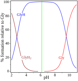
glycine pK = 2.72, 9.60
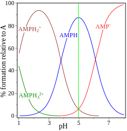
adenosine monophosphate pK = 0.9, 3.8, 6.1
In the two examples (on the right) the isoelectric point is shown by the green vertical line. In glycine the pK values are separated by nearly 7 units. Thus in the gas phase, the concentration of the neutral species, glycine (GlyH), is effectively 100% of the analytical glycine concentration.[7] Glycine may exist as a zwitterion at the isoelectric point, but the equilibrium constant for the isomerization reaction in solution
is not known.
The other example, adenosine monophosphate is shown to illustrate the fact that a third species may, in principle, be involved. In fact the concentration of (AMP)H2+3 is negligible at the isoelectric point in this case. If the pI is greater than the pH, the molecule will have a positive charge.
Peptides and proteins
A number of algorithms for estimating isoelectric points of peptides and proteins have been developed. Most of them use Henderson–Hasselbalch equation with different pK values. For instance, within the model proposed by Bjellqvist and co-workers, the pKs were determined between closely related immobilines by focusing the same sample in overlapping pH gradients.[8] Some improvements in the methodology (especially in the determination of the pK values for modified amino acids) have been also proposed.[9][10] More advanced methods take into account the effect of adjacent amino acids ±3 residues away from a charged aspartic or glutamic acid, the effects on free C terminus, as well as they apply a correction term to the corresponding pK values using genetic algorithm.[11] Other recent approaches are based on a support vector machine algorithm[12] and pKa optimization against experimentally known protein/peptide isoelectric points.[13]
Moreover, experimentally measured isoelectric point of proteins were aggregated into the databases.[14][15] Recently, a database of isoelectric points for all proteins predicted using most of the available methods had been also developed.[16]
In practice, a protein with an excess of basic aminoacids (arginine, lysine and/or histidine) will bear an isoelectric point roughly greater than 7 (basic), while a protein with an excess of acidic aminoacids (aspartic acid and/or glutamic acid) will often have an isoelectric point lower than 7 (acidic). The electrophoretic linear (horizontal) separation of proteins by Ip along a pH gradient in a polyacrylamide gel (also known as isoelectric focusing), followed by a standard molecular weight linear (vertical) separation in a second polyacrylamide gel (SDS-PAGE), constitutes the so called two-dimensional gel electrophoresis or PAGE 2D. This technique allows a thorough separation of proteins as distinct "spots", with proteins of high molecular weight and low Ip migrating to the upper-left part of the bidimensional gel, while proteins with low molecular weight and high Ip locate to the bottom-right region of the same gel.
Ceramic materials
The isoelectric points (IEP) of metal oxide ceramics are used extensively in material science in various aqueous processing steps (synthesis, modification, etc.). In the absence of chemisorbed or physisorbed species particle surfaces in aqueous suspension are generally assumed to be covered with surface hydroxyl species, M-OH (where M is a metal such as Al, Si, etc.).[17] At pH values above the IEP, the predominant surface species is M-O−, while at pH values below the IEP, M-OH2+ species predominate. Some approximate values of common ceramics are listed below:[18][19]
Note: The following list gives the isoelectric point at 25°C for selected materials in water. The exact value can vary widely, depending on material factors such as purity and phase as well as physical parameters such as temperature. Moreover, the precise measurement of isoelectric points can be difficult, thus many sources often cite differing values for isoelectric points of these materials.
Mixed oxides may exhibit isoelectric point values that are intermediate to those of the corresponding pure oxides. For example, a synthetically prepared amorphous aluminosilicate (Al2O3-SiO2) was initially measured as having IEP of 4.5 (the electrokinetic behavior of the surface was dominated by surface Si-OH species, thus explaining the relatively low IEP value).[27] Significantly higher IEP values (pH 6 to 8) have been reported for 3Al2O3-2SiO2 by others.[24] Similarly, also IEP of barium titanate, BaTiO3 was reported in the range 5–6[24] while others got a value of 3.[28] Mixtures of titania (TiO2) and zirconia (ZrO2) were studied and found to have an isoelectric point between 5.3–6.9, varying non-linearly with%(ZrO2).[29] The surface charge of the mixed oxides was correlated with acidity. Greater titania content led to increased Lewis acidity, whereas zirconia-rich oxides displayed Br::onsted acidity. The different types of acidities produced differences in ion adsorption rates and capacities.
Versus point of zero charge
The terms isoelectric point (IEP) and point of zero charge (PZC) are often used interchangeably, although under certain circumstances, it may be productive to make the distinction.
In systems in which H+/OH− are the interface potential-determining ions, the point of zero charge is given in terms of pH. The pH at which the surface exhibits a neutral net electrical charge is the point of zero charge at the surface. Electrokinetic phenomena generally measure zeta potential, and a zero zeta potential is interpreted as the point of zero net charge at the shear plane. This is termed the isoelectric point.[30] Thus, the isoelectric point is the value of pH at which the colloidal particle remains stationary in an electrical field. The isoelectric point is expected to be somewhat different from the point of zero charge at the particle surface, but this difference is often ignored in practice for so-called pristine surfaces, i.e., surfaces with no specifically adsorbed positive or negative charges.[17] In this context, specific adsorption is understood as adsorption occurring in a Stern layer or chemisorption. Thus, point of zero charge at the surface is taken as equal to isoelectric point in the absence of specific adsorption on that surface.
According to Jolivet,[21] in the absence of positive or negative charges, the surface is best described by the point of zero charge. If positive and negative charges are both present in equal amounts, then this is the isoelectric point. Thus, the PZC refers to the absence of any type of surface charge, while the IEP refers to a state of neutral net surface charge. The difference between the two, therefore, is the quantity of charged sites at the point of net zero charge. Jolivet uses the intrinsic surface equilibrium constants, pK− and pK+ to define the two conditions in terms of the relative number of charged sites:
For large ΔpK (>4 according to Jolivet), the predominant species is MOH while there are relatively few charged species – so the PZC is relevant. For small values of ΔpK, there are many charged species in approximately equal numbers, so one speaks of the IEP.
↑ Acceptable variants on pH(I) would include pHI, pHIEP, etc; the main point is that one cannot take the 'power' of I, rather one measures the pH subject to a nominated condition.
↑ Kastenholz B (2007). "New hope for the diagnosis and therapy of Alzheimer's disease". Protein and Peptide Letters. 14 (4): 389–93. doi:10.2174/092986607780363970. PMID17504097.
↑ Bjellqvist, B.; Hughes, G. J.; Pasquali, C.; Paquet, N.; Ravier, F.; Sanchez, J. C.; Frutiger, S.; Hochstrasser, D. (1993-10-01). "The focusing positions of polypeptides in immobilized pH gradients can be predicted from their amino acid sequences". Electrophoresis. 14 (10): 1023–1031. doi:10.1002/elps.11501401163. ISSN0173-0835. PMID8125050. S2CID38041111.
↑ Gauci, Sharon; van Breukelen, Bas; Lemeer, Simone M.; Krijgsveld, Jeroen; Heck, Albert J. R. (2008-12-01). "A versatile peptide pI calculator for phosphorylated and N-terminal acetylated peptides experimentally tested using peptide isoelectric focusing". Proteomics. 8 (23–24): 4898–4906. doi:10.1002/pmic.200800295. ISSN1615-9861. PMID19003858. S2CID21527631.
1 2 Hanaor, D.A.H.; Michelazzi, M.; Leonelli, C.; Sorrell, C.C. (2012). "The effects of carboxylic acids on the aqueous dispersion and electrophoretic deposition of ZrO2". Journal of the European Ceramic Society. 32 (1): 235–244. arXiv:1303.2754. doi:10.1016/j.jeurceramsoc.2011.08.015. S2CID98812224.
↑ Haruta, M (2004). "Nanoparticulate Gold Catalysts for Low-Temperature CO Oxidation". Journal of New Materials for Electrochemical Systems. 7: 163–172.
1 2 3 4 Jolivet J.P., Metal Oxide Chemistry and Synthesis. From Solution to Solid State, John Wiley & Sons Ltd. 2000, ISBN0-471-97056-5 (English translation of the original French text, De la Solution à l'Oxyde, InterEditions et CNRS Editions, Paris, 1994).
↑ Daido, T; Akaike, T (1993). "Electrochemistry of cytochrome c: influence of coulombic attraction with indium tin oxide electrode". Journal of Electroanalytical Chemistry. 344 (1–2): 91–106. doi:10.1016/0022-0728(93)80048-m.
↑ Vamvakaki, Maria; Billingham, Norman C.; Armes, Steven P.; Watts, John F.; Greaves, Stephen J. (2001). "Controlled structure copolymers for the dispersion of high-performance ceramics in aqueous media". Journal of Materials Chemistry. 11 (10): 2437–2444. doi:10.1039/b101728o. ISSN0959-9428.
↑ Drisko, Glenna L; Luca, Vittorio; Sizgek, Erden; Scales, Nicolas F.; Caruso, Rachel A. (2009). "Template Synthesis and Adsorption Properties of Hierarchically Porous Zirconium Titanium Oxides". Langmuir. 25 (9): 5286–5293. doi:10.1021/la804030h. ISSN0743-7463. PMID19397363.
↑ A.W. Adamson, A.P. Gast, "Physical Chemistry of Surfaces", John Wiley and Sons, 1997.
Further reading
Nelson DL, Cox MM (2004). Lehninger Principles of Biochemistry. W. H. Freeman; 4th edition (Hardcover). ISBN0-7167-4339-6
Kosmulski M. (2009). Surface Charging and Points of Zero Charge. CRC Press; 1st edition (Hardcover). ISBN978-1-4200-5188-9
prot pi – protein isoelectric point— an online program for calculating pI of proteins (include multiple subunits and posttranslational modifications)
CurTiPot— a suite of spreadsheets for computing acid-base equilibria (charge versus pH plot of amphoteric molecules e.g., amino acids)
pICalculax— Isoelectric point (pI) predictor for chemically modified peptides and proteins
SWISS-2DPAGEArchived 2016-12-10 at the Wayback Machine— a database of isoelectric points coming from two-dimensional polyacrylamide gel electrophoresis (~ 2,000 proteins)
PIP-DB— a Protein Isoelectric Point database (~ 5,000 proteins)
Proteome-pI— a proteome isoelectric point database (predicted isoelectric point for all proteins)
This page is based on this Wikipedia article Text is available under the CC BY-SA 4.0 license; additional terms may apply. Images, videos and audio are available under their respective licenses.