![]() Skeletal formula of L-glutamic acid | |||
| |||
![]() | |||
Names | |||
---|---|---|---|
IUPAC name Glutamic acid | |||
Systematic IUPAC name 2-Aminopentanedioic acid | |||
Other names 2-Aminoglutaric acid | |||
Identifiers | |||
3D model (JSmol) |
| ||
3DMet |
| ||
1723801 (L) 1723799 (rac) 1723800 (D) | |||
ChEBI |
| ||
ChEMBL |
| ||
ChemSpider |
| ||
DrugBank | |||
ECHA InfoCard | 100.009.567 | ||
EC Number |
| ||
E number | E620 (flavour enhancer) | ||
3502 (L) 101971 (rac) 201189 (D) | |||
KEGG | |||
PubChem CID | |||
UNII |
| ||
CompTox Dashboard (EPA) |
| ||
| |||
| |||
Properties | |||
C5H9NO4 | |||
Molar mass | 147.130 g·mol−1 | ||
Appearance | white crystalline powder | ||
Density | 1.4601 (20 °C) | ||
Melting point | 199 °C (390 °F; 472 K)decomposes | ||
7.5 g/L (20 °C) [1] | |||
Solubility | 0.00035 g/100 g ethanol (25 °C) [2] | ||
Acidity (pKa) | 2.10, 4.07, 9.47 [3] | ||
−78.5·10−6 cm3/mol | |||
Hazards | |||
GHS labelling: | |||
![]() | |||
Warning | |||
H315, H319, H335 | |||
P261, P264, P271, P280, P302+P352, P304+P340, P305+P351+P338, P312, P321, P332+P313, P337+P313, P362, P403+P233, P405, P501 | |||
NFPA 704 (fire diamond) | |||
Supplementary data page | |||
Glutamic acid (data page) | |||
Except where otherwise noted, data are given for materials in their standard state (at 25 °C [77 °F], 100 kPa). |
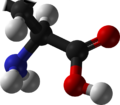
Glutamic acid (symbol Glu or E; [4] the ionic form is known as glutamate) is an α-amino acid that is used by almost all living beings in the biosynthesis of proteins. It is non-essential in humans, meaning that the body can synthesize it. It is also an excitatory neurotransmitter, in fact the most abundant one, in the vertebrate nervous system. It serves as the precursor for the synthesis of the inhibitory gamma-aminobutyric acid (GABA) in GABA-ergic neurons.
Its molecular formula is C
5H
9NO
4. Glutamic acid exists in three optically isomeric forms; the dextrorotatory L-form is usually obtained by hydrolysis of gluten or from the waste waters of beet-sugar manufacture or by fermentation. [5] Its molecular structure could be idealized as HOOC−CH(NH
2)−(CH
2)2−COOH, with two carboxyl groups −COOH and one amino group −NH
2. However, in the solid state and mildly acidic water solutions, the molecule assumes an electrically neutral zwitterion structure −OOC−CH(NH+
3)−(CH
2)2−COOH. It is encoded by the codons GAA or GAG.
The acid can lose one proton from its second carboxyl group to form the conjugate base, the singly-negative anion glutamate−OOC−CH(NH+
3)−(CH
2)2−COO−. This form of the compound is prevalent in neutral solutions. The glutamate neurotransmitter plays the principal role in neural activation. [6] This anion creates the savory umami flavor of foods and is found in glutamate flavorings such as MSG. In Europe it is classified as food additive E620. In highly alkaline solutions the doubly negative anion −OOC−CH(NH
2)−(CH
2)2−COO− prevails. The radical corresponding to glutamate is called glutamyl.
When glutamic acid is dissolved in water, the amino group (−NH
2) may gain a proton (H+
), and/or the carboxyl groups may lose protons, depending on the acidity of the medium.
In sufficiently acidic environments, the amino group gains a proton and the molecule becomes a cation with a single positive charge, HOOC−CH(NH+
3)−(CH
2)2−COOH. [7]
At pH values between about 2.5 and 4.1, [7] the carboxylic acid closer to the amine generally loses a proton, and the acid becomes the neutral zwitterion −OOC−CH(NH+
3)−(CH
2)2−COOH. This is also the form of the compound in the crystalline solid state. [8] [9] The change in protonation state is gradual; the two forms are in equal concentrations at pH 2.10. [10]
At even higher pH, the other carboxylic acid group loses its proton and the acid exists almost entirely as the glutamate anion −OOC−CH(NH+
3)−(CH
2)2−COO−, with a single negative charge overall. The change in protonation state occurs at pH 4.07. [10] This form with both carboxylates lacking protons is dominant in the physiological pH range (7.35–7.45).
At even higher pH, the amino group loses the extra proton, and the prevalent species is the doubly-negative anion −OOC−CH(NH
2)−(CH
2)2−COO−. The change in protonation state occurs at pH 9.47. [10]
The carbon atom adjacent to the amino group is chiral (connected to four distinct groups). Glutamic acid can exist in three [5] optical isomers, including the dextrorotatory L-form, [5] d(−), and l(+). The l form is the one most widely occurring in nature, but the d form occurs in some special contexts, such as the cell walls of the bacteria (which can manufacture it from the l form with the enzyme glutamate racemase) and the liver of mammals. [11] [12]
Although they occur naturally in many foods, the flavor contributions made by glutamic acid and other amino acids were only scientifically identified early in the 20th century. The substance was discovered and identified in the year 1866 by the German chemist Karl Heinrich Ritthausen, who treated wheat gluten (for which it was named) with sulfuric acid. [13] In 1908, Japanese researcher Kikunae Ikeda of the Tokyo Imperial University identified brown crystals left behind after the evaporation of a large amount of kombu broth as glutamic acid. These crystals, when tasted, reproduced the ineffable but undeniable flavor he detected in many foods, most especially in seaweed. Professor Ikeda termed this flavor umami. He then patented a method of mass-producing a crystalline salt of glutamic acid, monosodium glutamate. [14] [15]
Reactants | Products | Enzymes |
---|---|---|
Glutamine + H2O | → Glu + NH3 | GLS, GLS2 |
NAcGlu + H2O | → Glu + Acetate | N-acetyl-glutamate synthase |
α-ketoglutarate + NADPH + NH4+ | → Glu + NADP + + H2O | GLUD1, GLUD2 [16] |
α-ketoglutarate + α-amino acid | → Glu + α-keto acid | transaminase |
1-Pyrroline-5-carboxylate + NAD+ + H2O | → Glu + NADH | ALDH4A1 |
N-formimino-L-glutamate + FH4 | → Glu + 5-formimino-FH4 | FTCD |
NAAG | → Glu + NAA | GCPII |
Glutamic acid is produced on the largest scale of any amino acid, with an estimated annual production of about 1.5 million tons in 2006. [17] Chemical synthesis was supplanted by the aerobic fermentation of sugars and ammonia in the 1950s, with the organism Corynebacterium glutamicum (also known as Brevibacterium flavum) being the most widely used for production. [18] Isolation and purification can be achieved by concentration and crystallization; it is also widely available as its hydrochloride salt. [19]
Glutamate is a key compound in cellular metabolism. In humans, dietary proteins are broken down by digestion into amino acids, which serve as metabolic fuel for other functional roles in the body. A key process in amino acid degradation is transamination, in which the amino group of an amino acid is transferred to an α-ketoacid, typically catalysed by a transaminase. The reaction can be generalised as such:
A very common α-keto acid is α-ketoglutarate, an intermediate in the citric acid cycle. Transamination of α-ketoglutarate gives glutamate. The resulting α-ketoacid product is often a useful one as well, which can contribute as fuel or as a substrate for further metabolism processes. Examples are as follows:
Both pyruvate and oxaloacetate are key components of cellular metabolism, contributing as substrates or intermediates in fundamental processes such as glycolysis, gluconeogenesis, and the citric acid cycle.
Glutamate also plays an important role in the body's disposal of excess or waste nitrogen. Glutamate undergoes deamination, an oxidative reaction catalysed by glutamate dehydrogenase, [16] as follows:
Ammonia (as ammonium) is then excreted predominantly as urea, synthesised in the liver. Transamination can thus be linked to deamination, effectively allowing nitrogen from the amine groups of amino acids to be removed, via glutamate as an intermediate, and finally excreted from the body in the form of urea.
Glutamate is also a neurotransmitter (see below), which makes it one of the most abundant molecules in the brain. Malignant brain tumors known as glioma or glioblastoma exploit this phenomenon by using glutamate as an energy source, especially when these tumors become more dependent on glutamate due to mutations in the gene IDH1. [20] [21]
Glutamate is the most abundant excitatory neurotransmitter in the vertebrate nervous system. [22] At chemical synapses, glutamate is stored in vesicles. Nerve impulses trigger the release of glutamate from the presynaptic cell. Glutamate acts on ionotropic and metabotropic (G-protein coupled) receptors. [22] In the opposing postsynaptic cell, glutamate receptors, such as the NMDA receptor or the AMPA receptor, bind glutamate and are activated. Because of its role in synaptic plasticity, glutamate is involved in cognitive functions such as learning and memory in the brain. [23] The form of plasticity known as long-term potentiation takes place at glutamatergic synapses in the hippocampus, neocortex, and other parts of the brain. Glutamate works not only as a point-to-point transmitter, but also through spill-over synaptic crosstalk between synapses in which summation of glutamate released from a neighboring synapse creates extrasynaptic signaling/volume transmission. [24] In addition, glutamate plays important roles in the regulation of growth cones and synaptogenesis during brain development as originally described by Mark Mattson.
Extracellular glutamate in Drosophila brains has been found to regulate postsynaptic glutamate receptor clustering, via a process involving receptor desensitization. [25] A gene expressed in glial cells actively transports glutamate into the extracellular space, [25] while, in the nucleus accumbens-stimulating group II metabotropic glutamate receptors, this gene was found to reduce extracellular glutamate levels. [26] This raises the possibility that this extracellular glutamate plays an "endocrine-like" role as part of a larger homeostatic system.
Glutamate also serves as the precursor for the synthesis of the inhibitory gamma-aminobutyric acid (GABA) in GABA-ergic neurons. This reaction is catalyzed by glutamate decarboxylase (GAD), which is most abundant in the cerebellum and pancreas.[ citation needed ]
Stiff person syndrome is a neurologic disorder caused by anti-GAD antibodies, leading to a decrease in GABA synthesis and, therefore, impaired motor function such as muscle stiffness and spasm. Since the pancreas has abundant GAD, a direct immunological destruction occurs in the pancreas and the patients will have diabetes mellitus.[ citation needed ]
Glutamic acid, being a constituent of protein, is present in foods that contain protein, but it can only be tasted when it is present in an unbound form. Significant amounts of free glutamic acid are present in a wide variety of foods, including cheeses and soy sauce, and glutamic acid is responsible for umami, one of the five basic tastes of the human sense of taste. Glutamic acid often is used as a food additive and flavor enhancer in the form of its sodium salt, known as monosodium glutamate (MSG).
All meats, poultry, fish, eggs, dairy products, and kombu are excellent sources of glutamic acid. Some protein-rich plant foods also serve as sources. 30% to 35% of gluten (much of the protein in wheat) is glutamic acid. Ninety-five percent of the dietary glutamate is metabolized by intestinal cells in a first pass. [27]
Auxigro is a plant growth preparation that contains 30% glutamic acid.
In recent years,[ when? ] there has been much research into the use of residual dipolar coupling (RDC) in nuclear magnetic resonance spectroscopy (NMR). A glutamic acid derivative, poly-γ-benzyl-L-glutamate (PBLG), is often used as an alignment medium to control the scale of the dipolar interactions observed. [28]
The drug phencyclidine (more commonly known as PCP or 'Angel Dust') antagonizes glutamic acid non-competitively at the NMDA receptor. For the same reasons, dextromethorphan and ketamine also have strong dissociative and hallucinogenic effects. Acute infusion of the drug LY354740 (also known as eglumegad, an agonist of the metabotropic glutamate receptors 2 and 3) resulted in a marked diminution of yohimbine-induced stress response in bonnet macaques (Macaca radiata); chronic oral administration of LY354740 in those animals led to markedly reduced baseline cortisol levels (approximately 50 percent) in comparison to untreated control subjects. [29] LY354740 has also been demonstrated to act on the metabotropic glutamate receptor 3 (GRM3) of human adrenocortical cells, downregulating aldosterone synthase, CYP11B1, and the production of adrenal steroids (i.e. aldosterone and cortisol). [30] Glutamate does not easily pass the blood brain barrier, but, instead, is transported by a high-affinity transport system. [31] [32] It can also be converted into glutamine.
Amino acids are organic compounds that contain amino and carboxylate functional groups, along with a side chain specific to each amino acid. The elements present in every amino acid are carbon (C), hydrogen (H), oxygen (O), and nitrogen (N) (CHON); in addition sulfur (S) is present in the side chains of cysteine and methionine, and selenium (Se) in the less common amino acid selenocysteine. More than 500 naturally occurring amino acids are known to constitute monomer units of peptides, including proteins, as of 2020 although only 22 appear in the genetic code, 20 of which have their own designated codons and 2 of which have special coding mechanisms: Selenocysteine which is present in all eukaryotes and pyrrolysine which is present in some prokaryotes.
α-Ketoglutaric acid is one of two ketone derivatives of glutaric acid. The term "ketoglutaric acid," when not further qualified, almost always refers to the alpha variant. β-Ketoglutaric acid varies only by the position of the ketone functional group, and is much less common.
γ-Aminobutyric acid, or GABA, is the chief inhibitory neurotransmitter in the developmentally mature mammalian central nervous system. Its principal role is reducing neuronal excitability throughout the nervous system.
Decarboxylation is a chemical reaction that removes a carboxyl group and releases carbon dioxide (CO2). Usually, decarboxylation refers to a reaction of carboxylic acids, removing a carbon atom from a carbon chain. The reverse process, which is the first chemical step in photosynthesis, is called carboxylation, the addition of CO2 to a compound. Enzymes that catalyze decarboxylations are called decarboxylases or, the more formal term, carboxy-lyases (EC number 4.1.1).
Transamination is a chemical reaction that transfers an amino group to a ketoacid to form new amino acids. This pathway is responsible for the deamination of most amino acids. This is one of the major degradation pathways which convert essential amino acids to non-essential amino acids.
Aspartate transaminase (AST) or aspartate aminotransferase, also known as AspAT/ASAT/AAT or (serum) glutamic oxaloacetic transaminase, is a pyridoxal phosphate (PLP)-dependent transaminase enzyme that was first described by Arthur Karmen and colleagues in 1954. AST catalyzes the reversible transfer of an α-amino group between aspartate and glutamate and, as such, is an important enzyme in amino acid metabolism. AST is found in the liver, heart, skeletal muscle, kidneys, brain,red blood cells and gall bladder. Serum AST level, serum ALT level, and their ratio are commonly measured clinically as biomarkers for liver health. The tests are part of blood panels.
Glutamate dehydrogenase is an enzyme observed in both prokaryotes and eukaryotic mitochondria. The aforementioned reaction also yields ammonia, which in eukaryotes is canonically processed as a substrate in the urea cycle. Typically, the α-ketoglutarate to glutamate reaction does not occur in mammals, as glutamate dehydrogenase equilibrium favours the production of ammonia and α-ketoglutarate. Glutamate dehydrogenase also has a very low affinity for ammonia, and therefore toxic levels of ammonia would have to be present in the body for the reverse reaction to proceed. However, in brain, the NAD+/NADH ratio in brain mitochondria encourages oxidative deamination. In bacteria, the ammonia is assimilated to amino acids via glutamate and aminotransferases. In plants, the enzyme can work in either direction depending on environment and stress. Transgenic plants expressing microbial GLDHs are improved in tolerance to herbicide, water deficit, and pathogen infections. They are more nutritionally valuable.
Glutamate decarboxylase or glutamic acid decarboxylase (GAD) is an enzyme that catalyzes the decarboxylation of glutamate to gamma-aminobutyric acid (GABA) and carbon dioxide. GAD uses pyridoxal-phosphate (PLP) as a cofactor. The reaction proceeds as follows:
Molecular neuroscience is a branch of neuroscience that observes concepts in molecular biology applied to the nervous systems of animals. The scope of this subject covers topics such as molecular neuroanatomy, mechanisms of molecular signaling in the nervous system, the effects of genetics and epigenetics on neuronal development, and the molecular basis for neuroplasticity and neurodegenerative diseases. As with molecular biology, molecular neuroscience is a relatively new field that is considerably dynamic.
Transaminases or aminotransferases are enzymes that catalyze a transamination reaction between an amino acid and an α-keto acid. They are important in the synthesis of amino acids, which form proteins.
GABAB receptors (GABABR) are G-protein coupled receptors for gamma-aminobutyric acid (GABA), therefore making them metabotropic receptors, that are linked via G-proteins to potassium channels. The changing potassium concentrations hyperpolarize the cell at the end of an action potential. The reversal potential of the GABAB-mediated IPSP is –100 mV, which is much more hyperpolarized than the GABAA IPSP. GABAB receptors are found in the central nervous system and the autonomic division of the peripheral nervous system.
The metabotropic glutamate receptors, or mGluRs, are a type of glutamate receptor that are active through an indirect metabotropic process. They are members of the group C family of G-protein-coupled receptors, or GPCRs. Like all glutamate receptors, mGluRs bind with glutamate, an amino acid that functions as an excitatory neurotransmitter.
Glutamate receptors are synaptic and non synaptic receptors located primarily on the membranes of neuronal and glial cells. Glutamate is abundant in the human body, but particularly in the nervous system and especially prominent in the human brain where it is the body's most prominent neurotransmitter, the brain's main excitatory neurotransmitter, and also the precursor for GABA, the brain's main inhibitory neurotransmitter. Glutamate receptors are responsible for the glutamate-mediated postsynaptic excitation of neural cells, and are important for neural communication, memory formation, learning, and regulation.
Neurotransmitter transporters are a class of membrane transport proteins that span the cellular membranes of neurons. Their primary function is to carry neurotransmitters across these membranes and to direct their further transport to specific intracellular locations. There are more than twenty types of neurotransmitter transporters.
N-Acetylaspartylglutamic acid is a peptide neurotransmitter and the third-most-prevalent neurotransmitter in the mammalian nervous system. NAAG consists of N-acetylaspartic acid (NAA) and glutamic acid coupled via a peptide bond.
Branched-chain amino acid aminotransferase (BCAT), also known as branched-chain amino acid transaminase, is an aminotransferase enzyme (EC 2.6.1.42) which acts upon branched-chain amino acids (BCAAs). It is encoded by the BCAT2 gene in humans. The BCAT enzyme catalyzes the conversion of BCAAs and α-ketoglutarate into branched chain α-keto acids and glutamate.
Metabotropic glutamate receptor 3 (mGluR3) is an inhibitory Gi/G0-coupled G-protein coupled receptor (GPCR) generally localized to presynaptic sites of neurons in classical circuits. However, in higher cortical circuits in primates, mGluR3 are localized post-synaptically, where they strengthen rather than weaken synaptic connectivity. In humans, mGluR3 is encoded by the GRM3 gene. Deficits in mGluR3 signaling have been linked to impaired cognition in humans, and to increased risk of schizophrenia, consistent with their expanding role in cortical evolution.
The glutamate–glutamine cycle in biochemistry, is a sequence of events by which an adequate supply of the neurotransmitter glutamate is maintained in the central nervous system. Neurons are unable to synthesize either the excitatory neurotransmitter glutamate, or the inhibitory GABA from glucose. Discoveries of glutamate and glutamine pools within intercellular compartments led to suggestions of the glutamate–glutamine cycle working between neurons and astrocytes. The glutamate/GABA–glutamine cycle is a metabolic pathway that describes the release of either glutamate or GABA from neurons which is then taken up into astrocytes. In return, astrocytes release glutamine to be taken up into neurons for use as a precursor to the synthesis of either glutamate or GABA.
Dicarboxylic aminoaciduria is a rare form of aminoaciduria which is an autosomal recessive disorder of urinary glutamate and aspartate due to genetic errors related to transport of these amino acids. Mutations resulting in a lack of expression of the SLC1A1 gene, a member of the solute carrier family, are found to cause development of dicarboxylic aminoaciduria in humans. SLC1A1 encodes for EAAT3 which is found in the neurons, intestine, kidney, lung, and heart. EAAT3 is part of a family of high affinity glutamate transporters which transport both glutamate and aspartate across the plasma membrane.
In neuroscience, glutamate refers to the anion of glutamic acid in its role as a neurotransmitter: a chemical that nerve cells use to send signals to other cells. It is by a wide margin the most abundant excitatory neurotransmitter in the vertebrate nervous system. It is used by every major excitatory function in the vertebrate brain, accounting in total for well over 90% of the synaptic connections in the human brain. It also serves as the primary neurotransmitter for some localized brain regions, such as cerebellum granule cells.
This organization does not allow net glutamate entry to the brain; rather, it promotes the removal of glutamate and the maintenance of low glutamate concentrations in the ECF.
![]() | Wikimedia Commons has media related to Glutamic acid . |
![]() | Look up glutamic acid in Wiktionary, the free dictionary. |