Glutamate decarboxylase

Last updated
glutamate decarboxylase
Identifiers
EC no. 4.1.1.15
CAS no. 9024-58-2m
Databases
IntEnz IntEnz view
BRENDA BRENDA entry
ExPASy NiceZyme view
KEGG KEGG entry
MetaCyc metabolic pathway
PRIAM profile
PDB structures RCSB PDB PDBe PDBsum
Gene Ontology AmiGO / QuickGO
Search
PMC articles
PubMed articles
NCBI proteins
Glutamic acid decarboxylase 1
PDB GAD67.jpg
GAD67 derived from PDB: 2okj
Identifiers
Symbol GAD1
Alt. symbolsglutamate decarboxylase 1
(brain, 67kD); GAD67
NCBI gene 2571
HGNC 4092
OMIM 605363
RefSeq NM_000817
UniProt Q99259
Other data
EC number 4.1.1.15
Locus Chr. 2 q31
Search for
Structures Swiss-model
Domains InterPro
glutamic acid decarboxylase 2
Identifiers
Symbol GAD2
Alt. symbolsGAD65
NCBI gene 2572
HGNC 11284
OMIM 4093
RefSeq NM_001047
UniProt Q05329
Other data
EC number 4.1.1.15
Locus Chr. 10 p11.23
Search for
Structures Swiss-model
Domains InterPro

Glutamate decarboxylase or glutamic acid decarboxylase (GAD) is an enzyme that catalyzes the decarboxylation of glutamate to gamma-aminobutyric acid (GABA) and carbon dioxide (CO2). GAD uses pyridoxal-phosphate (PLP) as a cofactor. The reaction proceeds as follows:

Contents

HOOC−CH2−CH2−CH(NH2)−COOH → CO2 + HOOC−CH2−CH2−CH2NH2

In mammals, GAD exists in two isoforms with molecular weights of 67 and 65 kDa (GAD67 and GAD65), which are encoded by two different genes on different chromosomes (GAD1 and GAD2 genes, chromosomes 2 and 10 in humans, respectively). [1] [2] GAD67 and GAD65 are expressed in the brain where GABA is used as a neurotransmitter, and they are also expressed in the insulin-producing β-cells of the pancreas, in varying ratios depending upon the species. [3] Together, these two enzymes maintain the major physiological supply of GABA in mammals, [2] though it may also be synthesized from putrescine in the enteric nervous system, [4] brain, [5] [6] and elsewhere by the actions of diamine oxidase and aldehyde dehydrogenase 1a1. [4] [6]

Several truncated transcripts and polypeptides of GAD67 are detectable in the developing brain, [7] however their function, if any, is unknown.

Structure and mechanism

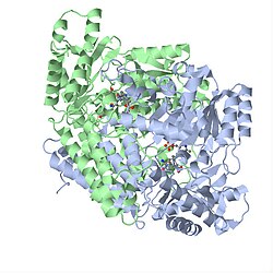
Both isoforms of GAD are homodimeric structures, consisting of 3 primary domains: the PLP, C-terminal and N-terminal domains. The PLP-binding domain of this enzyme adopts a type I PLP-dependent transferase-like fold. [8] The reaction proceeds via the canonical mechanism, involving Schiff base linkage between PLP and Lys405. PLP is held in place through base-stacking with an adjacent histidine residue, and GABA is positioned such that its carboxyl group forms a salt bridge with arginine and a hydrogen bond with glutamine.

GAD67 active site containing PLP-glutamate complex (shown in green), with Schiff base linkage at Lys405. Side chain residues shown in red. GAD67 active site.png
GAD67 active site containing PLP-glutamate complex (shown in green), with Schiff base linkage at Lys405. Side chain residues shown in red.

Dimerization is essential to maintaining function as the active site is found at this interface, and mutations interfering with optimal association between the 2 chains has been linked to pathology, such as schizophrenia. [9] [10] Interference of dimerization by GAD inhibitors such as 2-keto-4-pentenoic acid (KPA) and ethyl ketopentenoate (EKP) were also shown to lead to dramatic reductions in GABA production and incidence of seizures. [11] [8]

Catalytic activity is mediated by a short flexible loop at the dimer interface (residues 432–442 in GAD67, and 423–433 in GAD65). In GAD67 this loop remains tethered, covering the active site and providing a catalytic environment to sustain GABA production; its mobility in GAD65 promotes a side reaction that results in release of PLP, leading to autoinactivation. [12] The conformation of this loop is intimately linked to the C-terminal domain, which also affects the rate of autoinactivation. [13] Moreover, GABA-bound GAD65 is intrinsically more flexible and exists as an ensemble of states, thus providing more opportunities for autoantigenicity as seen in Type 1 diabetes. [14] [15] GAD derived from Escherichia coli shows additional structural intricacies, including a pH-dependent conformational change. This behavior is defined by the presence of a triple helical bundle formed by the N-termini of the hexameric protein in acidic environments. [16]

Hexameric E. coli GAD conformational transition: low-pH (left), neutral pH (right). PH conformational changes.png
Hexameric E. coli GAD conformational transition: low-pH (left), neutral pH (right).

Regulation of GAD65 and GAD67

Despite an extensive sequence similarity between the two genes, GAD65 and GAD67 fulfill very different roles within the human body. Additionally, research suggests that GAD65 and GAD67 are regulated by distinctly different cellular mechanisms.

GAD65 and GAD67 synthesize GABA at different locations in the cell, at different developmental times, and for functionally different purposes. [17] [18] GAD67 is spread evenly throughout the cell while GAD65 is localized to nerve terminals. [17] [19] [20] GAD67 synthesizes GABA for neuron activity unrelated to neurotransmission, such as synaptogenesis and protection from neural injury. [17] [18] This function requires widespread, ubiquitous presence of GABA. GAD65, however, synthesizes GABA for neurotransmission, [17] and therefore is only necessary at nerve terminals and synapses. In order to aid in neurotransmission, GAD65 forms a complex with heat shock cognate 70 (HSC70), cysteine string protein (CSP) and vesicular GABA transporter VGAT, which, as a complex, helps package GABA into vesicles for release during neurotransmission. [21] GAD67 is transcribed during early development, while GAD65 is not transcribed until later in life. [17] This developmental difference in GAD67 and GAD65 reflects the functional properties of each isoform; GAD67 is needed throughout development for normal cellular functioning, while GAD65 is not needed until slightly later in development when synaptic inhibition is more prevalent. [17]

Gad65 in red, Gad67 in green, and tyrosine hydroxylase (blue) in the ventral tegmental area of the mouse brain MouseGad65ThGad67.jpg
Gad65 in red, Gad67 in green, and tyrosine hydroxylase (blue) in the ventral tegmental area of the mouse brain

GAD67 and GAD65 are also regulated differently post-translationally. Both GAD65 and GAD67 are regulated via phosphorylation of a dynamic catalytic loop, [22] [12] but the regulation of these isoforms differs; GAD65 is activated by phosphorylation while GAD67 is inhibited by phosphorylation. GAD67 is predominantly found activated (~92%), whereas GAD65 is predominantly found inactivated (~72%). [23] GAD67 is phosphorylated at threonine 91 by protein kinase A (PKA), while GAD65 is phosphorylated, and therefore regulated by, protein kinase C (PKC). Both GAD67 and GAD65 are also regulated post-translationally by pyridoxal 5’-phosphate (PLP); GAD is activated when bound to PLP and inactive when not bound to PLP. [23] Majority of GAD67 is bound to PLP at any given time, whereas GAD65 binds PLP when GABA is needed for neurotransmission. [23] This reflects the functional properties of the two isoforms; GAD67 must be active at all times for normal cellular functioning, and is therefore constantly activated by PLP, while GAD65 must only be activated when GABA neurotransmission occurs, and is therefore regulated according to the synaptic environment.

Studies with mice also show functional differences between Gad67 and Gad65. GAD67−/− mice are born with cleft palate and die within a day after birth while GAD65−/− mice survive with a slightly increased tendency in seizures. Additionally, GAD65+/- have symptoms defined similarly to attention deficit hyperactivity disorder (ADHD) in humans. [24]

Role in the nervous system

Both GAD67 and GAD65 are present in all types of synapses within the human nervous system. This includes dendrodendritic, axosomatic, and axodendritic synapses. Preliminary evidence suggests that GAD65 is dominant in the visual and neuroendocrine systems, which undergo more phasic changes. It is also believed that GAD67 is present at higher amounts in tonically active neurons. [25]

Role in pathology

Autism

Both GAD65 and GAD67 experience significant downregulation in cases of autism. In a comparison of autistic versus control brains, GAD65 and GAD67 experienced a downregulation average of 50% in parietal and cerebellar cortices of autistic brains. [26] Cerebellar Purkinje cells also reported a 40% downregulation, suggesting that affected cerebellar nuclei may disrupt output to higher order motor and cognitive areas of the brain. [18]

Diabetes

Both GAD67 and GAD65 are targets of autoantibodies in people who later develop type 1 diabetes mellitus or latent autoimmune diabetes. [27] [28] Injections with GAD65 in ways that induce immune tolerance have been shown to prevent type 1 diabetes in rodent models. [29] [30] [31] In clinical trials, injections with GAD65 have been shown to preserve some insulin production for 30 months in humans with type 1 diabetes. [32] [33] A Cochrane systematic review also examined 1 study showing improvement of C-peptide levels in cases of Latent Autoimmune Diabetes in adults, 5 years following treatment with GAD65 .Still, it is important to highlight that the studies available to be included in this review presented considerable flaws in quality and design. [34]

Stiff person syndrome

Healthy human cerebellum stained with a reference anti-GAD65 monoclonal antibody. Thin arrows show presynaptic terminals staining with the anti-GAD65 monoclonal antibody Stiff man human cerebellum.JPG
Healthy human cerebellum stained with a reference anti-GAD65 monoclonal antibody. Thin arrows show presynaptic terminals staining with the anti-GAD65 monoclonal antibody

High titers of autoantibodies to glutamic acid decarboxylase (GAD) are well documented in association with stiff person syndrome (SPS). [35] Glutamic acid decarboxylase is the rate-limiting enzyme in the synthesis of γ-aminobutyric acid (GABA), and impaired function of GABAergic neurons has been implicated in the pathogenesis of SPS. Autoantibodies to GAD might be the causative agent or a disease marker. [36]

Schizophrenia and bipolar disorder

Substantial dysregulation of GAD mRNA expression, coupled with downregulation of reelin, is observed in schizophrenia and bipolar disorder. [37] [38] The most pronounced downregulation of GAD67 was found in hippocampal stratum oriens layer in both disorders and in other layers and structures of hippocampus with varying degrees. [39]

GAD67 is a key enzyme involved in the synthesis of inhibitory neurotransmitter GABA and people with schizophrenia have been shown to express lower amounts of GAD67 in the dorsolateral prefrontal cortex compared to healthy controls. [40] The mechanism underlying the decreased levels of GAD67 in people with schizophrenia remains unclear. [41] Some have proposed that an immediate early gene, Zif268, which normally binds to the promoter region of GAD67 and increases transcription of GAD67, is lower in schizophrenic patients, thus contributing to decreased levels of GAD67. [40] Since the dorsolateral prefrontal cortex (DLPFC) is involved in working memory, and GAD67 and Zif268 mRNA levels are lower in the DLPFC of schizophrenic patients, this molecular alteration may account, at least in part, for the working memory impairments associated with the disease.

Parkinson disease

The bilateral delivery of glutamic acid decarboxylase (GAD) by an adeno-associated viral vector into the subthalamic nucleus of patients between 30 and 75 years of age with advanced, progressive, levodopa-responsive Parkinson disease resulted in significant improvement over baseline during the course of a six-month study. [42]

Cerebellar disorders

Intracerebellar administration of GAD autoantibodies to animals increases the excitability of motoneurons and impairs the production of nitric oxide (NO), a molecule involved in learning. Epitope recognition contributes to cerebellar involvement. [43] Reduced GABA levels increase glutamate levels as a consequence of lower inhibition of subtypes of GABA receptors. Higher glutamate levels activate microglia and activation of xc(−) increases the extracellular glutamate release. [44]

Neuropathic pain

Peripheral nerve injury of the sciatic nerve (a neuropathic pain model) induces a transient loss of GAD65 immunoreactive terminals in the spinal cord dorsal horn and suggests a potential involvement for these alterations in the development and amelioration of pain behaviour. [45]

Other anti-GAD-associated neurologic disorders

Antibodies directed against glutamic acid decarboxylase (GAD) are increasingly found in patients with other symptoms indicative of central nervous system (CNS) dysfunction, such as ataxia, progressive encephalomyelitis with rigidity and myoclonus (PERM), limbic encephalitis, and epilepsy. [46] The pattern of anti-GAD antibodies in epilepsy differs from type 1 diabetes and stiff-person syndrome. [47]

Role of glutamate decarboxylase in other organisms

Besides the synthesis of GABA, GAD has additional functions and structural variations that are organism-dependent. In Saccharomyces cerevisiae , GAD binds the Ca2+ regulatory protein calmodulin (CaM) and is also involved in responding to oxidative stress. [48] Similarly, GAD in plants binds calmodulin as well. [49] This interaction occurs at the 30-50bp CAM-binding domain (CaMBD) in its C terminus and is necessary for proper regulation of GABA production. [50] Unlike vertebrates and invertebrates, the GABA produced by GAD is used in plants to signal abiotic stress by controlling levels of intracellular Ca2+ via CaM. Binding to CaM opens Ca2+ channels and leads to an increase in Ca2+ concentrations in the cytosol, allowing Ca2+ to act as a secondary messenger and activate downstream pathways. When GAD is not bound to CaM, the CaMBD acts as an autoinhibitory domain, thus deactivating GAD in the absence of stress. [50] Interesting, in two plant species, rice and apples, Ca2+ /CAM-independent GAD isoforms have been discovered. [51] [52] The C-terminus of these isoforms contain substitutions at key residues necessary to interact with CaM in the CaMBD, preventing the protein from binding to GAD. Whereas CaMBD of the isoform in rice still functions as an autoinhibitory domain, [51] the C-terminus in the isoform in apples does not. [52] Finally, the structure of plant GAD is a hexamer and has pH-dependent activity, with the optimal pH of 5.8 in multiple species. [50] [53] but also significant activity at pH 7.3 in the presence of CaM [16]

It is also believed that the control of glutamate decarboxylase has the prospect of improving citrus produce quality post-harvest. In Citrus plants, research has shown that glutamate decarboxylase plays a key role in citrate metabolism. With the increase of glutamate decarboxylase via direct exposure, citrate levels have been seen to significantly increase within plants, and in conjunction post-harvest quality maintenance was significantly improved, and rot rates decreased. [54]

Just like GAD in plants, GAD in E. coli has a hexamer structure and is more active under acidic pH; the pH optimum for E. coli GAD is 3.8-4.6. However, unlike plants and yeast, GAD in E. coli does not require calmodulin binding to function. There are also two isoforms of GAD, namely GadA and GadB, encoded by separate genes in E. coli, [55] although both isoforms are biochemically identical. [56] The enzyme plays a major role in conferring acid resistance and allows bacteria to temporarily survive in highly acidic environments (pH < 2.5) like the stomach. [57] This is done by GAD decarboxylating glutamate to GABA, which requires H+ to be uptaken as a reactant and raises the pH inside the bacteria. GABA can then be exported out of E. coli cells and contribute to increasing the pH of the nearby extracellular environments. [16]

Related Research Articles

Ataxia is a neurological sign consisting of lack of voluntary coordination of muscle movements that can include gait abnormality, speech changes, and abnormalities in eye movements, that indicates dysfunction of parts of the nervous system that coordinate movement, such as the cerebellum.

<span class="mw-page-title-main">Glutamic acid</span> Amino acid and neurotransmitter

Glutamic acid is an α-amino acid that is used by almost all living beings in the biosynthesis of proteins. It is a non-essential nutrient for humans, meaning that the human body can synthesize enough for its use. It is also the most abundant excitatory neurotransmitter in the vertebrate nervous system. It serves as the precursor for the synthesis of the inhibitory gamma-aminobutyric acid (GABA) in GABAergic neurons.

<span class="mw-page-title-main">GABA receptor</span> Receptors that respond to gamma-aminobutyric acid

The GABA receptors are a class of receptors that respond to the neurotransmitter gamma-aminobutyric acid (GABA), the chief inhibitory compound in the mature vertebrate central nervous system. There are two classes of GABA receptors: GABAA and GABAB. GABAA receptors are ligand-gated ion channels ; whereas GABAB receptors are G protein-coupled receptors, also called metabotropic receptors.

Aromatic <small>L</small>-amino acid decarboxylase Class of enzymes

Aromatic L-amino acid decarboxylase, also known as DOPA decarboxylase (DDC), tryptophan decarboxylase, and 5-hydroxytryptophan decarboxylase, is a lyase enzyme, located in region 7p12.2-p12.1.

<span class="mw-page-title-main">Pyridoxal phosphate</span> Active form of vitamin B6

Pyridoxal phosphate (PLP, pyridoxal 5'-phosphate, P5P), the active form of vitamin B6, is a coenzyme in a variety of enzymatic reactions. The International Union of Biochemistry and Molecular Biology has catalogued more than 140 PLP-dependent activities, corresponding to ~4% of all classified activities. The versatility of PLP arises from its ability to covalently bind the substrate, and then to act as an electrophilic catalyst, thereby stabilizing different types of carbanionic reaction intermediates.

<span class="mw-page-title-main">Calcineurin</span> Class of enzymes

Calcineurin (CaN) is a calcium and calmodulin dependent serine/threonine protein phosphatase. It activates the T cells of the immune system and can be blocked by drugs. Calcineurin activates nuclear factor of activated T cell cytoplasmic (NFATc), a transcription factor, by dephosphorylating it. The activated NFATc is then translocated into the nucleus, where it upregulates the expression of interleukin 2 (IL-2), which, in turn, stimulates the growth and differentiation of the T cell response. Calcineurin is the target of a class of drugs called calcineurin inhibitors, which include ciclosporin, voclosporin, pimecrolimus and tacrolimus.

<span class="mw-page-title-main">Aspartate transaminase</span> Enzyme involved in amino acid metabolism

Aspartate transaminase (AST) or aspartate aminotransferase, also known as AspAT/ASAT/AAT or (serum) glutamic oxaloacetic transaminase, is a pyridoxal phosphate (PLP)-dependent transaminase enzyme that was first described by Arthur Karmen and colleagues in 1954. AST catalyzes the reversible transfer of an α-amino group between aspartate and glutamate and, as such, is an important enzyme in amino acid metabolism. AST is found in the liver, heart, skeletal muscle, kidneys, brain, red blood cells and gall bladder. Serum AST level, serum ALT level, and their ratio are commonly measured clinically as biomarkers for liver health. The tests are part of blood panels.

<span class="mw-page-title-main">Glutamate dehydrogenase</span> Hexameric enzyme

Glutamate dehydrogenase is an enzyme observed in both prokaryotes and eukaryotic mitochondria. The aforementioned reaction also yields ammonia, which in eukaryotes is canonically processed as a substrate in the urea cycle. Typically, the α-ketoglutarate to glutamate reaction does not occur in mammals, as glutamate dehydrogenase equilibrium favours the production of ammonia and α-ketoglutarate. Glutamate dehydrogenase also has a very low affinity for ammonia, and therefore toxic levels of ammonia would have to be present in the body for the reverse reaction to proceed. However, in brain, the NAD+/NADH ratio in brain mitochondria encourages oxidative deamination. In bacteria, the ammonia is assimilated to amino acids via glutamate and aminotransferases. In plants, the enzyme can work in either direction depending on environment and stress. Transgenic plants expressing microbial GLDHs are improved in tolerance to herbicide, water deficit, and pathogen infections. They are more nutritionally valuable.

Slowly evolving immune-mediated diabetes, or latent autoimmune diabetes in adults (LADA), is a form of diabetes that exhibits clinical features similar to both type 1 diabetes (T1D) and type 2 diabetes (T2D), and is sometimes referred to as type 1.5 diabetes. It is an autoimmune form of diabetes, similar to T1D, but patients with LADA often show insulin resistance, similar to T2D, and share some risk factors for the disease with T2D. Studies have shown that LADA patients have certain types of antibodies against the insulin-producing cells, and that these cells stop producing insulin more slowly than in T1D patients. Since many people develop the disease later in life, it is often misdiagnosed as type 2 diabetes.

<i>N</i>-Acetylaspartylglutamic acid Peptide neurotransmitter

N-Acetylaspartylglutamic acid is a peptide neurotransmitter and the third-most-prevalent neurotransmitter in the mammalian nervous system. NAAG consists of N-acetylaspartic acid (NAA) and glutamic acid coupled via a peptide bond.

<span class="mw-page-title-main">Branched-chain amino acid aminotransferase</span> Aminotransferase enzyme

Branched-chain amino acid aminotransferase (BCAT), also known as branched-chain amino acid transaminase, is an aminotransferase enzyme (EC 2.6.1.42) which acts upon branched-chain amino acids (BCAAs). It is encoded by the BCAT2 gene in humans. The BCAT enzyme catalyzes the conversion of BCAAs and α-ketoglutarate into branched chain α-keto acids and glutamate.

<span class="mw-page-title-main">Arginine decarboxylase</span>

The enzyme Acid-Induced Arginine Decarboxylase (AdiA), also commonly referred to as arginine decarboxylase, catalyzes the conversion of L-arginine into agmatine and carbon dioxide. The process consumes a proton in the decarboxylation and employs a pyridoxal-5'-phosphate (PLP) cofactor, similar to other enzymes involved in amino acid metabolism, such as ornithine decarboxylase and glutamine decarboxylase. It is found in bacteria and virus, though most research has so far focused on forms of the enzyme in bacteria. During the AdiA catalyzed decarboxylation of arginine, the necessary proton is consumed from the cell cytoplasm which helps to prevent the over-accumulation of protons inside the cell and serves to increase the intracellular pH. Arginine decarboxylase is part of an enzymatic system in Escherichia coli, Salmonella Typhimurium, and methane-producing bacteria Methanococcus jannaschii that makes these organisms acid resistant and allows them to survive under highly acidic medium.

<span class="mw-page-title-main">4-aminobutyrate transaminase</span> Class of enzymes

In enzymology, 4-aminobutyrate transaminase, also called GABA transaminase or 4-aminobutyrate aminotransferase, or GABA-T, is an enzyme that catalyzes the chemical reaction:

<span class="mw-page-title-main">PTPRN</span> Protein-coding gene in the species Homo sapiens

Receptor-type tyrosine-protein phosphatase-like N, also called "IA-2", is an enzyme that in humans is encoded by the PTPRN gene.

<span class="mw-page-title-main">GABA transporter type 1</span> Protein-coding gene in the species Homo sapiens

GABA transporter 1 (GAT1) also known as sodium- and chloride-dependent GABA transporter 1 is a protein that in humans is encoded by the SLC6A1 gene and belongs to the solute carrier 6 (SLC6) family of transporters. It mediates gamma-aminobutyric acid's translocation from the extracellular to intracellular spaces within brain tissue and the central nervous system as a whole.

<span class="mw-page-title-main">GAD2</span> Protein-coding gene in the species Homo sapiens

Glutamate decarboxylase 2 is an enzyme that in humans is encoded by the GAD2 gene.

<span class="mw-page-title-main">GAD1</span> Protein-coding gene in the species Homo sapiens

Glutamate decarboxylase 1 (GAD67), also known as GAD1, is a human gene.

In biochemistry, the glutamate–glutamine cycle is a cyclic metabolic pathway which maintains an adequate supply of the neurotransmitter glutamate in the central nervous system. Neurons are unable to synthesize either the excitatory neurotransmitter glutamate, or the inhibitory GABA from glucose. Discoveries of glutamate and glutamine pools within intercellular compartments led to suggestions of the glutamate–glutamine cycle working between neurons and astrocytes. The glutamate/GABA–glutamine cycle is a metabolic pathway that describes the release of either glutamate or GABA from neurons which is then taken up into astrocytes. In return, astrocytes release glutamine to be taken up into neurons for use as a precursor to the synthesis of either glutamate or GABA.

<span class="mw-page-title-main">Autoimmune encephalitis</span> Type of encephalitis

Autoimmune encephalitis (AIE) is a type of encephalitis, and one of the most common causes of noninfectious encephalitis. It can be triggered by tumors, infections, or it may be cryptogenic. The neurological manifestations can be either acute or subacute and usually develop within six weeks. The clinical manifestations include behavioral and psychiatric symptoms, autonomic disturbances, movement disorders, and seizures.

Fernando Garcia de Mello is a renowned neurochemist from Brazil. He obtained his degree in Biochemistry in 1968 from the State University of Rio de Janeiro. Fernando Mello started his scientific training as an undergraduate student at the Brazilian National Institute of Cancer, and later at the Institute of Biophysics from the Federal University of Rio de Janeiro, being mentored by dr. Firmino de Castro, which greatly influenced him to have a more humanistic approach towards the students that he would train. It was only during his post-doc period (1973-1976) at the National Institutes of Health under supervision of dr. Marshall Warren Nirenberg that Mello began his research in Neurochemistry, using the embryonary Retina as a model for his investigations.

References

  1. Erlander MG, Tillakaratne NJ, Feldblum S, Patel N, Tobin AJ (July 1991). "Two genes encode distinct glutamate decarboxylases". Neuron. 7 (1): 91–100. doi:10.1016/0896-6273(91)90077-D. PMID   2069816. S2CID   15863479.
  2. 1 2 Langendorf CG, Tuck KL, Key TL, Fenalti G, Pike RN, Rosado CJ, et al. (January 2013). "Structural characterization of the mechanism through which human glutamic acid decarboxylase auto-activates". Bioscience Reports. 33 (1): 137–44. doi:10.1042/BSR20120111. PMC   3546353 . PMID   23126365.
  3. Kim J, Richter W, Aanstoot HJ, Shi Y, Fu Q, Rajotte R, et al. (December 1993). "Differential expression of GAD65 and GAD67 in human, rat, and mouse pancreatic islets". Diabetes. 42 (12): 1799–808. doi:10.2337/diab.42.12.1799. PMID   8243826. S2CID   29615710.
  4. 1 2 Krantis A (December 2000). "GABA in the Mammalian Enteric Nervous System". News in Physiological Sciences. 15 (6): 284–290. doi:10.1152/physiologyonline.2000.15.6.284. PMID   11390928.
  5. Sequerra EB, Gardino P, Hedin-Pereira C, de Mello FG (May 2007). "Putrescine as an important source of GABA in the postnatal rat subventricular zone". Neuroscience. 146 (2): 489–93. doi:10.1016/j.neuroscience.2007.01.062. PMID   17395389. S2CID   43003476.
  6. 1 2 Kim JI, Ganesan S, Luo SX, Wu YW, Park E, Huang EJ, et al. (October 2015). "Aldehyde dehydrogenase 1a1 mediates a GABA synthesis pathway in midbrain dopaminergic neurons" (PDF). Science. 350 (6256): 102–6. Bibcode:2015Sci...350..102K. doi:10.1126/science.aac4690. PMC   4725325 . PMID   26430123.
  7. Szabo G, Katarova Z, Greenspan R (November 1994). "Distinct protein forms are produced from alternatively spliced bicistronic glutamic acid decarboxylase mRNAs during development". Molecular and Cellular Biology. 14 (11): 7535–45. doi:10.1128/mcb.14.11.7535. PMC   359290 . PMID   7935469.
  8. 1 2 Reingold DF, Orlowski M (Mar 1979). "Inhibition of brain glutamate decarboxylase by 2-keto-4-pentenoic acid, a metabolite of allylglycine". J Neurochem. 32 (3): 907–13. doi:10.1111/j.1471-4159.1979.tb04574.x. PMID   430066. S2CID   31823191.
  9. Magri C, Giacopuzzi E, La Via L, Bonini D, Ravasio V, Elhussiny ME, Orizio F, Gangemi F, Valsecchi P, Bresciani R, Barbon A, Vita A, Gennarelli M (Oct 2018). "A novel homozygous mutation in GAD1 gene described in a schizophrenic patient impairs activity and dimerization of GAD67 enzyme". Sci Rep. 8 (1): 15470. Bibcode:2018NatSR...815470M. doi:10.1038/s41598-018-33924-8. PMC   6195539 . PMID   30341396.
  10. Giacopuzzi E, Gennarelli M, Minelli A, Gardella R, Valsecchi P, Traversa M, Bonvicini C, Vita A, Sacchetti E, Magri C (Aug 2017). "Exome sequencing in schizophrenic patients with high levels of homozygosity identifies novel and extremely rare mutations in the GABA/glutamatergic pathways". PLOS ONE. 12 (8): e0182778. Bibcode:2017PLoSO..1282778G. doi: 10.1371/journal.pone.0182778 . PMC   5546675 . PMID   28787007.
  11. Zhang Y, Vanmeert M, Siekierska A, Ny A, John J, Callewaert G, Lescrinier E, Dehaen W, de Witte PA, Kaminski RM (Aug 2017). "Inhibition of glutamate decarboxylase (GAD) by ethyl ketopentenoate (EKP) induces treatment-resistant epileptic seizures in zebrafish". Sci Rep. 7 (1): 7195. Bibcode:2017NatSR...7.7195Z. doi:10.1038/s41598-017-06294-w. PMC   5543107 . PMID   28775328.
  12. 1 2 Fenalti G, Law RH, Buckle AM, Langendorf C, Tuck K, Rosado CJ, et al. (April 2007). "GABA production by glutamic acid decarboxylase is regulated by a dynamic catalytic loop". Nature Structural & Molecular Biology. 14 (4): 280–6. doi:10.1038/nsmb1228. PMID   17384644. S2CID   20265911.
  13. Langendorf CG, Tuck KL, Key TL, Fenalti G, Pike RN, Rosado CJ, Wong AS, Buckle AM, Law RH, Whisstock JC (Jan 2013). "Structural characterization of the mechanism through which human glutamic acid decarboxylase auto-activates". Biosci Rep. 33 (1): 137–44. doi:10.1042/BSR20120111. PMC   3546353 . PMID   23126365.
  14. Kass I, Hoke DE, Costa MG, Reboul CF, Porebski BT, Cowieson NP, Leh H, Pennacchietti E, McCoey J, Kleifeld O, Borri Voltattorni C, Langley D, Roome B, Mackay IR, Christ D, Perahia D, Buckle M, Paiardini A, De Biase D, Buckle AM (Jun 2019). "Cofactor-dependent conformational heterogeneity of GAD65 and its role in autoimmunity and neurotransmitter homeostasis". Proc Natl Acad Sci U S A. 111 (25): E2524-9. doi: 10.1073/pnas.1403182111 . PMC   4078817 . PMID   24927554.
  15. Ellis TM, Atkinson MA (Feb 1996). "The clinical significance of an autoimmune response against glutamic acid decarboxylase". Nat Med. 2 (2): 148–53. doi:10.1038/nm0296-148. PMID   8574952. S2CID   12788084.
  16. 1 2 3 Capitani G, De Biase D, Aurizi C, Gut H, Bossa F, Grütter MG (Aug 2003). "Crystal structure and functional analysis of Escherichia coli glutamate decarboxylase". EMBO J. 22 (16): 4027–37. doi:10.1093/emboj/cdg403. PMC   175793 . PMID   12912902.
  17. 1 2 3 4 5 6 Pinal CS, Tobin AJ (1998). "Uniqueness and redundancy in GABA production". Perspectives on Developmental Neurobiology. 5 (2–3): 109–18. PMID   9777629.
  18. 1 2 3 Soghomonian JJ, Martin DL (December 1998). "Two isoforms of glutamate decarboxylase: why?". Trends in Pharmacological Sciences. 19 (12): 500–5. doi:10.1016/s0165-6147(98)01270-x. PMID   9871412.
  19. Kaufman DL, Houser CR, Tobin AJ (February 1991). "Two forms of the gamma-aminobutyric acid synthetic enzyme glutamate decarboxylase have distinct intraneuronal distributions and cofactor interactions". Journal of Neurochemistry. 56 (2): 720–3. doi:10.1111/j.1471-4159.1991.tb08211.x. PMC   8194030 . PMID   1988566. S2CID   35743434.
  20. Kanaani J, Cianciaruso C, Phelps EA, Pasquier M, Brioudes E, Billestrup N, Baekkeskov S (2015). "Compartmentalization of GABA synthesis by GAD67 differs between pancreatic beta cells and neurons". PLOS ONE. 10 (2): e0117130. Bibcode:2015PLoSO..1017130K. doi: 10.1371/journal.pone.0117130 . PMC   4315522 . PMID   25647668.
  21. Jin H, Wu H, Osterhaus G, Wei J, Davis K, Sha D, et al. (April 2003). "Demonstration of functional coupling between gamma -aminobutyric acid (GABA) synthesis and vesicular GABA transport into synaptic vesicles". Proceedings of the National Academy of Sciences of the United States of America. 100 (7): 4293–8. Bibcode:2003PNAS..100.4293J. doi: 10.1073/pnas.0730698100 . PMC   153086 . PMID   12634427.
  22. Wei J, Davis KM, Wu H, Wu JY (May 2004). "Protein phosphorylation of human brain glutamic acid decarboxylase (GAD)65 and GAD67 and its physiological implications". Biochemistry. 43 (20): 6182–9. doi:10.1021/bi0496992. PMID   15147202.
  23. 1 2 3 Battaglioli G, Liu H, Martin DL (August 2003). "Kinetic differences between the isoforms of glutamate decarboxylase: implications for the regulation of GABA synthesis". Journal of Neurochemistry. 86 (4): 879–87. doi:10.1046/j.1471-4159.2003.01910.x. PMID   12887686. S2CID   23640198.
  24. Ueno H (October 2000). "Enzymatic and structural aspects on glutamate decarboxylase". Journal of Molecular Catalysis B: Enzymatic. 10 (1–3): 67–79. doi:10.1016/S1381-1177(00)00114-4.
  25. Feldblum S, Erlander MG, Tobin AJ (April 1993). "Different distributions of GAD65 and GAD67 mRNAs suggest that the two glutamate decarboxylases play distinctive functional roles". Journal of Neuroscience Research. 34 (6): 689–706. doi:10.1002/jnr.490340612. PMID   8315667. S2CID   19314092.
  26. Fatemi SH, Halt AR, Stary JM, Kanodia R, Schulz SC, Realmuto GR (October 2002). "Glutamic acid decarboxylase 65 and 67 kDa proteins are reduced in autistic parietal and cerebellar cortices". Biological Psychiatry. 52 (8): 805–10. doi:10.1016/S0006-3223(02)01430-0. PMID   12372652. S2CID   30140735.
  27. Baekkeskov S, Aanstoot HJ, Christgau S, Reetz A, Solimena M, Cascalho M, Folli F, Richter-Olesen H, De Camilli P, Camilli PD (September 1990). "Identification of the 64K autoantigen in insulin-dependent diabetes as the GABA-synthesizing enzyme glutamic acid decarboxylase". Nature. 347 (6289): 151–6. Bibcode:1990Natur.347..151B. doi:10.1038/347151a0. PMID   1697648. S2CID   4317318.
  28. Kaufman DL, Erlander MG, Clare-Salzler M, Atkinson MA, Maclaren NK, Tobin AJ (January 1992). "Autoimmunity to two forms of glutamate decarboxylase in insulin-dependent diabetes mellitus". The Journal of Clinical Investigation. 89 (1): 283–92. doi:10.1172/JCI115573. PMC   442846 . PMID   1370298.
  29. Tisch R, Yang XD, Singer SM, Liblau RS, Fugger L, McDevitt HO (November 1993). "Immune response to glutamic acid decarboxylase correlates with insulitis in non-obese diabetic mice". Nature. 366 (6450): 72–5. Bibcode:1993Natur.366...72T. doi:10.1038/366072a0. PMID   8232539. S2CID   4273636.
  30. Kaufman DL, Clare-Salzler M, Tian J, Forsthuber T, Ting GS, Robinson P, Atkinson MA, Sercarz EE, Tobin AJ, Lehmann PV (November 1993). "Spontaneous loss of T-cell tolerance to glutamic acid decarboxylase in murine insulin-dependent diabetes". Nature. 366 (6450): 69–72. Bibcode:1993Natur.366...69K. doi:10.1038/366069a0. PMC   8216222 . PMID   7694152. S2CID   4370149.
  31. Tian J, Clare-Salzler M, Herschenfeld A, Middleton B, Newman D, Mueller R, Arita S, Evans C, Atkinson MA, Mullen Y, Sarvetnick N, Tobin AJ, Lehmann PV, Kaufman DL (December 1996). "Modulating autoimmune responses to GAD inhibits disease progression and prolongs islet graft survival in diabetes-prone mice". Nature Medicine. 2 (12): 1348–53. doi:10.1038/nm1296-1348. PMID   8946834. S2CID   27692555.
  32. Ludvigsson J, Faresjö M, Hjorth M, Axelsson S, Chéramy M, Pihl M, Vaarala O, Forsander G, Ivarsson S, Johansson C, Lindh A, Nilsson NO, Aman J, Ortqvist E, Zerhouni P, Casas R (October 2008). "GAD treatment and insulin secretion in recent-onset type 1 diabetes". The New England Journal of Medicine. 359 (18): 1909–20. doi: 10.1056/NEJMoa0804328 . PMID   18843118.
  33. "Diamyd announces completion of type 1 diabetes vaccine trial with long term efficacy demonstrated at 30 months". Press Release. Diamyd Medical AB. 2008-01-28. Retrieved 2010-01-13.
  34. Brophy, Sinead; Davies, Helen; Mannan, Sopna; Brunt, Huw; Williams, Rhys (2011-09-07). "Interventions for latent autoimmune diabetes (LADA) in adults". Cochrane Database of Systematic Reviews. 2011 (9): CD006165. doi:10.1002/14651858.cd006165.pub3. ISSN   1465-1858. PMC   6486159 . PMID   21901702.
  35. Dalakas MC, Fujii M, Li M, Lutfi B, Kyhos J, McElroy B (December 2001). "High-dose intravenous immune globulin for stiff-person syndrome". The New England Journal of Medicine. 345 (26): 1870–6. doi: 10.1056/NEJMoa01167 . PMID   11756577.
  36. Chang T, Alexopoulos H, McMenamin M, Carvajal-González A, Alexander SK, Deacon R, Erdelyi F, Szabó G, Gabor S, Lang B, Blaes F, Brown P, Vincent A (September 2013). "Neuronal surface and glutamic acid decarboxylase autoantibodies in Nonparaneoplastic stiff person syndrome". JAMA Neurology. 70 (9): 1140–9. doi:10.1001/jamaneurol.2013.3499. PMC   6055982 . PMID   23877118.
  37. Guidotti, Alessandro; Auta, James; Davis, John M.; Gerevini, Valeria DiGiorgi; Dwivedi, Yogesh; Grayson, Dennis R.; Impagnatiello, Francesco; Pandey, Ghanshyam; Pesold, Christine; Sharma, Rajiv; Uzunov, Doncho; Costa, Erminio (2000). "Decrease in reelin and glutamic acid decarboxylase67 (GAD67) expression in schizophrenia and bipolar disorder: a postmortem brain study". Archives of General Psychiatry. 57 (11): 1061–1069. doi:10.1001/archpsyc.57.11.1061. PMID   11074872.
  38. Akbarian, Schahram; Huang, Hsien-Sung (2006). "Molecular and cellular mechanisms of altered GAD1/GAD67 expression in schizophrenia and related disorders". Brain Research Reviews. 52 (2): 293–304. doi:10.1016/j.brainresrev.2006.04.001. PMID   16759710. S2CID   25771139.
  39. Benes FM, Lim B, Matzilevich D, Walsh JP, Subburaju S, Minns M (June 2007). "Regulation of the GABA cell phenotype in hippocampus of schizophrenics and bipolars". Proceedings of the National Academy of Sciences of the United States of America. 104 (24): 10164–9. Bibcode:2007PNAS..10410164B. doi: 10.1073/pnas.0703806104 . PMC   1888575 . PMID   17553960.
  40. 1 2 Kimoto S, Bazmi HH, Lewis DA (September 2014). "Lower expression of glutamic acid decarboxylase 67 in the prefrontal cortex in schizophrenia: contribution of altered regulation by Zif268". The American Journal of Psychiatry. 171 (9): 969–78. doi:10.1176/appi.ajp.2014.14010004. PMC   4376371 . PMID   24874453.
  41. Georgiev, Danko; Yoshihara, Toru; Kawabata, Rika; Matsubara, Takurou; Tsubomoto, Makoto; Minabe, Yoshio; Lewis, David A.; Hashimoto, Takanori (2016). "Cortical gene expression after a conditional knockout of 67 kDa glutamic acid decarboxylase in parvalbumin neurons". Schizophrenia Bulletin. 42 (4): 992–1002. doi:10.1093/schbul/sbw022. PMC   4903066 . PMID   26980143. S2CID   24197087.
  42. LeWitt PA, Rezai AR, Leehey MA, Ojemann SG, Flaherty AW, Eskandar EN, Kostyk SK, Thomas K, Sarkar A, Siddiqui MS, Tatter SB, Schwalb JM, Poston KL, Henderson JM, Kurlan RM, Richard IH, Van Meter L, Sapan CV, During MJ, Kaplitt MG, Feigin A (April 2011). "AAV2-GAD gene therapy for advanced Parkinson's disease: a double-blind, sham-surgery controlled, randomised trial". The Lancet. Neurology. 10 (4): 309–19. doi:10.1016/S1474-4422(11)70039-4. PMID   21419704. S2CID   37154043.
  43. Manto MU, Hampe CS, Rogemond V, Honnorat J (February 2011). "Respective implications of glutamate decarboxylase antibodies in stiff person syndrome and cerebellar ataxia". Orphanet Journal of Rare Diseases. 6 (3): 3. doi: 10.1186/1750-1172-6-3 . PMC   3042903 . PMID   21294897.
  44. Mitoma H, Manto M, Hampe CS (2017-03-12). "Pathogenic Roles of Glutamic Acid Decarboxylase 65 Autoantibodies in Cerebellar Ataxias". Journal of Immunology Research. 2017: 2913297. doi: 10.1155/2017/2913297 . PMC   5366212 . PMID   28386570.
  45. Lorenzo LE, Magnussen C, Bailey AL, St Louis M, De Koninck Y, Ribeiro-da-Silva A (September 2014). "Spatial and temporal pattern of changes in the number of GAD65-immunoreactive inhibitory terminals in the rat superficial dorsal horn following peripheral nerve injury". Molecular Pain. 10 (1): 1744-8069–10-57. doi: 10.1186/1744-8069-10-57 . PMC   4164746 . PMID   25189404.
  46. Dayalu P, Teener JW (November 2012). "Stiff Person syndrome and other anti-GAD-associated neurologic disorders". Seminars in Neurology. 32 (5): 544–9. doi:10.1055/s-0033-1334477. PMID   23677666. S2CID   35562171.
  47. Liimatainen S, Honnorat J, Pittock SJ, McKeon A, Manto M, Radtke JR, Hampe CS (April 2018). "GAD65 autoantibody characteristics in patients with co-occurring type 1 diabetes and epilepsy may help identify underlying epilepsy etiologies". Orphanet Journal of Rare Diseases. 13 (1): 55. doi: 10.1186/s13023-018-0787-5 . PMC   5892043 . PMID   29636076.
  48. Coleman ST, Fang TK, Rovinsky SA, Turano FJ, Moye-Rowley WS (Jan 2001). "Expression of a glutamate decarboxylase homologue is required for normal oxidative stress tolerance in Saccharomyces cerevisiae". J Biol Chem. 276 (1): 244–50. doi: 10.1074/jbc.M007103200 . PMID   11031268.
  49. Baum G, Lev-Yadun S, Fridmann Y, Arazi T, Katsnelson H, Zik M, Fromm H (Jun 1996). "Calmodulin binding to glutamate decarboxylase is required for regulation of glutamate and GABA metabolism and normal development in plants". EMBO J. 15 (12): 2988–96. doi:10.1002/j.1460-2075.1996.tb00662.x. PMC   450240 . PMID   8670800.
  50. 1 2 3 Baum G, Chen Y, Arazi T, Takatsuji H, Fromm H (Sep 1993). "A plant glutamate decarboxylase containing a calmodulin binding domain. Cloning, sequence, and functional analysis". J Biol Chem. 268 (26): 19610–7. doi: 10.1016/S0021-9258(19)36560-3 . PMID   8366104.
  51. 1 2 Akama K, Akihiro T, Kitagawa M, Takaiwa F (Dec 2001). "Rice (Oryza sativa) contains a novel isoform of glutamate decarboxylase that lacks an authentic calmodulin-binding domain at the C-terminus". Biochim Biophys Acta. 1522 (3): 143–50. doi:10.1016/s0167-4781(01)00324-4. PMID   11779628.
  52. 1 2 Trobacher CP, Zarei A, Liu J, Clark SM, Bozzo GG, Shelp (Sep 2013). "Calmodulin-dependent and calmodulin-independent glutamate decarboxylases in apple fruit". BMC Plant Biol. 144 (13): 144. doi: 10.1186/1471-2229-13-144 . PMC   3849887 . PMID   24074460.
  53. Zik M, Arazi T, Snedden WA, Fromm H (Aug 1998). "Two isoforms of glutamate decarboxylase in Arabidopsis are regulated by calcium/calmodulin and differ in organ distribution". Plant Mol Biol. 37 (6): 967–75. doi:10.1023/a:1006047623263. PMID   9700069. S2CID   28501096.
  54. Sheng L, Shen D, Luo Y, Sun X, Wang J, Luo T, Zeng Y, Xu J, Deng X, Cheng Y (February 2017). "Exogenous γ-aminobutyric acid treatment affects citrate and amino acid accumulation to improve fruit quality and storage performance of postharvest citrus fruit". Food Chemistry. 216: 138–45. doi:10.1016/j.foodchem.2016.08.024. PMID   27596402.
  55. Smith DK, Kassam T, Singh B, Elliott JF (Sep 1992). "Escherichia coli has two homologous glutamate decarboxylase genes that map to distinct loci". J Bacteriol. 174 (18): 5820–6. doi:10.1128/jb.174.18.5820-5826.1992. PMC   207112 . PMID   1522060.
  56. De Biase D, Tramonti A, John RA, Bossa F (Dec 1996). "Isolation, overexpression, and biochemical characterization of the two isoforms of glutamic acid decarboxylase from Escherichia coli". Protein Expr Purif. 8 (4): 430–8. doi:10.1006/prep.1996.0121. PMID   8954890.
  57. Lin J, Lee IS, Frey J, Slonczewski JL, Foster JW (Jul 1995). "Comparative analysis of extreme acid survival in Salmonella typhimurium, Shigella flexneri, and Escherichia coli". J Bacteriol. 177 (14): 4097–104. doi:10.1128/jb.177.14.4097-4104.1995. PMC   177142 . PMID   7608084.