Arginine decarboxylase

Last updated
arginine decarboxylase
Decamer.png
A 3d depiction of the arginine decarboxylase pentamer of homodimers
Identifiers
EC no. 4.1.1.19
CAS no. 9024-77-5
Databases
IntEnz IntEnz view
BRENDA BRENDA entry
ExPASy NiceZyme view
KEGG KEGG entry
MetaCyc metabolic pathway
PRIAM profile
PDB structures RCSB PDB PDBe PDBsum
Gene Ontology AmiGO / QuickGO
Search
PMC articles
PubMed articles
NCBI proteins

The enzyme Acid-Induced Arginine Decarboxylase (AdiA) (EC 4.1.1.19), also commonly referred to as arginine decarboxylase, catalyzes the conversion of L-arginine into agmatine and carbon dioxide. The process consumes a proton in the decarboxylation and employs a pyridoxal-5'-phosphate (PLP) cofactor, similar to other enzymes involved in amino acid metabolism, such as ornithine decarboxylase and glutamine decarboxylase. [1] It is found in bacteria and virus, though most research has so far focused on forms of the enzyme in bacteria. During the AdiA catalyzed decarboxylation of arginine, the necessary proton is consumed from the cell cytoplasm which helps to prevent the over-accumulation of protons inside the cell and serves to increase the intracellular pH. [2] Arginine decarboxylase is part of an enzymatic system in Escherichia coli (E. coli), [3] Salmonella Typhimurium, [4] and methane-producing bacteria Methanococcus jannaschii [5] that makes these organisms acid resistant and allows them to survive under highly acidic medium.

Contents

Structure

Arginine decarboxylase is a multimer of protein subunits. For instance, the form of this enzyme in E. coli is a ca. 800 kDa decamer of identical subunits, and is composed as a pentamer of dimers. [6] Each subunit can be divided into five domains: (1) the amino-terminal wing domain, (2) the linker domain, (3) the PLP-binding domain, (4) the aspartate aminotransferase- (AspAT-) like small domain, and (5) the carboxy-terminal domain. [3] The AspAT-like small domain, the PLP-binding domain, and the carboxy-terminal domain form an open bowl-like structure. The wing domain extends from the other three domains like a handle of the bowl, and the linker domain connects these two parts together. Altogether, the five domains associate with one another through hydrogen bonds and electrostatic interactions. [3]

Arginine Decarboxylase Monomer showing: (A) Wing domain (purple); (B) linker domain (red); (C) PLP-binding-domain (orange); (D) AspAT-like small domain (blue); (E) carboxy-terminal domain (green). Generated from 2VYC. Arginine Decarboxylase Monomer.png
Arginine Decarboxylase Monomer showing: (A) Wing domain (purple); (B) linker domain (red); (C) PLP-binding-domain (orange); (D) AspAT-like small domain (blue); (E) carboxy-terminal domain (green). Generated from 2VYC.

In E. coli arginine decarboxylase, each homodimer has two active sites that are buried about 30 Å from the dimer surface. The active site, found in the PLP-binding domain, consists of the PLP cofactor bound to a lysine residue in the form of a Schiff base. The phosphate group of PLP is held in place through hydrogen bonding with the alcoholic side chains of several serine and threonine residues, as well as through hydrogen bonding with the imidazole side chain of a histidine residue. The protonated nitrogen on the PLP aromatic ring is hydrogen bonded to a carboxylate on an aspartic side chain. [3]

Key residues that interact with PLP within the active site. Generated from 2VYC. AdiA active site.png
Key residues that interact with PLP within the active site. Generated from 2VYC.

Mechanism

The mechanism of arginine decarboxylase is analogous to other deaminating and decarboxylating PLP enzymes in its use of a Schiff base intermediate. [7] Initially, Lys386 residue is displaced in a transamination reaction by the L-arginine substate, forming an arginine Schiff base with the PLP cofactor. [8] Decarboxylation of arginine carboxylate group then occurs, where it is hypothesized that the C-C bond broken is perpendicular to the PLP pyridine ring. [9] The pyridine nitrogen group acts as an electron-withdrawing group that facilitates the C-C bond breaking. Protonation of the amino acid leads to the formation of a new Schiff base that subsequently undergoes a transamination reaction by the lysine reside of arginine decarboxylase, regenerating the catalytically active PLP and releasing agmantine as a product. Though it has been hypothesized that a protonated histidine residue involves in the protonation step as a proton source, [10] the identity of the proton-donating residue in arginine decarboxylase has yet to be confirmed.

Mechanism of arginine decarboxylase (AdiA) AdiA mechanism.png
Mechanism of arginine decarboxylase (AdiA)

Function

Arginine decarboxylase is one of the main components of arginine-dependent acid resistance (AR3) [11] that allows E. coli to survive long enough in the highly acidic environment of the stomach to pass through the digestive tract and infect a human host. The enzyme consumes a cytoplasmic proton in the decarboxylation reaction, preventing the pH of the cell from becoming too acidic. The activity of the enzyme is dependent upon the surrounding pH. At more basic cellular pH levels, the enzyme exists in an inactive homodimer form, as electrostatic repulsion between negatively-charged acidic residues in the wing domains prevent homodimers from assembling into the catalytically active decamer. As the cellular environment becomes more acidic, these residues become neutrally charged through protonation. With less electrostatic repulsion between homodimers, the enzyme is allowed to assemble as the catalytically active decamer. [12] This particular assembling strategy used by E. coli arginine decarboxylase is also commonly used by other acidophilic organisms to cope with acidic growth conditions. [13] Overall, the acid resistance activity of arginine decarboxylase is two-fold. The surface protein residues of the homodimer consume protons, leading to the formation of active decamers which further increase proton consumption via the decarboxylation reaction. Arginine decarboxylase works in tandem with arginine decarboxylase antiporters (AdiC), another component of AR3, that exchange the extracellular arginine substrate for the intracellular by-product of decarboxylation. [14] [15]

Related Research Articles

<span class="mw-page-title-main">Ornithine decarboxylase</span>

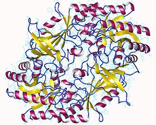
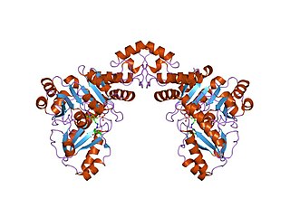
The enzyme ornithine decarboxylase catalyzes the decarboxylation of ornithine to form putrescine. This reaction is the committed step in polyamine synthesis. In humans, this protein has 461 amino acids and forms a homodimer.

<span class="mw-page-title-main">Aminolevulinic acid synthase</span> Class of enzymes

Aminolevulinic acid synthase (ALA synthase, ALAS, or delta-aminolevulinic acid synthase) is an enzyme (EC 2.3.1.37) that catalyzes the synthesis of δ-aminolevulinic acid (ALA) the first common precursor in the biosynthesis of all tetrapyrroles such as hemes, cobalamins and chlorophylls. The reaction is as follows:

Aromatic <small>L</small>-amino acid decarboxylase Class of enzymes

Aromatic L-amino acid decarboxylase, also known as DOPA decarboxylase (DDC), tryptophan decarboxylase, and 5-hydroxytryptophan decarboxylase, is a lyase enzyme, located in region 7p12.2-p12.1.

<span class="mw-page-title-main">Pyridoxal phosphate</span> Active form of vitamin B6

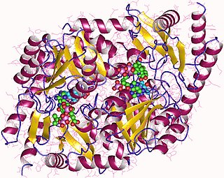
Pyridoxal phosphate (PLP, pyridoxal 5'-phosphate, P5P), the active form of vitamin B6, is a coenzyme in a variety of enzymatic reactions. The International Union of Biochemistry and Molecular Biology has catalogued more than 140 PLP-dependent activities, corresponding to ~4% of all classified activities. The versatility of PLP arises from its ability to covalently bind the substrate, and then to act as an electrophilic catalyst, thereby stabilizing different types of carbanionic reaction intermediates.

<span class="mw-page-title-main">Aspartate transaminase</span> Enzyme involved in amino acid metabolism

Aspartate transaminase (AST) or aspartate aminotransferase, also known as AspAT/ASAT/AAT or (serum) glutamic oxaloacetic transaminase, is a pyridoxal phosphate (PLP)-dependent transaminase enzyme that was first described by Arthur Karmen and colleagues in 1954. AST catalyzes the reversible transfer of an α-amino group between aspartate and glutamate and, as such, is an important enzyme in amino acid metabolism. AST is found in the liver, heart, skeletal muscle, kidneys, brain, red blood cells and gall bladder. Serum AST level, serum ALT level, and their ratio are commonly measured clinically as biomarkers for liver health. The tests are part of blood panels.

<span class="mw-page-title-main">Isocitrate dehydrogenase</span> Class of enzymes

Isocitrate dehydrogenase (IDH) (EC 1.1.1.42) and (EC 1.1.1.41) is an enzyme that catalyzes the oxidative decarboxylation of isocitrate, producing alpha-ketoglutarate (α-ketoglutarate) and CO2. This is a two-step process, which involves oxidation of isocitrate (a secondary alcohol) to oxalosuccinate (a ketone), followed by the decarboxylation of the carboxyl group beta to the ketone, forming alpha-ketoglutarate. In humans, IDH exists in three isoforms: IDH3 catalyzes the third step of the citric acid cycle while converting NAD+ to NADH in the mitochondria. The isoforms IDH1 and IDH2 catalyze the same reaction outside the context of the citric acid cycle and use NADP+ as a cofactor instead of NAD+. They localize to the cytosol as well as the mitochondrion and peroxisome.

<span class="mw-page-title-main">Histidine decarboxylase</span> Enzyme that converts histidine to histamine

The enzyme histidine decarboxylase is transcribed on chromosome 15, region q21.1-21.2, and catalyzes the decarboxylation of histidine to form histamine. In mammals, histamine is an important biogenic amine with regulatory roles in neurotransmission, gastric acid secretion and immune response. Histidine decarboxylase is the sole member of the histamine synthesis pathway, producing histamine in a one-step reaction. Histamine cannot be generated by any other known enzyme. HDC is therefore the primary source of histamine in most mammals and eukaryotes. The enzyme employs a pyridoxal 5'-phosphate (PLP) cofactor, in similarity to many amino acid decarboxylases. Eukaryotes, as well as gram-negative bacteria share a common HDC, while gram-positive bacteria employ an evolutionarily unrelated pyruvoyl-dependent HDC. In humans, histidine decarboxylase is encoded by the HDC gene.

<span class="mw-page-title-main">Oxaloacetate decarboxylase</span> Enzyme

Oxaloacetate decarboxylase is a carboxy-lyase involved in the conversion of oxaloacetate into pyruvate.

<span class="mw-page-title-main">Serine hydroxymethyltransferase</span>

Serine hydroxymethyltransferase (SHMT) is a pyridoxal phosphate (PLP) (Vitamin B6) dependent enzyme (EC 2.1.2.1) which plays an important role in cellular one-carbon pathways by catalyzing the reversible, simultaneous conversions of L-serine to glycine and tetrahydrofolate (THF) to 5,10-Methylenetetrahydrofolate (5,10-CH2-THF). This reaction provides the largest part of the one-carbon units available to the cell.

<span class="mw-page-title-main">Serine dehydratase</span>

Serine dehydratase or L-serine ammonia lyase (SDH) is in the β-family of pyridoxal phosphate-dependent (PLP) enzymes. SDH is found widely in nature, but its structural and properties vary among species. SDH is found in yeast, bacteria, and the cytoplasm of mammalian hepatocytes. SDH catalyzes is the deamination of L-serine to yield pyruvate, with the release of ammonia.

<span class="mw-page-title-main">Branched-chain amino acid aminotransferase</span> Aminotransferase enzyme

Branched-chain amino acid aminotransferase (BCAT), also known as branched-chain amino acid transaminase, is an aminotransferase enzyme (EC 2.6.1.42) which acts upon branched-chain amino acids (BCAAs). It is encoded by the BCAT2 gene in humans. The BCAT enzyme catalyzes the conversion of BCAAs and α-ketoglutarate into branched chain α-keto acids and glutamate.

<span class="mw-page-title-main">Alanine racemase</span>

In enzymology, an alanine racemase is an enzyme that catalyzes the chemical reaction

In enzymology, an aspartate racemase is an enzyme that catalyzes the following chemical reaction:

<span class="mw-page-title-main">Cystathionine beta-lyase</span> Enzyme

Cystathionine beta-lyase, also commonly referred to as CBL or β-cystathionase, is an enzyme that primarily catalyzes the following α,β-elimination reaction

<span class="mw-page-title-main">Threonine ammonia-lyase</span>

Threonine ammonia-lyase (EC 4.3.1.19, systematic name L-threonine ammonia-lyase (2-oxobutanoate-forming), also commonly referred to as threonine deaminase or threonine dehydratase, is an enzyme responsible for catalyzing the conversion of L-threonine into α-ketobutyrate and ammonia:

<span class="mw-page-title-main">Diaminopimelate decarboxylase</span>

The enzyme diaminopimelate decarboxylase (EC 4.1.1.20) catalyzes the cleavage of carbon-carbon bonds in meso 2,6 diaminoheptanedioate to produce CO2 and L-lysine, the essential amino acid. It employs the cofactor pyridoxal phosphate, also known as PLP, which participates in numerous enzymatic transamination, decarboxylation and deamination reactions.

<span class="mw-page-title-main">Serine C-palmitoyltransferase</span>

In enzymology, a serine C-palmitoyltransferase (EC 2.3.1.50) is an enzyme that catalyzes the chemical reaction:

<span class="mw-page-title-main">Cys/Met metabolism PLP-dependent enzyme family</span>

In molecular biology, the Cys/Met metabolism PLP-dependent enzyme family is a family of proteins including enzymes involved in cysteine and methionine metabolism which use PLP (pyridoxal-5'-phosphate) as a cofactor.

<span class="mw-page-title-main">Group III pyridoxal-dependent decarboxylases</span> Class of enzymes

In molecular biology, group III pyridoxal-dependent decarboxylases are a family of bacterial enzymes comprising ornithine decarboxylase EC 4.1.1.17, lysine decarboxylase EC 4.1.1.18 and arginine decarboxylase EC 4.1.1.19.

<span class="mw-page-title-main">Group IV pyridoxal-dependent decarboxylases</span> Family of enzymes

In molecular biology, group IV pyridoxal-dependent decarboxylases are a family of enzymes comprising ornithine decarboxylase EC 4.1.1.17, lysine decarboxylase EC 4.1.1.18, arginine decarboxylase EC 4.1.1.19 and diaminopimelate decarboxylaseEC 4.1.1.20. It is also known as the Orn/Lys/Arg decarboxylase class-II family.

References

  1. Paiardini A, Contestabile R, Buckle AM, Cellini B (2014). "PLP-dependent enzymes". BioMed Research International. 2014: 856076. doi: 10.1155/2014/856076 . PMC   3914556 . PMID   24527459.
  2. Boeker EA, Snell EE (January 1971). "[225] Arginine decarboxylase (Escherichia coli B)". [225] Arginine decarboxylase (Escherichia coli B). Methods in Enzymology. Vol. 17. pp. 657–662. doi:10.1016/0076-6879(71)17114-5. ISBN   9780121818777.
  3. 1 2 3 4 Andréll J, Hicks MG, Palmer T, Carpenter EP, Iwata S, Maher MJ (May 2009). "Crystal structure of the acid-induced arginine decarboxylase from Escherichia coli: reversible decamer assembly controls enzyme activity". Biochemistry. 48 (18): 3915–27. doi:10.1021/bi900075d. PMID   19298070.
  4. Deka G, Bharath SR, Savithri HS, Murthy MR (September 2017). "Structural studies on the decameric S. Typhimurium arginine decarboxylase (ADC): Pyridoxal 5'-phosphate binding induces conformational changes". Biochemical and Biophysical Research Communications. 490 (4): 1362–1368. doi:10.1016/j.bbrc.2017.07.032. PMID   28694189.
  5. PDB: 1MT1 ; Tolbert WD, Graham DE, White RH, Ealick SE (March 2003). "Pyruvoyl-dependent arginine decarboxylase from Methanococcus jannaschii: crystal structures of the self-cleaved and S53A proenzyme forms". Structure. 11 (3): 285–94. doi: 10.1016/S0969-2126(03)00026-1 . PMID   12623016.
  6. Boeker EA, Snell EE (April 1968). "Arginine decarboxylase from Escherichia coli. II. Dissociation and reassociation of subunits". The Journal of Biological Chemistry. 243 (8): 1678–84. PMID   4870600.
  7. Eliot AC, Kirsch JF (2004). "Pyridoxal phosphate enzymes: mechanistic, structural, and evolutionary considerations". Annual Review of Biochemistry. 73: 383–415. doi:10.1146/annurev.biochem.73.011303.074021. PMID   15189147.
  8. John RA (April 1995). "Pyridoxal phosphate-dependent enzymes". Biochimica et Biophysica Acta (BBA) - Protein Structure and Molecular Enzymology. 1248 (2): 81–96. doi:10.1016/0167-4838(95)00025-p. PMID   7748903.
  9. Toney MD (January 2005). "Reaction specificity in pyridoxal phosphate enzymes". Archives of Biochemistry and Biophysics. 433 (1): 279–87. doi:10.1016/j.abb.2004.09.037. PMID   15581583.
  10. Akhtar M, Stevenson DE, Gani D (August 1990). "Fern L-methionine decarboxylase: kinetics and mechanism of decarboxylation and abortive transamination". Biochemistry. 29 (33): 7648–60. doi:10.1021/bi00485a014. PMID   2271524.
  11. Lin J, Smith MP, Chapin KC, Baik HS, Bennett GN, Foster JW (September 1996). "Mechanisms of acid resistance in enterohemorrhagic Escherichia coli". Applied and Environmental Microbiology. 62 (9): 3094–100. doi:10.1128/AEM.62.9.3094-3100.1996. PMC   168100 . PMID   8795195.
  12. Nowak S, Boeker EA (March 1981). "The inducible arginine decarboxylase of Escherichia coli B: activity of the dimer and the decamer". Archives of Biochemistry and Biophysics. 207 (1): 110–6. doi:10.1016/0003-9861(81)90015-1. PMID   7016035.
  13. Richard H, Foster JW (September 2004). "Escherichia coli glutamate- and arginine-dependent acid resistance systems increase internal pH and reverse transmembrane potential". Journal of Bacteriology. 186 (18): 6032–41. doi:10.1128/jb.186.18.6032-6041.2004. PMC   515135 . PMID   15342572.
  14. Gong S, Richard H, Foster JW (August 2003). "YjdE (AdiC) is the arginine:agmatine antiporter essential for arginine-dependent acid resistance in Escherichia coli". Journal of Bacteriology. 185 (15): 4402–9. doi:10.1128/jb.185.15.4402-4409.2003. PMC   165756 . PMID   12867448.
  15. Iyer R, Williams C, Miller C (November 2003). "Arginine-agmatine antiporter in extreme acid resistance in Escherichia coli". Journal of Bacteriology. 185 (22): 6556–61. doi:10.1128/jb.185.22.6556-6561.2003. PMC   262112 . PMID   14594828.