Limbic encephalitis

Last updated
Limbic encephalitis
Brain limbicsystem.svg
The limbic system within the brain.
Specialty Neurology   OOjs UI icon edit-ltr-progressive.svg

Limbic encephalitis is a form of encephalitis, a disease characterized by inflammation of the brain. [1] Limbic encephalitis is caused by autoimmunity: an abnormal state where the body produces antibodies against itself. Some cases are associated with cancer and some are not. [1] Although the disease is known as "limbic" encephalitis, it is seldom limited to the limbic system and post-mortem studies usually show involvement of other parts of the brain. [2] [3] [4] The disease was first described by Brierley and others in 1960 as a series of three cases. The link to cancer was first noted in 1968 [3] and confirmed by later investigators. [5]

Contents

The majority of cases of limbic encephalitis are associated with a tumor (diagnosed or undiagnosed). In cases caused by tumor, recovery can only occur following complete removal of the tumor, which is not always possible. Limbic encephalitis is classified according to the auto-antibody that causes the disease. The most common types are:

Since 1999, following the publication of a case report of a 15-year-old teenager of Indian descent from South Africa who developed subacute memory loss subsequent to herpes simplex type 1 encephalitis, [6] similar cases of non-paraneoplastic LE have been described, as has its association with auto-antibodies and response to steroid. [7] [8] Limbic encephalitis associated with voltage-gated potassium channel antibodies (VGKC-Abs) [9] may frequently be non-paraneoplastic. [10] A recent study of 15 cases of limbic encephalitis found raised VGKC-Abs associated with non-paraneoplastic disorders and remission following immunosuppressive treatment. [11]

Classification

Limbic encephalitis is broadly grouped into two types: paraneoplastic limbic encephalitis and non-paraneoplastic limbic encephalitis.[ citation needed ]

Symptoms and signs

Symptoms develop over days or weeks. The subacute development of short-term memory deficits is considered the hallmark of this disease, [1] but this symptom is often overlooked, because it is overshadowed by other more obvious symptoms such as headache, irritability, sleep disturbance, delusions, hallucinations, agitation, seizures and psychosis, or because the other symptoms mean the patient has to be sedated, and it is not possible to test memory in a sedated patient.[ citation needed ]

Cause

Limbic encephalitis is associated with an autoimmune reaction. [1] In non-paraneoplastic limbic encephalitis, this is typically due to infection (commonly herpes simplex virus) or as a systemic autoimmune disorder. [13] Limbic encephalitis associated with cancer or tumors is called paraneoplastic limbic encephalitis.[ citation needed ]

Diagnosis

The diagnosis of limbic encephalitis is extremely difficult and it is usual for the diagnosis to be delayed for weeks. The key diagnostic test (detection of specific auto-antibodies in cerebrospinal fluid) is not routinely offered by most immunology laboratories. Some of the rarer auto-antibodies (e.g., NMDAR) have no commercially available assay and can only be measured by a very small number of research laboratories worldwide, further delaying diagnosis by weeks or months. Most patients with limbic encephalitis are initially diagnosed with herpes simplex encephalitis, because the two syndromes cannot be distinguished clinically. [1] HHV-6 (human herpes virus 6) encephalitis is also clinically indistinguishable from limbic encephalitis. [1]

There are two sets of diagnostic criteria used. The oldest are those proposed by Gultekin et al. in 2000. [14]

Gultekin criteria
EITHER, Pathological demonstration of limbic encephalitis
OR, All four of:
  • Short-term memory loss, seizures, or psychiatric symptoms suggestive of limbic system involvement
  • <4 years between onset of neurological symptoms and cancer diagnosis
  • Exclusion of metastases, infection, metabolic and nutritional deficits, stroke and side-effects of therapy that may cause limbic encephalopathy
  • At least one of
    • CSF with inflammatory findings
    • Hyperintensity of the temporal lobes bilaterally on MRI FLAIR or T2
    • EEG with epilepsy or slow activity involving the temporal lobes focally

A revised set of criteria were proposed by Graus and Saiz in 2005. [15]

Graus and Saiz criteria
All four of
  • Subacute onset (<12 weeks) of seizures, short-term memory loss, confusion, and psychiatric symptoms
  • Neuropathologic or radiologic evidence (MRI, SPECT, PET) of involvement of the limbic system
  • Exclusion of other possible aetiologies of limbic dysfunction
  • Demonstration of a cancer within 5 years of the diagnosis of neurologic symptoms, or the development of classic symptoms of limbic dysfunction in association with a well-characterized paraneoplastic antibody (Hu, Ma2, CV2, amphiphysin, Ri)

The main distinction between the two sets of criteria is whether or not the detection of a paraneoplastic antibody is needed for diagnosis.

Antibodies against intracellular neuronal antigens

The main antibodies within this group are those against Hu, Ma2, CV2, amphiphysin and Ri. The syndrome of anti-Ma2 encephalitis may be clinically mistaken for Whipple's disease. [16]

Antibodies against cell membrane antigens

The main antibodies within this group are those against N-methyl-D-aspartate receptors (NMDAR) and the voltage-gated potassium channel-complex (VGKC-complex). Anti-NMDAR encephalitis is strongly associated with benign tumours of the ovary (usually teratomata or dermoid cysts). Anti-VGKC-complex encephalitis is most often not associated with tumours.[ citation needed ]

Patients with NMDAR encephalitis are frequently young women who present with fever, headache and fatigue. This is often misdiagnosed as influenza, but progresses to severe behavioural and personality disturbance, delusions, paranoia and hallucinations. [17] Patients may therefore initially be admitted to a psychiatric ward for acute psychosis or schizophrenia. The disease then progresses to catatonia, seizures and loss of consciousness. The next stage is hypoventilation (inadequate breathing) requiring intubation, orofacial dyskinesia and autonomic instability (dramatic fluctuations in blood pressure, temperature and heart rate). [18]

Investigation

Cerebrospinal fluid (CSF)

Examination of cerebrospinal fluid (CSF) shows elevated numbers of lymphocytes (but usually < 100 cells/μl); elevated CSF protein (but usually <1.5 g/L), normal glucose, elevated IgG index and oligoclonal bands. Patients with antibodies to voltage-gated potassium channels may have a completely normal CSF examination. [19] [20] [21]

Neuroimaging

Brain MRI is the mainstay of initial investigation pointing to limbic lobe pathology revealing increased T2 signal involving one or both temporal lobes in most cases. [22] [14]

Serial MRI in LE starts as an acute disease with uni- or bilateral swollen temporomesial structures that are hyperintense on fluid attenuation inversion recovery and T2-weighted sequences. Swelling and hyperintensity may persist over months to years, but in most cases progressive temporomesial atrophy develops. [23]

PET-CT is not an essential investigation but can help in suspected cases with MRI negative for early diagnosis. [24]

Neuro-electrophysiology

EEG is mostly nonspecific slowing and epileptiform activity arising from temporal lobes. [14]

Treatment

Limbic encephalitis is a rare condition with no randomised-controlled trials to guide treatment. Treatments that have been tried include intravenous immunoglobulin, plasmapheresis, corticosteroids, cyclophosphamide and rituximab. [1]

If an associated tumour is found, then recovery is not possible until the tumour is removed. Unfortunately, this is not always possible, especially if the tumour is malignant and advanced.

History

Clinical FeaturesPathologyYearSyndromes
Subacute onset of episodic memory impairment, disorientation and agitation, commonly associated with seizures, hallucinations, sleep disturbanceMedial temporal lobe inflammation1960Subacute encephalitis of later adult life mainly affecting the limbic areas [2]
1968Limbic Encephalitis association with Carcinoma [25] [2]
2008Paraneoplastic LE with Lung Carcinoma, thymoma, [26]
2002Non Paraneoplastic LE [6]

Related Research Articles

Neuromyotonia (NMT) is a form of peripheral nerve hyperexcitability that causes spontaneous muscular activity resulting from repetitive motor unit action potentials of peripheral origin. NMT along with Morvan's syndrome are the most severe types in the Peripheral Nerve Hyperexciteability spectrum. Example of two more common and less severe syndromes in the spectrum are Cramp Fasciculation Syndrome and Benign Fasciculation Syndrome. NMT can have both hereditary and acquired forms. The prevalence of NMT is unknown.

<span class="mw-page-title-main">Encephalitis</span> Inflammation of the brain

Encephalitis is inflammation of the brain. The severity can be variable with symptoms including reduction or alteration in consciousness, headache, fever, confusion, a stiff neck, and vomiting. Complications may include seizures, hallucinations, trouble speaking, memory problems, and problems with hearing.

Morvan's syndrome is a rare, life-threatening autoimmune disease named after the nineteenth century French physician Augustin Marie Morvan. "La chorée fibrillaire" was first coined by Morvan in 1890 when describing patients with multiple, irregular contractions of the long muscles, cramping, weakness, pruritus, hyperhidrosis, insomnia and delirium. It normally presents with a slow insidious onset over months to years. Approximately 90% of cases spontaneously go into remission, while the other 10% of cases lead to death.

Opsoclonus myoclonus syndrome (OMS), also known as opsoclonus-myoclonus-ataxia (OMA), is a rare neurological disorder of unknown cause which appears to be the result of an autoimmune process involving the nervous system. It is an extremely rare condition, affecting as few as 1 in 10,000,000 people per year. It affects 2 to 3% of children with neuroblastoma and has been reported to occur with celiac disease and diseases of neurologic and autonomic dysfunction.

Neuromyelitis optica spectrum disorders (NMOSD), including neuromyelitis optica (NMO), are autoimmune diseases characterized by acute inflammation of the optic nerve and the spinal cord (myelitis). Episodes of ON and myelitis can be simultaneous or successive. A relapsing disease course is common, especially in untreated patients. In more than 80% of cases, NMO is caused by immunoglobulin G autoantibodies to aquaporin 4 (anti-AQP4), the most abundant water channel protein in the central nervous system. A subset of anti-AQP4-negative cases is associated with antibodies against myelin oligodendrocyte glycoprotein (anti-MOG). Rarely, NMO may occur in the context of other autoimmune diseases or infectious diseases. In some cases, the etiology remains unknown.

<span class="mw-page-title-main">Stiff-person syndrome</span> Human medical condition

Stiff-person syndrome (SPS), also known as stiff-man syndrome, is a rare neurologic disorder of unclear cause characterized by progressive muscular rigidity and stiffness. The stiffness primarily affects the truncal muscles and is superimposed by spasms, resulting in postural deformities. Chronic pain, impaired mobility, and lumbar hyperlordosis are common symptoms.

<span class="mw-page-title-main">Viral encephalitis</span> Medical condition

Viral encephalitis is inflammation of the brain parenchyma, called encephalitis, by a virus. The different forms of viral encephalitis are called viral encephalitides. It is the most common type of encephalitis and often occurs with viral meningitis. Encephalitic viruses first cause infection and replicate outside of the central nervous system (CNS), most reaching the CNS through the circulatory system and a minority from nerve endings toward the CNS. Once in the brain, the virus and the host's inflammatory response disrupt neural function, leading to illness and complications, many of which frequently are neurological in nature, such as impaired motor skills and altered behavior.

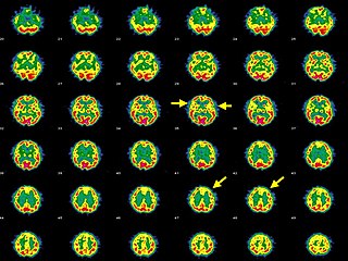
Paraneoplastic cerebellar degeneration (PCD) is a paraneoplastic syndrome associated with a broad variety of tumors including lung cancer, ovarian cancer, breast cancer, Hodgkin’s lymphoma and others. PCD is a rare condition that occurs in less than 1% of cancer patients.

Inflammatory demyelinating diseases (IDDs), sometimes called Idiopathic (IIDDs) due to the unknown etiology of some of them, are a heterogenous group of demyelinating diseases - conditions that cause damage to myelin, the protective sheath of nerve fibers - that occur against the background of an acute or chronic inflammatory process. IDDs share characteristics with and are often grouped together under Multiple Sclerosis. They are sometimes considered different diseases from Multiple Sclerosis, but considered by others to form a spectrum differing only in terms of chronicity, severity, and clinical course.

<span class="mw-page-title-main">Hashimoto's encephalopathy</span> Human disease (neurological condition)

Hashimoto's encephalopathy, also known as steroid-responsive encephalopathy associated with autoimmune thyroiditis (SREAT), is a neurological condition characterized by encephalopathy, thyroid autoimmunity, and good clinical response to corticosteroids. It is associated with Hashimoto's thyroiditis, and was first described in 1966. It is sometimes referred to as a neuroendocrine disorder, although the condition's relationship to the endocrine system is widely disputed. It is recognized as a rare disease by the NIH Genetic and Rare Diseases Information Center.

A paraneoplastic syndrome is a syndrome that is the consequence of a tumor in the body. It is specifically due to the production of chemical signaling molecules by tumor cells or by an immune response against the tumor. Unlike a mass effect, it is not due to the local presence of cancer cells.

Anti-glutamate receptor antibodies are autoantibodies detected in serum and/or cerebrospinal fluid samples of a variety of disorders such as encephalitis, epilepsy and ataxia. Clinical and experimental studies starting around the year 2000 suggest that these antibodies are not simply epiphenomena and are involved in autoimmune disease pathogenesis.

<span class="mw-page-title-main">PNMA2</span> Mammalian protein found in Homo sapiens

Paraneoplastic antigen Ma2 is a protein that in humans is encoded by the PNMA2 gene.

<span class="mw-page-title-main">Tumefactive multiple sclerosis</span> Medical condition

Tumefactive multiple sclerosis is a condition in which the central nervous system of a person has multiple demyelinating lesions with atypical characteristics for those of standard multiple sclerosis (MS). It is called tumefactive as the lesions are "tumor-like" and they mimic tumors clinically, radiologically and sometimes pathologically.

Paraneoplastic pemphigus (PNP) is an autoimmune disorder stemming from an underlying tumor. It is hypothesized that antigens associated with the tumor trigger an immune response resulting in blistering of the skin and mucous membranes.

<span class="mw-page-title-main">Anti-NMDA receptor encephalitis</span> Rare disease which results in brain inflammation

Anti-NMDA receptor encephalitis is a type of brain inflammation caused by antibodies. Early symptoms may include fever, headache, and feeling tired. This is then typically followed by psychosis which presents with false beliefs (delusions) and seeing or hearing things that others do not see or hear (hallucinations). People are also often agitated or confused. Over time, seizures, decreased breathing, and blood pressure and heart rate variability typically occur. In some cases, patients may develop catatonia.

Bickerstaff brainstem encephalitis is a rare inflammatory disorder of the central nervous system, first described by Edwin Bickerstaff in 1951. It may also affect the peripheral nervous system, and has features in common with both Miller Fisher syndrome and Guillain–Barré syndrome.

<span class="mw-page-title-main">Autoimmune encephalitis</span> Type of encephalitis

Autoimmune encephalitis (AIE) is a type of encephalitis, and one of the most common causes of noninfectious encephalitis. It can be triggered by tumors, infections, or it may be cryptogenic. The neurological manifestations can be either acute or subacute and usually develop within six weeks. The clinical manifestations include behavioral and psychiatric symptoms, autonomic disturbances, movement disorders, and seizures.

Anti-VGKC-complex encephalitis are caused by antibodies against the voltage gated potassium channel-complex (VGKC-complex) and are implicated in several autoimmune conditions including limbic encephalitis, epilepsy and neuromyotonia.

Anti-Hu associated encephalitis, also known as Anti-ANNA1 associated encephalitis, is an uncommon form of brain inflammation that is associated with an underlying cancer. It can cause psychiatric symptoms such as depression, anxiety, and hallucinations. It can also produce neurological symptoms such as confusion, memory loss, weakness, sensory loss, pain, seizures, and problems coordinating the movement of the body.

References

  1. 1 2 3 4 5 6 7 Tüzün E, Dalmau J (2007). "Limbic encephalitis and variants: classification, diagnosis and treatment". The Neurologist. 13 (5): 261–271. doi:10.1097/NRL.0b013e31813e34a5. PMID   17848866. S2CID   44987647.
  2. 1 2 3 Brierley JB, Corsellis JA, Hierons R, et al. (1960). "Subacute encephalitis of later adult life. Mainly affecting the limbic areas". Brain. 83 (3): 357–368. doi:10.1093/brain/83.3.357.
  3. 1 2 Corsellis JA, Goldberg GJ, Norton AR (1968). ""Limbic encephalitis" and its association with carcinoma". Brain. 91 (3): 481–496. doi:10.1093/brain/91.3.481. PMID   5723018.
  4. Bakheit AM, Kennedy PG, Behan PO (1990). "Paraneoplastic limbic encephalitis: clinico-pathological correlations". J Neurol Neurosurg Psychiatry. 53 (12): 1084–1088. doi:10.1136/jnnp.53.12.1084. PMC   488321 . PMID   1963440.
  5. Henson RA, Hoffman HL, Urich H (1965). "Encephalomyelitis with carcinoma". Brain. 88 (3): 449–464. doi:10.1093/brain/88.3.449. PMID   5890519.
  6. 1 2 Mori, Masahiro; Kuwabara, Satoshi; Yoshiyama, Mitsuharu; Kanesaka, Toshihide; Ogata, Tsuyoshi; Hattori, Takamichi (2002-09-15). "Successful immune treatment for non-paraneoplastic limbic encephalitis". Journal of the Neurological Sciences. 201 (1–2): 85–88. doi:10.1016/s0022-510x(02)00188-0. ISSN   0022-510X. PMID   12163199. S2CID   43558497.
  7. Watanabe, Yasuhiro; Shimizu, Yasutaka; Ooi, Shinji; Tanaka, Keiko; Inuzuka, Takashi; Nakashima, Kenji (2003-05-01). "Steroid-responsive limbic encephalitis". Internal Medicine. 42 (5): 428–432. doi: 10.2169/internalmedicine.42.428 . ISSN   0918-2918. PMID   12793715.
  8. Lang, Pierre Olivier; Sellal, François (2008-05-01). "[Non-paraneoplastic limbic encephalitis revealed by anterograde amnesia]". Presse Médicale. 37 (5 Pt 1): 775–782. doi:10.1016/j.lpm.2007.11.013. ISSN   0755-4982. PMID   18261871.
  9. Buckley, C.; Oger, J.; Clover, L.; Tüzün, E.; Carpenter, K.; Jackson, M.; Vincent, A. (2001-07-01). "Potassium channel antibodies in two patients with reversible limbic encephalitis". Annals of Neurology. 50 (1): 73–78. doi:10.1002/ana.1097. ISSN   0364-5134. PMID   11456313. S2CID   38993448.
  10. Schott, J. M.; Harkness, K.; Barnes, J.; della Rocchetta, A. Incisa; Vincent, A.; Rossor, M. N. (2003-04-12). "Amnesia, cerebral atrophy, and autoimmunity". Lancet. 361 (9365): 1266. doi:10.1016/s0140-6736(03)12983-2. ISSN   0140-6736. PMID   12699955. S2CID   36303221.
  11. Pozo-Rosich, Patricia; Clover, Linda; Saiz, Albert; Vincent, Angela; Graus, Francesc (2003-10-01). "Voltage-gated potassium channel antibodies in limbic encephalitis". Annals of Neurology. 54 (4): 530–533. doi:10.1002/ana.10713. ISSN   0364-5134. PMID   14520669. S2CID   10535157.
  12. "Limbic encephalitis". The Encephalitis Society. Retrieved 2017-12-23.
  13. Anderson, N. E.; Barber, P. A. (September 2008). "Limbic encephalitis - a review". Journal of Clinical Neuroscience. 15 (9): 961–971. doi:10.1016/j.jocn.2007.12.003. ISSN   0967-5868. PMID   18411052. S2CID   6545575.
  14. 1 2 3 Gultekin SH, Rosenfeld MR, Voltz R, et al. (2000). "Paraneoplastic limbic encephalitis: neurological symptoms, immunological findings and tumour association in 50 patients". Brain. 123 (7): 1481–1494. doi:10.1093/brain/123.7.1481. PMID   10869059.
  15. Graus F, Saiz A (2005). "Limbic encephalitis: a probably under-recognized syndrome". Neurologia. 20 (1): 24–30. PMID   15704018.
  16. Castle J, Sakonju A, Dalmau J, et al. (2006). "Anti-Ma2-associated encephalitis with normal FDG-PET: a case of pseudo-Whipple's disease". Nat Clin Pract Neurol. 2 (10): 566–572. doi:10.1038/ncpneuro0287. PMID   16990830. S2CID   10797381.
  17. Koide R, Shimizu T, Koike K, et al. (2007). "EFA6A-like antibodies in paraneoplastic encephalitis associated with immature ovarian teratoma: a case report". J Neurooncol. 81 (1): 71–74. doi:10.1007/s11060-006-9200-7. PMID   16807779. S2CID   6010024.
  18. Dalmau J, Tuzun E, Wu HY, et al. (2007). "Paraneoplastic anti-N-methyl-D-aspartate receptor encephalitis associated with ovarian teratoma". Ann Neurol. 61 (1): 25–36. doi:10.1002/ana.21050. PMC   2430743 . PMID   17262855.
  19. Buckley C, Oger J, Clover L, et al. (2001). "Potassium channel antibodies in two patients with reversible limbic encephalitis". Ann Neurol. 50 (1): 73–78. doi:10.1002/ana.1097. PMID   11456313. S2CID   38993448.
  20. Vincent A, Buckley C, Schott JM, et al. (2004). "Potassium channel antibody-associated encephalopathy: a potentially immunotherapy-responsive form of limbic encephalitis". Brain. 127 (3): 701–712. doi: 10.1093/brain/awh077 . PMID   14960497.
  21. Thieben MJ, Lennon VA, Boeve BF, et al. (2004). "Potentially reversible auto-immune limbic encephalitis with neuronal potassium channel antibody". Neurology. 62 (7): 1177–1182. doi:10.1212/01.WNL.0000122648.19196.02. PMID   15079019. S2CID   30883641.
  22. Nicholas, D (2003). "Clinical, Magnetic Resonance Imaging, and 7Electroencephalographic Findings in Paraneoplastic Limbic Encephalitis". Mayo Clinic Proceedings. 78 (11): 1363–1368. doi:10.4065/78.11.1363. PMID   14601695.
  23. Horst, Urbach (2006). "Serial MRI of limbic encephalitis". Neuroradiology. 48 (6): 380–386. doi:10.1007/s00234-006-0069-0. PMID   16586118. S2CID   7993234.
  24. Cózar Santiago, Maria Del Puig; Sanchez Jurado, Raul; Sanz Llorens, Rut; Aguilar Barrios, Jose Enrique; Ferrer Rebolleda, Jose (2016-02-01). "Limbic Encephalitis Diagnosed With 18F-FDG PET/CT". Clinical Nuclear Medicine. 41 (2): e101–103. doi:10.1097/RLU.0000000000001076. ISSN   1536-0229. PMID   26571448.
  25. Corsellis, JA (1968). "Limbic encephalitis" and its association with carcinoma". Brain. 91 (3): 481–96. doi:10.1093/brain/91.3.481. PMID   5723018.
  26. Dalmau, Josep; Gleichman, Amy J.; Hughes, Ethan G.; Rossi, Jeffrey E.; Peng, Xiaoyu; Lai, Meizan; Dessain, Scott K.; Rosenfeld, Myrna R.; Balice-Gordon, Rita (2008-12-01). "Anti-NMDA-receptor encephalitis: case series and analysis of the effects of antibodies". The Lancet. Neurology. 7 (12): 1091–1098. doi:10.1016/S1474-4422(08)70224-2. ISSN   1474-4422. PMC   2607118 . PMID   18851928.