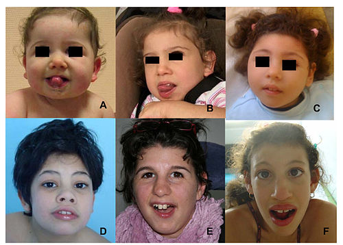
Pontocerebellar hypoplasia

Last updated
Pontocerebellar hypoplasia
Other namesNon-syndromic pontocerebellar hypoplasia
Autosomal recessive - en.svg
Pontocerebellar hypoplasia is inherited in an autosomal recessive manner
Specialty Neurology
TreatmentUnknown

Pontocerebellar hypoplasia (PCH) is a heterogeneous group of rare neurodegenerative disorders caused by genetic mutations and characterised by progressive atrophy of various parts of the brain such as the cerebellum or brainstem (particularly the pons). [1] Where known, these disorders are inherited in an autosomal recessive fashion. There is no known cure for PCH. [2]

Contents

Signs and symptoms

There are different signs and symptoms for different forms of pontocerebellar hypoplasia, at least six of which have been described by researchers. All forms involve abnormal development of the brain, leading to slow development, movement problems, and intellectual impairment. [2]

The following values seem to be aberrant in children with CASK gene defects: lactate, pyruvate, 2-ketoglutaric acid, adipic acid, and suberic acid which seems to support the thesis that CASK affects mitochondrial function. [3]

Causes

Pontocerebellar hypoplasia is caused by mutations in genes including Sepsecs gene, VRK1 (PCH1); TSEN2, TSEN34 (PCH2); RARS2 (PCH6); and TSEN54 (PCH2 and PCH4). The genes associated with PCH3 and PCH5 have not yet been identified. [2]

The mutated genes in PCH are autosomal recessive, which means that parents of an affected child each carry only one copy of the damaged gene. In each parent the other copy performs its proper function and they display no signs of PCH. A child inheriting two damaged copies of the gene will be affected by PCH. [2]

Mechanism

Mutations in the genes that cause PCH produce faults in the production of chemicals, usually enzymes, that are required for the development of nerve cells (neurons) and for properly processing RNA, which is needed for any cell to function normally. The exact mechanism by which PCH affects the development of the cerebellum and pons is not well understood. [2]

Diagnosis

Classification

Pontocerebellar hypoplasia is classified as follows: [4]

Type OMIM Gene Locus Distinctive featuresAlternate names
PCH1A 607596 VRK1 14q32Infantile onset anterior horn cell degeneration resulting in progressive muscle atrophy; resembles infantile spinal muscular atrophy [5] Spinal muscular atrophy with pontocerebellar hypoplasia (SMA-PCH)
PCH1B 614678 EXOSC3 9p13.2Cerebellar and spinal motor neuron degeneration beginning at birth and resulting in decreased body tone, respiratory insufficiency, muscle atrophy, progressive microcephaly and global developmental delay [6]
PCH2A 277470 TSEN54 17q25.1Dyskinetic movements, seizures (frequently)Volendam neurodegenerative disease
PCH2B 612389 TSEN2 3p25.2
PCH2C 612390 TSEN34 19q13.42
PCH2D 613811 SEPSECS 4p15.2Progressive cerebello-cerebral atrophy (PCCA)
PCH2E 615851 VPS53 17p13.3Profound mental retardation, progressive microcephaly, spasticity, and early-onset epilepsy [7]
PCH2F 617026 TSEN15 1q25.3Variable neurologic signs and symptoms, including cognitive and motor delay, poor or absent speech, seizures, and spasticity
PCH3 608027 PCLO 7q11–q21Seizures, short stature, optic atrophy, progressive microcephaly, severe developmental delay; described only in a handful of cases. [8] CLAM-PCH, cerebellar atrophy with progressive microcephaly
PCH4 225753 TSEN54 17q25.1Severe prenatal form of PCH2 with excess fluid in the amniotic sac, muscle contractures, brief involuntary muscle twitching, brief episodes without breathing, and early death following birth
PCH5 610204 TSEN54 17q25.1Severe prenatal form, described in one familyOlivopontocerebellar hypoplasia (OPCH)
PCH6 611523 RARS2 6q15Severe encephalopathy in the newborn with hypotonia, and inconstantly: intractable seizures, edema, increased lactate blood levels, mitochondrial respiratory chain defects
PCH7 614969 TOE1 1p34.1 Hypotonia, apneic episodes, seizures, vanishing testis [9] [10]
PCH8 614961 CHMP1A 16q24.3Severe psychomotor retardation, abnormal movements, hypotonia, spasticity, and variable visual defects [11]
PCH9 615809 AMPD2 1p13.3Severely delayed psychomotor development, progressive microcephaly, spasticity, seizures, and brain abnormalities, including brain atrophy, thin corpus callosum, and delayed myelination [12]
PCH10 615803 CLP1 11q12.1Severely delayed psychomotor development, progressive microcephaly, spasticity, seizures, and brain abnormalities, including brain atrophy and delayed myelination [13]

Pontine and cerebellar hypoplasia is also observed in certain phenotypes of X-linked mental retardation – so called MICPCH.

Another gene that has been associated with this condition is coenzyme A synthase (COASY). [14]

Treatment

Outcomes

The severity of different forms of PCH varies, but many children inheriting the mutated gene responsible do not survive infancy [15] or childhood; nevertheless, some individuals born with PCH have reached adulthood. [2]

See also

Related Research Articles

<span class="mw-page-title-main">Joubert syndrome</span> Medical condition

Joubert syndrome is a rare autosomal recessive genetic disorder that affects the cerebellum, an area of the brain that controls balance and coordination.

<span class="mw-page-title-main">Cerebellar hypoplasia</span> Medical condition

Cerebellar hypoplasia is characterized by reduced cerebellar volume, even though cerebellar shape is (near) normal. It consists of a heterogeneous group of disorders of cerebellar maldevelopment presenting as early-onset non–progressive congenital ataxia, hypotonia and motor learning disability.

<span class="mw-page-title-main">Aniridia</span> Absence of the iris, usually involving both eyes

Aniridia is the absence of the iris, a muscular structure that opens and closes the pupil to allow light into the eye. It is also responsible for eye color. Without it, the central eye appears all black. It can be congenital, in which both eyes are usually involved, or caused by a penetrant injury. Isolated aniridia is a congenital disorder that is not limited to a defect in iris development, but is a panocular condition with macular and optic nerve hypoplasia, cataract, and corneal changes. Vision may be severely compromised and the disorder is frequently associated with some ocular complications: nystagmus, amblyopia, buphthalmos, and cataract. Aniridia in some individuals occurs as part of a syndrome, such as WAGR syndrome, or Gillespie syndrome.

<span class="mw-page-title-main">Immunodeficiency–centromeric instability–facial anomalies syndrome</span> Medical condition

ICF syndrome is a very rare autosomal recessive immune disorder.

<span class="mw-page-title-main">Cerebellar vermis</span> Structure connecting the two cerebellar hemispheres

The cerebellar vermis is located in the medial, cortico-nuclear zone of the cerebellum, which is in the posterior fossa of the cranium. The primary fissure in the vermis curves ventrolaterally to the superior surface of the cerebellum, dividing it into anterior and posterior lobes. Functionally, the vermis is associated with bodily posture and locomotion. The vermis is included within the spinocerebellum and receives somatic sensory input from the head and proximal body parts via ascending spinal pathways.

<span class="mw-page-title-main">Spinocerebellar ataxia</span> Medical condition

Spinocerebellar ataxia (SCA) is a progressive, degenerative, genetic disease with multiple types, each of which could be considered a neurological condition in its own right. An estimated 150,000 people in the United States have a diagnosis of spinocerebellar ataxia at any given time. SCA is hereditary, progressive, degenerative, and often fatal. There is no known effective treatment or cure. SCA can affect anyone of any age. The disease is caused by either a recessive or dominant gene. In many cases people are not aware that they carry a relevant gene until they have children who begin to show signs of having the disorder.

<span class="mw-page-title-main">Flocculus</span>

The flocculus is a small lobe of the cerebellum at the posterior border of the middle cerebellar peduncle anterior to the biventer lobule. Like other parts of the cerebellum, the flocculus is involved in motor control. It is an essential part of the vestibulo-ocular reflex, and aids in the learning of basic motor skills in the brain.

<span class="mw-page-title-main">Dandy–Walker malformation</span> Congenital malformation of the cerebellar vermis

Dandy–Walker malformation (DWM), also known as Dandy–Walker syndrome (DWS), is a rare congenital brain malformation in which the part joining the two hemispheres of the cerebellum does not fully form, and the fourth ventricle and space behind the cerebellum are enlarged with cerebrospinal fluid. Most of those affected develop hydrocephalus within the first year of life, which can present as increasing head size, vomiting, excessive sleepiness, irritability, downward deviation of the eyes and seizures. Other, less common symptoms are generally associated with comorbid genetic conditions and can include congenital heart defects, eye abnormalities, intellectual disability, congenital tumours, other brain defects such as agenesis of the corpus callosum, skeletal abnormalities, an occipital encephalocele or underdeveloped genitalia or kidneys. It is sometimes discovered in adolescents or adults due to mental health problems.

<span class="mw-page-title-main">Middle cerebellar peduncle</span> Structure in the brain connecting the pons to the cerebellum

The middle cerebellar peduncle is a paired structure of the brain. It connects the pons to the cerebellum, with fibres originating from the pontine nucleus and travelling to the opposite hemisphere of the cerebellar cortex. It is supplied by the anterior inferior cerebellar artery (AICA) and branches from the basilar artery. It conveys information from the cerebrum and the pons to the cerebellum.

<span class="mw-page-title-main">3C syndrome</span> Medical condition

3C syndrome is a rare condition whose symptoms include heart defects, cerebellar hypoplasia, and cranial dysmorphism. It was first described in the medical literature in 1987 by Ritscher and Schinzel, for whom the disorder is sometimes named.

Ohtahara syndrome (OS), also known as early infantile epileptic encephalopathy (EIEE) is a progressive epileptic encephalopathy. The syndrome is outwardly characterized by tonic spasms and partial seizures within the first few months of life, and receives its more elaborate name from the pattern of burst activity on an electroencephalogram (EEG). It is an extremely debilitating progressive neurological disorder, involving intractable seizures and severe intellectual disabilities. No single cause has been identified, although in many cases structural brain damage is present.

<span class="mw-page-title-main">Gillespie syndrome</span> Medical condition

Gillespie syndrome, also called aniridia, cerebellar ataxia and mental deficiency, is a rare genetic disorder. The disorder is characterized by partial aniridia, ataxia, and, in most cases, intellectual disability. It is heterogeneous, inherited in either an autosomal dominant or autosomal recessive manner. Gillespie syndrome was first described by American ophthalmologist Fredrick Gillespie in 1965.

<span class="mw-page-title-main">Anatomy of the cerebellum</span> Structures in the cerebellum, a part of the brain

The anatomy of the cerebellum can be viewed at three levels. At the level of gross anatomy, the cerebellum consists of a tightly folded and crumpled layer of cortex, with white matter underneath, several deep nuclei embedded in the white matter, and a fluid-filled ventricle in the middle. At the intermediate level, the cerebellum and its auxiliary structures can be broken down into several hundred or thousand independently functioning modules or compartments known as microzones. At the microscopic level, each module consists of the same small set of neuronal elements, laid out with a highly stereotyped geometry.

Cerebellar cognitive affective syndrome (CCAS), also called Schmahmann's syndrome is a condition that follows from lesions (damage) to the cerebellum of the brain. It refers to a constellation of deficits in the cognitive domains of executive function, spatial cognition, language, and affect resulting from damage to the cerebellum. Impairments of executive function include problems with planning, set-shifting, abstract reasoning, verbal fluency, and working memory, and there is often perseveration, distractibility and inattention. Language problems include dysprosodia, agrammatism and mild anomia. Deficits in spatial cognition produce visual–spatial disorganization and impaired visual–spatial memory. Personality changes manifest as blunting of affect or disinhibited and inappropriate behavior. These cognitive impairments result in an overall lowering of intellectual function. CCAS challenges the traditional view of the cerebellum being responsible solely for regulation of motor functions. It is now thought that the cerebellum is responsible for monitoring both motor and nonmotor functions. The nonmotor deficits described in CCAS are believed to be caused by dysfunction in cerebellar connections to the cerebral cortex and limbic system.

<span class="mw-page-title-main">Kohlschütter-Tönz syndrome</span> Medical condition

Kohlschütter-Tönz syndrome (KTS), also called amelo-cerebro-hypohidrotic syndrome, is a rare inherited syndrome characterized by epilepsy, psychomotor delay or regression, intellectual disability, and yellow teeth caused by amelogenesis imperfecta. It is a type A ectodermal dysplasia.

tRNA-intron lyase is an enzyme. As an endonuclease enzyme, tRNA-intron lyase is responsible for splicing phosphodiester bonds within non-coding ribonucleic acid chains. These non-coding RNA molecules form tRNA molecules after being processed, and this is dependent on tRNA-intron lyase to splice the pretRNA. tRNA processing is an important post-transcriptional modification necessary for tRNA maturation because it locates and removes introns in the pretRNA. This enzyme catalyses the following chemical reaction:

<span class="mw-page-title-main">Mental retardation and microcephaly with pontine and cerebellar hypoplasia</span> Rare X-linked dominant genetic disorder

Mental retardation and microcephaly with pontine and cerebellar hypoplasia (MICPCH) – also known as mental retardation, X-linked, syndromic, Najm type (MRXSNA); X-linked intellectual deficit, Najm type; intellectual developmental disorder, X-linked, syndromic, Najm type; X-linked intellectual disability–microcephaly–pontocerebellar hypoplasia syndrome; and by variations of these terms – is a rare X-linked dominant genetic disorder of infants characterised by intellectual disability and pontocerebellar hypoplasia. It usually affects females; many males die before birth or not long after.

<span class="mw-page-title-main">TSEN54 (gene)</span> Protein-coding gene in the species Homo sapiens

TRNA splicing endonuclease subunit 54 is a protein that in humans is encoded by the TSEN54 gene.

<span class="mw-page-title-main">X-linked complicated corpus callosum dysgenesis</span> Medical condition

X-linked complicated corpus callosum dysgenesis is a genetic disorder characterized by dysplasia, hypoplasia or agenesis of the corpus callosum alongside variable intellectual disability and spastic paraplegia. Only 13 cases have been described in medical literature. Transmission is X-linked recessive. It is the mildest subtype of L1 syndrome.

<span class="mw-page-title-main">Porencephaly-cerebellar hypoplasia-internal malformations syndrome</span> Medical condition

Porencephaly-cerebellar hypoplasia-internal malformations syndrome is a rare autosomal recessive syndrome that mainly affects the central nervous system. It causes cardiac defects, brain anomalies, and craniofacial dysmorphisms. It has been reported in a pair of German siblings of the opposite sex born to consanguineous Turkish parents.

References

  1. Millen KJ, Gleeson JG (February 2008). "Cerebellar development and disease". Curr Opin Neurobiol. 18 (1): 12–9. doi:10.1016/j.conb.2008.05.010. PMC   2474776 . PMID   18513948.
  2. 1 2 3 4 5 6 "Pontocerebellar hypoplasia". Genetics Home Reference. U.S. National Library of Medicine. December 2009. Retrieved 20 September 2014.
  3. Mukherjee, K; Slawson, JB; Christmann, BL; Griffith, LC (2014). "Neuron-specific protein interactions of Drosophila CASK-β are revealed by mass spectrometry". Frontiers in Molecular Neuroscience. 7: 58. doi: 10.3389/fnmol.2014.00058 . PMC   4075472 . PMID   25071438.
  4. Online Mendelian Inheritance in Man (OMIM):
  5. Online Mendelian Inheritance in Man (OMIM): 607596
  6. Online Mendelian Inheritance in Man (OMIM): 614678
  7. Online Mendelian Inheritance in Man (OMIM): 615851
  8. Online Mendelian Inheritance in Man (OMIM): 608027
  9. Anderson, C; Davies, JH; Lamont, L; Foulds, N (April 2011). "Early pontocerebellar hypoplasia with vanishing testes: A new syndrome?". American Journal of Medical Genetics Part A. 155A (4): 667–72. doi:10.1002/ajmg.a.33897. PMID   21594990. S2CID   37977323.
  10. Namavar, Y; Barth, PG; Poll-The, BT; Baas, F (2011). "Classification, diagnosis and potential mechanisms in pontocerebellar hypoplasia". Orphanet Journal of Rare Diseases. 6: 50. doi: 10.1186/1750-1172-6-50 . PMC   3159098 . PMID   21749694.
  11. Online Mendelian Inheritance in Man (OMIM): 614961
  12. Online Mendelian Inheritance in Man (OMIM): 615809
  13. Online Mendelian Inheritance in Man (OMIM): 615803
  14. van Dijk T, Ferdinandusse S, Ruiter JPN, Alders M, Mathijssen IB, Parboosingh JS, Innes AM, Meijers-Heijboer H, Poll-The BT, Bernier FP, Wanders RJA, Lamont RE, Baas F (2018) Biallelic loss of function variants in COASY cause prenatal onset pontocerebellar hypoplasia, microcephaly, and arthrogryposis. Eur J Hum Genet doi: 10.1038/s41431-018-0233-0
  15. Basson MA, Wingate RJ (September 2013). "Congenital hypoplasia of the cerebellum: developmental causes and behavioral consequences". Front Neuroanat. 7: 29. doi: 10.3389/fnana.2013.00029 . PMC   3759752 . PMID   24027500.