Athetosis

Last updated
Athetosis
Medical diseases of infancy and childhood (1900) (14580019148).jpg
Bilateral athetosis
Specialty Neurology

Athetosis is a symptom characterized by slow, involuntary, convoluted, writhing movements of the fingers, hands, toes, and feet and in some cases, arms, legs, neck and tongue. [1] Movements typical of athetosis are sometimes called athetoid movements. Lesions to the brain are most often the direct cause of the symptoms, particularly to the corpus striatum. [2] This symptom does not occur alone and is often accompanied by the symptoms of cerebral palsy, as it is often a result of this physical disability. Treatments for athetosis are not very effective, and in most cases are simply aimed at managing the uncontrollable movement, rather than the cause itself.[ citation needed ]

Contents

Signs and symptoms

Athetosis can vary from mild to severe motor dysfunction; it is generally characterized by unbalanced, involuntary movements of muscle and a difficulty maintaining a symmetrical posture. The associated motor dysfunction can be restricted to a part of the body or present throughout the body, depending on the individual and the severity of the symptom. One of the pronounced signs can be observed in the extremities in particular, as the writhing, convoluted movement of the digits. [1] Athetosis can appear as early as 18 months from birth with first signs including difficulty feeding, hypotonia, spasm, and involuntary writhing movements of the hands, feet, and face, which progressively worsen through adolescence and at times of emotional distress. [3] Athetosis is caused by lesions in several brain areas such as the hippocampus and the motor thalamus, as well as the corpus striatum; [2] therefore children during the developmental age could possibly suffer from severe communication deficits such as speech impairment, hearing loss, and failed or delayed acquirement of sitting balance, although most people with athetosis have normal or near-normal intelligence. [3]

Causes

Athetosis is a symptom primarily caused by the marbling, or degeneration of the basal ganglia.[ citation needed ] This degeneration is most commonly caused by complications at birth or by Huntington's disease, in addition to rare cases in which the damage may also arise later in life due to stroke or trauma.[ citation needed ] The two complications of particular interest are intranatal asphyxia and neonatal jaundice.

Asphyxia

Asphyxia directly causes basal ganglia damage due to lack of oxygen and therefore, insufficient nutrient supply.[ citation needed ] The lesions caused by asphyxia are most prominent on the caudate nucleus and the putamen. [4] However, a less-studied consequence of the resulting hypoxia is its effect on the concentrations of the neurotransmitter dopamine within the synapses of neurons in the basal ganglia. Hypoxia leads to an increase in the extracellular dopamine levels and therefore, an increase in the activity of the dopaminergic neurons. Furthermore, this increase in extracellular concentration is not caused by an increase in the neurotransmitter synthesis, but instead on inhibiting its reuptake back into the neurons and glial cells. [5] Therefore, there is an increased dopaminergic effect as dopamine remains in the synapse at higher concentrations leading to additional post-synaptic response. As a result, the uncontrollable writhing motions witnessed with athetosis deal with the over-activity of synapses within the basal ganglia.[ citation needed ]

Neonatal jaundice

Neonatal jaundice is the other chief complication that leads to the basal ganglia damage associated with this condition. Jaundice is caused by hyperbilirubinemia, or abnormally high levels of bilirubin in the blood. Bilirubin is usually bound to albumin immediately and sent to the liver. However, in neonatal jaundice, the concentration of bilirubin overwhelms that of albumin and some of the bilirubin remains unconjugated and can enter the brain through the blood–brain barrier. [6] Normally bilirubin would not be able to diffuse across the blood–brain barrier, but in infants, the barrier is immature and has higher permeability. Bilirubin is toxic as it prevents the phosphorylation of many proteins, including synapsin I which binds vesicles in the presynaptic terminal. [7] Therefore, it directly inhibits the exocytosis of neurotransmitters and severely hinders the synapses it affects. In autopsies of children who suffered from neonatal jaundice, chronic changes of neuronal loss, gliosis and demyelination were observed in the basal ganglia and more specifically within the globus pallidus. [6]

Thalamic stroke

Another study was done where the onset of athetoid movement followed a thalamic stroke.[ citation needed ] The thalamus is part of a pathway that is involved with the cortical feedback loop in which signals from the cortex are relayed through the striatum, pallidus and thalamus before making it back to the cortex. [8] The striatum receives excitatory inputs from the cortex and inhibits the pallidum. By doing so it frees the thalamus from pallidal inhibition allowing the thalamus to send excitatory outputs to the cortex. Therefore, the lesions to the thalamus or any other part of this feedback loop can result in movement disorders as they can alter the reactivity of one towards the other. [8] Also, in a case of people with thalamic stroke, a majority suffered severe sensory deficits and ataxia. It is proposed that this loss of proprioception and the ensuing loss of synergic stabilization may also lead to abnormal movements, such as those dealt with in athetosis. [8]

Fahr's syndrome

Treatments

There are several different treatment approaches to dealing with athetosis. The most common methods are the use of drugs, surgical intervention, and retraining movements of the afflicted person. It is suggested that training a person to relearn movements can be helpful in select situations. Though, generally, this type of treatment will not work, in certain cases it can be found to be very helpful in treating the symptom of athetosis. [9]

Drugs can also be used in the treatment of athetosis, however their collective effectiveness is not very convincing. [10] There is not a single drug that is a standard among treatment. Many different medicines can be used, including:

Most instances of drug use where the symptoms seem to be lessened tend to be in more mild cases of athetosis. [10]

Treatment by surgical intervention can obviously have the most immediate impact, again however, it is not a cure-all. In patients that have cerebral palsy as the cause of their athetosis, it has been demonstrated that a subthalamotomy tends to help relieve the extent of athetosis in approximately half of patients. Additionally, late 19th and early 20th century surgical accounts state that athetosis can be relieved by the removal of a part of the cerebral motor cortex or by cutting a part of the posterior spinal roots. [12] Patients who undergo surgical treatment to relieve the athetosis often see significant improvement in the control of their limbs and digits. [9] While surgery is often very beneficial in the short term and can produce near immediate results, in the long term it has been seen that its effects are not incredibly long lasting. [4]

Choreoathetosis

Chorea is another condition which results from damage to the basal ganglia. Similar to athetosis, it results from mutations affecting the pallidum inhibition of the thalamus as well as increased dopaminergic activity at the level of the striatum. [13] Considering the etiology of both disorders are fairly similar, it comes as no surprise that chorea and athetosis can and usually do occur together in a condition called choreoathetosis.

Cerebral palsy

Athetosis is a commonly occurring symptom in the disease cerebral palsy. [14] Of all people with the disease, between 16% [15] and 25% [4] of them actually exhibit the symptom of athetosis. A component of this is the finding that most often the symptoms that involve athetosis occur as a part of choreoathetosis as opposed to athetosis alone. [16]

It is also noteworthy that the presence of athetosis in cerebral palsy (as well as other conditions) causes a significant increase in a person's basal resting metabolic rate. It has been observed that those who have cerebral palsy with athetosis require approximately 500 more Calories per day than their non-cerebral palsy non-athetoid counterpart. [15]

Pseudoathetosis

Pseudoathetosis is a movement disorder, very similar to athetosis, in which the symptoms are not differentiable from those of actual athetosis, however the underlying cause is different. While actual athetosis is caused by damage to the brain, specifically in the basal ganglia, [4] pseudoathetosis is caused by the loss of proprioception. [17] The loss in proprioception is caused by damage to the area between the primary somatosensory cortex and the muscle spindles and joint receptors. Additionally, when observing an MRI, it can be seen that in the brain of a pseudoathetoid patient, lesions on the brain are not seen in the basal ganglia, [18] the area that is oftentimes the cause of athetosis. [4]

Social implications

Athetosis is characterized as a symptom that is present in many forms of central nervous system disorders that affect the body movement coordination, such as cerebral palsy. Children may struggle to engage in social communication, since the poor coordination of the tongue and mouth muscles can reduce their speech ability and hinder their social interaction to a greater degree. [19] The caregivers of the affected children are encouraged to closely monitor their nutrition and growth and to provide them with hearing aids in order to relieve their symptoms as well as support their academic plans. [20] A growing number of patients is shown to benefit from communication devices such as shorthand typing programs and computer speech devices, as well as simple picture boards. [19]

Patients living with the disorder into their adulthood often have trouble being involved in daily activities such as eating, walking, dressing, as well as performing everyday tasks. They are consistently faced with challenges that limit their ability to live on their own. They are more reluctant to be involved in social activities and romantic relationships and more likely to develop poor self-esteem and self-image related to their physical limitations as well as cognitive disabilities, though such habitual thinking is shown to decline when they feel they are accepted and supported by their peers. [21] Patients are also inclined to associate themselves with people who tend not to be engaged in physical activities, according to the September 2008 issue of “Journal of Physical Activity and Health.” [22]

History

The first noted case of athetosis was discovered by W. A. Hammond and described in his book Diseases of the Nervous System in 1871. [9] Hammond was also the person who created the term "athetosis", Greek for "without position". [23] In his initial description of athetosis, the extent of the uncontrolled movement was limited to the fingers and toes. In association with this, he noted that the patients' calves and forearms were oftentimes flexed and that movements were generally slow. Over the period of time leading into the late 20th century, the definition of athetosis was expanded to include movements of the neck, tongue, face, and even the trunk. Along with the expansion of the symptoms came the recognition that it was a part of many medical conditions, including cerebral palsy and stroke. [23]

Research directions

As athetosis is relatively difficult to treat, efforts are being made to help those with the condition live and perform tasks more effectively and more efficiently. One such example of work that has been recently undertaken is a project to help those affected with athetosis to use a computer with more ease. Software for the control of the computer uses joysticks that perform linear filtering to aid in control. [24]

An additional possible treatment option for those afflicted with the symptom is neurostimulation. Studies have begun, and in cerebral palsy patients affected with dystonia-choreoathetosis, it has been demonstrated that neurostimulation has been an effective treatment in lessening symptoms in patients. There has not been a tremendous amount of experimentation, though, in this as a possible treatment option. [25]

See also

Related Research Articles

<span class="mw-page-title-main">Putamen</span> Round structure at the base of the forebrain

The putamen is a round structure located at the base of the forebrain (telencephalon). The putamen and caudate nucleus together form the dorsal striatum. It is also one of the structures that compose the basal nuclei. Through various pathways, the putamen is connected to the substantia nigra, the globus pallidus, the claustrum, and the thalamus, in addition to many regions of the cerebral cortex. A primary function of the putamen is to regulate movements at various stages and influence various types of learning. It employs GABA, acetylcholine, and enkephalin to perform its functions. The putamen also plays a role in degenerative neurological disorders, such as Parkinson's disease.

<span class="mw-page-title-main">Substantia nigra</span> Structure in the basal ganglia of the brain

The substantia nigra (SN) is a basal ganglia structure located in the midbrain that plays an important role in reward and movement. Substantia nigra is Latin for "black substance", reflecting the fact that parts of the substantia nigra appear darker than neighboring areas due to high levels of neuromelanin in dopaminergic neurons. Parkinson's disease is characterized by the loss of dopaminergic neurons in the substantia nigra pars compacta.

<span class="mw-page-title-main">Basal ganglia</span> Group of subcortical nuclei involved in the motor and reward systems

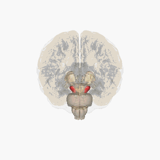
The basal ganglia (BG) or basal nuclei are a group of subcortical nuclei found in the brains of vertebrates. In humans and other primates, differences exist, primarily in the division of the globus pallidus into external and internal regions, and in the division of the striatum. Positioned at the base of the forebrain and the top of the midbrain, they have strong connections with the cerebral cortex, thalamus, brainstem and other brain areas. The basal ganglia are associated with a variety of functions, including regulating voluntary motor movements, procedural learning, habit formation, conditional learning, eye movements, cognition, and emotion.

<span class="mw-page-title-main">Kernicterus</span> Medical condition

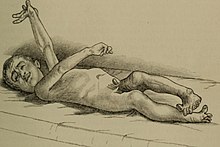
Kernicterus is a bilirubin-induced brain dysfunction. The term was coined in 1904 by Christian Georg Schmorl. Bilirubin is a naturally occurring substance in the body of humans and many other animals, but it is neurotoxic when its concentration in the blood is too high, a condition known as hyperbilirubinemia. Hyperbilirubinemia may cause bilirubin to accumulate in the grey matter of the central nervous system, potentially causing irreversible neurological damage. Depending on the level of exposure, the effects range from clinically unnoticeable to severe brain damage and even death.

<span class="mw-page-title-main">Globus pallidus</span> Structure of the basal ganglia of the brain

The globus pallidus (GP), also known as paleostriatum or dorsal pallidum, is a subcortical structure of the brain. It consists of two adjacent segments, one external, known in rodents simply as the globus pallidus, and one internal, known in rodents as the entopeduncular nucleus. It is part of the telencephalon, but retains close functional ties with the subthalamus in the diencephalon – both of which are part of the extrapyramidal motor system. The globus pallidus is a major component of the basal ganglia, with principal inputs from the striatum, and principal direct outputs to the thalamus and the substantia nigra. The latter is made up of similar neuronal elements, has similar afferents from the striatum, similar projections to the thalamus, and has a similar synaptology. Neither receives direct cortical afferents, and both receive substantial additional inputs from the intralaminar thalamus.

<span class="mw-page-title-main">Nigrostriatal pathway</span> Bilateral pathway in the brain

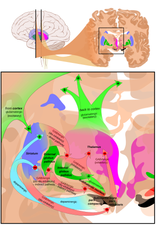
The nigrostriatal pathway is a bilateral dopaminergic pathway in the brain that connects the substantia nigra pars compacta (SNc) in the midbrain with the dorsal striatum in the forebrain. It is one of the four major dopamine pathways in the brain, and is critical in the production of movement as part of a system called the basal ganglia motor loop. Dopaminergic neurons of this pathway release dopamine from axon terminals that synapse onto GABAergic medium spiny neurons (MSNs), also known as spiny projection neurons (SPNs), located in the striatum.

Chorea is an abnormal involuntary movement disorder, one of a group of neurological disorders called dyskinesias. The term chorea is derived from the Ancient Greek: χορεία, as the quick movements of the feet or hands are comparable to dancing.

Choreoathetosis is the occurrence of involuntary movements in a combination of chorea and athetosis.

<span class="mw-page-title-main">Hyperkinesia</span> Excessive movements due to basal ganglia dysfunction

Hyperkinesia refers to an increase in muscular activity that can result in excessive abnormal movements, excessive normal movements, or a combination of both. Hyperkinesia is a state of excessive restlessness which is featured in a large variety of disorders that affect the ability to control motor movement, such as Huntington's disease. It is the opposite of hypokinesia, which refers to decreased bodily movement, as commonly manifested in Parkinson's disease.

Hemiballismus or hemiballism is a basal ganglia syndrome resulting from damage to the subthalamic nucleus in the basal ganglia. Hemiballismus is a rare hyperkinetic movement disorder, that is characterized by violent involuntary limb movements, on one side of the body, and can cause significant disability. Ballismus affects both sides of the body and is much rarer. Symptoms can decrease during sleep.

<span class="mw-page-title-main">Hypokinesia</span> Decreased movement due to basal ganglia dysfunction

Hypokinesia is one of the classifications of movement disorders, and refers to decreased bodily movement. Hypokinesia is characterized by a partial or complete loss of muscle movement due to a disruption in the basal ganglia. Hypokinesia is a symptom of Parkinson's disease shown as muscle rigidity and an inability to produce movement. It is also associated with mental health disorders and prolonged inactivity due to illness, amongst other diseases.

<span class="mw-page-title-main">Medium spiny neuron</span> Type of GABAergic neuron in the striatum

Medium spiny neurons (MSNs), also known as spiny projection neurons (SPNs), are a special type of GABAergic inhibitory cell representing 95% of neurons within the human striatum, a basal ganglia structure. Medium spiny neurons have two primary phenotypes : D1-type MSNs of the direct pathway and D2-type MSNs of the indirect pathway. Most striatal MSNs contain only D1-type or D2-type dopamine receptors, but a subpopulation of MSNs exhibit both phenotypes.

<span class="mw-page-title-main">Internal globus pallidus</span>

The internal globus pallidus and the external globus pallidus (GPe) make up the globus pallidus. The GPi is one of the output nuclei of the basal ganglia. The GABAergic neurons of the GPi send their axons to the ventral anterior nucleus (VA) and the ventral lateral nucleus (VL) in the dorsal thalamus, to the centromedian complex, and to the pedunculopontine complex.

<span class="mw-page-title-main">Paroxysmal kinesigenic choreoathetosis</span> Medical condition

Paroxysmal kinesigenic choreoathetosis (PKC) also called paroxysmal kinesigenic dyskinesia (PKD) is a hyperkinetic movement disorder characterized by attacks of involuntary movements, which are triggered by sudden voluntary movements. The number of attacks can increase during puberty and decrease in a person's 20s to 30s. Involuntary movements can take many forms such as ballism, chorea or dystonia and usually only affect one side of the body or one limb in particular. This rare disorder only affects about 1 in 150,000 people, with PKD accounting for 86.8% of all the types of paroxysmal dyskinesias, and occurs more often in males than females. There are two types of PKD, primary and secondary. Primary PKD can be further broken down into familial and sporadic. Familial PKD, which means the individual has a family history of the disorder, is more common, but sporadic cases are also seen. Secondary PKD can be caused by many other medical conditions such as multiple sclerosis (MS), stroke, pseudohypoparathyroidism, hypocalcemia, hypoglycemia, hyperglycemia, central nervous system trauma, or peripheral nervous system trauma. PKD has also been linked with infantile convulsions and choreoathetosis (ICCA) syndrome, in which patients have afebrile seizures during infancy and then develop paroxysmal choreoathetosis later in life. This phenomenon is actually quite common, with about 42% of individuals with PKD reporting a history of afebrile seizures as a child.

Subcortical dementias includes those diseases which predominantly affects the basal ganglia along with features of cognitive decline.

<span class="mw-page-title-main">Basal ganglia disease</span> Group of physical problems resulting from basal ganglia dysfunction

Basal ganglia disease is a group of physical problems that occur when the group of nuclei in the brain known as the basal ganglia fail to properly suppress unwanted movements or to properly prime upper motor neuron circuits to initiate motor function. Research indicates that increased output of the basal ganglia inhibits thalamocortical projection neurons. Proper activation or deactivation of these neurons is an integral component for proper movement. If something causes too much basal ganglia output, then the ventral anterior (VA) and ventral lateral (VL) thalamocortical projection neurons become too inhibited, and one cannot initiate voluntary movement. These disorders are known as hypokinetic disorders. However, a disorder leading to abnormally low output of the basal ganglia leads to reduced inhibition, and thus excitation, of the thalamocortical projection neurons which synapse onto the cortex. This situation leads to an inability to suppress unwanted movements. These disorders are known as hyperkinetic disorders.

<span class="mw-page-title-main">Blocq's disease</span> Loss of memory of specialized movements causing the inability to maintain an upright posture

Blocq's disease was first considered by Paul Blocq (1860–1896), who described this phenomenon as the loss of memory of specialized movements causing the inability to maintain an upright posture, despite normal function of the legs in the bed. The patient is able to stand up, but as soon as the feet are on the ground, the patient cannot hold himself upright nor walk; however when lying down, the subject conserved the integrity of muscular force and the precision of movements of the lower limbs. The motivation of this study came when a fellow student Georges Marinesco (1864) and Paul published a case of parkinsonian tremor (1893) due to a tumor located in the substantia nigra.

<span class="mw-page-title-main">Neuroferritinopathy</span> Medical condition

Neuroferritinopathy is a genetic neurodegenerative disorder characterized by the accumulation of iron in the basal ganglia, cerebellum, and motor cortex of the human brain. Symptoms, which are extrapyramidal in nature, progress slowly and generally do not become apparent until adulthood. These symptoms include chorea, dystonia, and cognitive deficits which worsen with age.

Dyskinetic cerebral palsy (DCP) is a subtype of cerebral palsy (CP) and is characterized by impaired muscle tone regulation, coordination and movement control. Dystonia and choreoathetosis are the two most dominant movement disorders in patients with DCP.

<span class="mw-page-title-main">Athetoid cerebral palsy</span> Type of cerebral palsy associated with basal ganglia damage

Athetoid cerebral palsy, or dyskinetic cerebral palsy, is a type of cerebral palsy primarily associated with damage, like other forms of CP, to the basal ganglia in the form of lesions that occur during brain development due to bilirubin encephalopathy and hypoxic–ischemic brain injury. Unlike spastic or ataxic cerebral palsies, ADCP is characterized by both hypertonia and hypotonia, due to the affected individual's inability to control muscle tone. Clinical diagnosis of ADCP typically occurs within 18 months of birth and is primarily based upon motor function and neuroimaging techniques. While there are no cures for ADCP, some drug therapies as well as speech, occupational therapy, and physical therapy have shown capacity for treating the symptoms.

References

  1. 1 2 Walker, Kenneth H (1990). "Ch. 70 Involuntary Movements". Clinical Methods: The History, Physical, and Laboratory (3rd ed.). PMID   21250235.
  2. 1 2 "Athetosis". Health Database - Medical Ailments & Diseases. Retrieved March 22, 2011.
  3. 1 2 McNeil, Malcolm R. (2009). Clinical Management of Sensorimotor Speech Disorders. Thieme. ISBN   978-1-58890-514-7.
  4. 1 2 3 4 5 Foley J (April 1983). "The athetoid syndrome. A review of a personal series". J. Neurol. Neurosurg. Psychiatry. 46 (4): 289–98. doi:10.1136/jnnp.46.4.289. PMC   1027350 . PMID   6341510.
  5. Akiyama Y, Koshimura K, Ohue T, Lee K, Miwa S, Yamagata, S (September 1991). "Effects of hypoxia on the activity of the dopaminergic neuron system in the rat striatum as studied by in vivo brain microdialysis". Journal of Neurochemistry. 57 (3): 997–1002. doi:10.1111/j.1471-4159.1991.tb08249.x. PMID   1861163. S2CID   42453096.
  6. 1 2 Martich-Kriss, V; Kollias, SS; Ball WS, Jr (April 1995). "MR findings in kernicterus". American Journal of Neuroradiology. 16 (4 Suppl): 819–21. PMC   8332263 . PMID   7611048.
  7. Hansen, TW (December 2001). "Bilirubin brain toxicity". Journal of Perinatology. 21 Suppl 1: S48–51, discussion S59–62. doi: 10.1038/sj.jp.7210634 . PMID   11803417.
  8. 1 2 3 Handley, A; Medcalf, P; Hellier, K; Dutta, D (May 2009). "Movement disorders after stroke". Age and Ageing. 38 (3): 260–6. doi: 10.1093/ageing/afp020 . PMID   19276093.
  9. 1 2 3 4 Putnam T (May 1939). "Athetosis". Yale J. Biol. Med. 11 (5): 459–465. PMC   2602263 . PMID   21433835.
  10. 1 2 Mawdsley C (December 1975). "Diseases of the central nervous system. Involuntary movements". Br. Med. J. 4 (5996): 572–4. doi:10.1136/bmj.4.5996.572. PMC   1675885 . PMID   1203674.
  11. Polzin, Scott J., MS, and Teresa G. Odle. "Cerebral palsy." Gale Encyclopedia of Medicine. Thomson Gale, 2006. NA. Health Reference Center Academic. Gale.
  12. Laitinen LV (August 1970). "Neurosurgery in cerebral palsy". J. Neurol. Neurosurg. Psychiatry. 33 (4): 513–8. doi:10.1136/jnnp.33.4.513. PMC   493511 . PMID   4918461.
  13. Bhidayasiri, R; Truong, DD (September 2004). "Chorea and related disorders". Postgraduate Medical Journal. 80 (947): 527–34. doi:10.1136/pgmj.2004.019356. PMC   1743104 . PMID   15356354.
  14. Rodriguez SP, Ding D, Riviere SP (September 2010). "Algorithms for target prediction for computer users with athetosis". 2010 Annual International Conference of the IEEE Engineering in Medicine and Biology. Vol. 2010. pp. 491–494. doi:10.1109/iembs.2010.5627118. ISBN   978-1-4244-4123-5. PMC   3253859 . PMID   21096307.
  15. 1 2 Johnson RK, Goran MI, Ferrara MS, Poehlman ET (February 1996). "Athetosis increases resting metabolic rate in adults with cerebral palsy". J. Am. Diet. Assoc. 96 (2): 145–8. doi:10.1016/S0002-8223(96)00043-0. PMID   8557940.
  16. O'Shea TM (December 2008). "Diagnosis, treatment, and prevention of cerebral palsy". Clin. Obstet. Gynecol. 51 (4): 816–28. doi:10.1097/GRF.0b013e3181870ba7. PMC   3051278 . PMID   18981805.
  17. Lo YL, See S (November 2010). "Images in clinical medicine. Pseudoathetosis". N. Engl. J. Med. 363 (19): e29. doi:10.1056/NEJMicm0907786. PMID   21047218.
  18. Salih F, Zimmer C, Meierkord H (March 2007). "Parietal proprioceptive loss with pseudoathetosis". J. Neurol. 254 (3): 396–7. doi:10.1007/s00415-006-0382-x. PMID   17345036. S2CID   32393176.
  19. 1 2 Jeff Brody (2005). "Social Problems with Cerebral Palsy". Cerebral Palsy Lawyer - Birth Injury Attorney. Cerebral Palsy Source. Retrieved March 21, 2011.
  20. T. Michael O’Shea (December 2009). "Diagnosis, Treatment, and Prevention of Cerebral Palsy in Near-Term/Term Infants". Clinical Obstetrics and Gynecology. 51 (4): 816–28. doi:10.1097/GRF.0b013e3181870ba7. PMC   3051278 . PMID   18981805.
  21. Lehrman, Mary (10 May 2010). "The Social, Emotional, & Psychological Effects Of Adults With Cerebral Palsy". Demand Media, Inc. Retrieved March 22, 2011.
  22. Gaskin, Cadeyrn; Morris, Tony (September 2008). "Physical Activity, Health-Related Quality of Life, and Psychosocial Functioning of Adults With Cerebral Palsy". Journal of Physical Activity and Health. 5 (1): 146–157. doi:10.1123/jpah.5.1.146. PMID   18209260.
  23. 1 2 Morris JG, Jankelowitz SK, Fung VS, Clouston PD, Hayes MW, Grattan-Smith P (November 2002). "Athetosis I: historical considerations". Mov. Disord. 17 (6): 1278–80. doi:10.1002/mds.10267. PMID   12465068. S2CID   13359301.
  24. Lopez JV, Sibenaller S, Ding D, Riviere CN (2007). "Toward filtering of athetoid movement with neural networks". Engineering in Medicine and Biology Society. 29th Annual International Conference of the IEEE. doi:10.1109/iembs.2007.4352569.
  25. Vidailhet M, Yelnik J, Lagrange C, et al. (August 2009). "Bilateral pallidal deep brain stimulation for the treatment of patients with dystonia-choreoathetosis cerebral palsy: a prospective pilot study". Lancet Neurol. 8 (8): 709–17. doi:10.1016/S1474-4422(09)70151-6. PMID   19576854. S2CID   24345609.