
Cranial nerves are the nerves that emerge directly from the brain, of which there are conventionally considered twelve pairs. Cranial nerves relay information between the brain and parts of the body, primarily to and from regions of the head and neck, including the special senses of vision, taste, smell, and hearing.
Spasticity is a feature of altered skeletal muscle performance with a combination of paralysis, increased tendon reflex activity, and hypertonia. It is also colloquially referred to as an unusual "tightness", stiffness, or "pull" of muscles.

Tetraplegia, also known as quadriplegia, is defined as the dysfunction or loss of motor and/or sensory function in the cervical area of the spinal cord. A loss of motor function can present as either weakness or paralysis leading to partial or total loss of function in the arms, legs, trunk, and pelvis; paraplegia is similar but affects the thoracic, lumbar, and sacral segments of the spinal cord and arm function is spared. The paralysis may be flaccid or spastic. A loss of sensory function can present as an impairment or complete inability to sense light touch, pressure, heat, pinprick/pain, and proprioception. In these types of spinal cord injury, it is common to have a loss of both sensation and motor control.
A muscle relaxant is a drug that affects skeletal muscle function and decreases the muscle tone. It may be used to alleviate symptoms such as muscle spasms, pain, and hyperreflexia. The term "muscle relaxant" is used to refer to two major therapeutic groups: neuromuscular blockers and spasmolytics. Neuromuscular blockers act by interfering with transmission at the neuromuscular end plate and have no central nervous system (CNS) activity. They are often used during surgical procedures and in intensive care and emergency medicine to cause temporary paralysis. Spasmolytics, also known as "centrally acting" muscle relaxant, are used to alleviate musculoskeletal pain and spasms and to reduce spasticity in a variety of neurological conditions. While both neuromuscular blockers and spasmolytics are often grouped together as muscle relaxant, the term is commonly used to refer to spasmolytics only.

Muscle spindles are stretch receptors within the body of a skeletal muscle that primarily detect changes in the length of the muscle. They convey length information to the central nervous system via afferent nerve fibers. This information can be processed by the brain as proprioception. The responses of muscle spindles to changes in length also play an important role in regulating the contraction of muscles, for example, by activating motor neurons via the stretch reflex to resist muscle stretch.

Afferent nerve fibers are the axons carried by a sensory nerve that relay sensory information from sensory receptors to regions of the brain. Afferent projections arrive at a particular brain region. Efferent nerve fibers are carried by efferent nerves and exit a region to act on muscles and glands.

The pyramidal tracts include both the corticobulbar tract and the corticospinal tract. These are aggregations of efferent nerve fibers from the upper motor neurons that travel from the cerebral cortex and terminate either in the brainstem (corticobulbar) or spinal cord (corticospinal) and are involved in the control of motor functions of the body.
The withdrawal reflex is a spinal reflex intended to protect the body from damaging stimuli. The reflex rapidly coordinates the contractions of all the flexor muscles and the relaxations of the extensors in that limb causing sudden withdrawal from the potentially damaging stimulus. Spinal reflexes are often monosynaptic and are mediated by a simple reflex arc. A withdrawal reflex is mediated by a polysynaptic reflex resulting in the stimulation of many motor neurons in order to give a quick response.
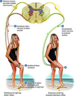
The crossed extensor reflex or crossed extensor response or crossed extension reflex is a reflex in which the contralateral limb compensates for loss of support when the ipsilateral limb withdraws from painful stimulus in a withdrawal reflex. During a withdrawal reflex, the flexors in the withdrawing limb contract and the extensors relax, while in the other limb, the opposite occurs as part of the crossed extensor reflex. Besides shifting the body weight to the other side, the reflex pathway is also associated with leg coordination when walking by flexing muscle on one side, while extending muscle on the other side. This crossed extensor response is properly part of the withdrawal reflex.
Astasis is a lack of motor coordination marked by an inability to stand, walk or even sit without assistance due to disruption of muscle coordination.

Acanthocyte, in biology and medicine, refers to an abnormal form of red blood cell that has a spiked cell membrane, due to thorny projections. A similar term is spur cells. Often they may be confused with echinocytes or schistocytes.

Tizanidine, sold under the brand name Zanaflex among others, is an alpha-2 (α2) adrenergic receptor agonist, similar to clonidine, that is used to treat muscle spasticity due to spinal cord injury, multiple sclerosis, and spastic cerebral palsy. Effectiveness appears similar to baclofen or diazepam. It is taken by mouth.

Foot drop is a gait abnormality in which the dropping of the forefoot happens due to weakness, irritation or damage to the deep fibular nerve, including the sciatic nerve, or paralysis of the muscles in the anterior portion of the lower leg. It is usually a symptom of a greater problem, not a disease in itself. Foot drop is characterized by inability or impaired ability to raise the toes or raise the foot from the ankle (dorsiflexion). Foot drop may be temporary or permanent, depending on the extent of muscle weakness or paralysis and it can occur in one or both feet. In walking, the raised leg is slightly bent at the knee to prevent the foot from dragging along the ground.
Astasia-abasia refers to the inability to either stand or walk in a normal manner. Astasia refers to the inability to stand upright unassisted. Abasia refers to lack of motor coordination in walking. The term abasia literally means that the base of gait is inconstant or unmeasurable. When seen in conversion disorder, the gait is bizarre and is not suggestive of a specific organic lesion: often the patient sways wildly and nearly falls, recovering at the last moment.

Sjögren–Larsson syndrome is a rare autosomal recessive form of ichthyosis with neurological symptoms. It can be identified by a triad of medical disorders. The first is ichthyosis, which is a buildup of skin to form a scale-like covering that causes dry skin and other problems. The second identifier is paraplegia which is characterized by leg spasms. The final identifier is intellectual delay.
Spastic diplegia is a form of cerebral palsy (CP) that is a chronic neuromuscular condition of hypertonia and spasticity—manifested as an especially high and constant "tightness" or "stiffness"—in the muscles of the lower extremities of the human body, usually those of the legs, hips and pelvis. Doctor William John Little's first recorded encounter with cerebral palsy is reported to have been among children who displayed signs of spastic diplegia.
Paul Oscar Blocq (1860–1896) was a French pathologist who is remembered for his neuropathological work done with Jean-Martin Charcot (1825-1893) and Gheorghe Marinescu (1863-1938) at the Salpêtrière in Paris.
The Golgi tendon reflex (also called inverse stretch reflex, autogenic inhibition, tendon reflex) is an inhibitory effect on the muscle resulting from the muscle tension stimulating Golgi tendon organs (GTO) of the muscle, and hence it is self-induced. The reflex arc is a negative feedback mechanism preventing too much tension on the muscle and tendon. When the tension is extreme, the inhibition can be so great it overcomes the excitatory effects on the muscle's alpha motoneurons causing the muscle to suddenly relax. This reflex is also called the inverse myotatic reflex, because it is the inverse of the stretch reflex.

Spastic cerebral palsy is the type of cerebral palsy characterized by spasticity or high muscle tone often resulting in stiff, jerky movements. Cases of spastic CP are further classified according to the part or parts of the body that are most affected. Such classifications include spastic diplegia, spastic hemiplegia, spastic quadriplegia, and in cases of single limb involvement, spastic monoplegia.

The Golgi tendon organ (GTO) is a proprioceptor – a type of sensory receptor that senses changes in muscle tension. It lies at the interface between a muscle and its tendon known as the musculotendinous junction also known as the myotendinous junction. It provides the sensory component of the Golgi tendon reflex.