Related Research Articles

Gastric dilatation volvulus (GDV), also known as gastric dilation, twisted stomach, or gastric torsion, is a medical condition that affects dogs in which the stomach becomes overstretched and rotated by excessive gas content. The word bloat is often used as a general term to mean gas distension without stomach torsion, or to refer to GDV.
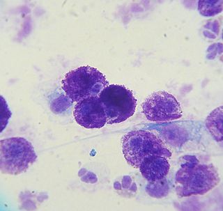
A mastocytoma or mast cell tumor is a type of round-cell tumor consisting of mast cells. It is found in humans and many animal species; it also can refer to an accumulation or nodule of mast cells that resembles a tumor.

Dirofilaria immitis, also known as heartworm or dog heartworm, is a parasitic roundworm that is a type of filarial worm, a small thread-like worm, that causes dirofilariasis. It is spread from host to host through the bites of mosquitoes. There are four genera of mosquitoes that transmit dirofilariasis, Aedes, Culex, Anopheles, and Mansonia. The definitive host is the dog, but it can also infect cats, wolves, coyotes, jackals, foxes, ferrets, bears, seals, sea lions and, under rare circumstances, humans.

The health of dogs is a well studied area in veterinary medicine.
Panosteitis, sometimes shortened to pano among breeders, is an occasionally seen long bone condition in large breed dogs. It manifests with sudden, unexplained pain and lameness that may shift from leg to leg, usually between 5 and 14 months of age, earning the nickname "growing pains. " Signs such as fever, weight loss, anorexia, and lethargy can also be seen. The cause is unknown, but genetics, stress, infection, metabolism, or an autoimmune component may be factors. It has also been suggested that rapid growth and high-protein food are involved in the pathogenesis. Whole blood analysis may show an elevated white blood cell count; this finding lends support to the theory that panosteitis is due to an infection.

Protothecosis, otherwise known as Algaemia, is a disease found in dogs, cats, cattle, and humans caused by a type of green alga known as Prototheca that lacks chlorophyll and enters the human or animal bloodstream. It and its close relative Helicosporidium are unusual in that they are actually green algae that have become parasites. The two most common species are Prototheca wickerhamii and Prototheca zopfii. Both are known to cause disease in dogs, while most human cases are caused by P. wickerhami. Prototheca is found worldwide in sewage and soil. Infection is rare despite high exposure, and can be related to a defective immune system. In dogs, females and Collies are most commonly affected.
Phycomycosis is an uncommon condition of the gastrointestinal tract and skin most commonly found in dogs and horses. The condition is caused by a variety of molds and fungi, and individual forms include pythiosis, zygomycosis, and lagenidiosis. Pythiosis is the most common type and is caused by Pythium, a type of water mould. Zygomycosis can also be caused by two types of zygomycetes, Entomophthorales and Mucorales. The latter type of zygomycosis is also referred to as mucormycosis. Lagenidiosis is caused by a Lagenidium species, which like Pythium is a water mould. Since both pythiosis and lagenidiosis are caused by organisms from the class Oomycetes, they are sometimes collectively referred to as oomycosis.

Pythiosis is a rare and deadly tropical disease caused by the oomycete Pythium insidiosum. Long regarded as being caused by a fungus, the causative agent was not discovered until 1987. It occurs most commonly in horses, dogs, and humans, with isolated cases in other large mammals. The disease is contracted after exposure to stagnant fresh water such as swamps, ponds, lakes, and rice paddies. P. insidiosum is different from other members of the genus in that human and horse hair, skin, and decaying animal and plant tissue are chemoattractants for its zoospores. Additionally, it is the only member in the genus known to infect mammals, while other members are pathogenic to plants and are responsible for some well-known plant diseases.
Craniomandibular osteopathy, also known as lion's jaw, is a developmental disease in dogs causing extensive bony changes in the mandible and skull. In this disease, a cyclical resorption of normal bone and replacement by immature bone occurs along the inner and outer surfaces of the affected bones. It usually occurs between the ages of 3 and 8 months. Breeds most commonly affected include the West Highland White Terrier, Scottish Terrier, Cairn Terrier, and Boston Terrier. It is rare in large-breed dogs, but it has been reported. Symptoms include firm swelling of the jaw, drooling, pain, and difficulty eating.
Hypertrophic Osteodystrophy (HOD) is a bone disease that occurs most often in fast-growing large and giant breed dogs; however, it also affects medium breed animals like the Australian Shepherd. The disorder is sometimes referred to as metaphyseal osteopathy, and typically first presents between the ages of 2 and 7 months. HOD is characterized by decreased blood flow to the metaphysis leading to a failure of ossification and necrosis and inflammation of cancellous bone. The disease is usually bilateral in the limb bones, especially the distal radius, ulna, and tibia.
Wobbler disease is a catchall term referring to several possible malformations of the cervical vertebrae that cause an unsteady (wobbly) gait and weakness in dogs and horses. A number of different conditions of the cervical (neck) spinal column cause similar clinical signs. These conditions may include malformation of the vertebrae, intervertebral disc protrusion, and disease of the interspinal ligaments, ligamenta flava, and articular facets of the vertebrae. Wobbler disease is also known as cervical vertebral instability, cervical spondylomyelopathy (CSM), and cervical vertebral malformation (CVM). In dogs, the disease is most common in large breeds, especially Great Danes and Doberman Pinschers. In horses, it is not linked to a particular breed, though it is most often seen in tall, race-bred horses of Thoroughbred or Standardbred ancestry. It is most likely inherited to at least some extent in dogs and horses.

A mammary tumor is a neoplasm originating in the mammary gland. It is a common finding in older female dogs and cats that are not spayed, but they are found in other animals as well. The mammary glands in dogs and cats are associated with their nipples and extend from the underside of the chest to the groin on both sides of the midline. There are many differences between mammary tumors in animals and breast cancer in humans, including tumor type, malignancy, and treatment options. The prevalence in dogs is about three times that of women. In dogs, mammary tumors are the second most common tumor over all and the most common tumor in female dogs with a reported incidence of 3.4%. Multiple studies have documented that spaying female dogs when young greatly decreases their risk of developing mammary neoplasia when aged. Compared with female dogs left intact, those spayed before puberty have 0.5% of the risk, those spayed after one estrous cycle have 8.0% of the risk, and dogs spayed after two estrous cycles have 26.0% of the risk of developing mammary neoplasia later in life. Overall, unspayed female dogs have a seven times greater risk of developing mammary neoplasia than do those that are spayed. While the benefit of spaying decreases with each estrous cycle, some benefit has been demonstrated in female dogs even up to 9 years of age. There is a much lower risk in male dogs and a risk in cats about half that of dogs.

An anal sac adenocarcinoma is an uncommon and aggressive malignant tumor found in dogs that arises from the apocrine glandular tissue of anal sac. The disease exists in cats as well, but is much less common in that species. They are the second most common cancerous cause of hypercalcaemia in dogs, following T-cell lymphoma.
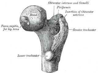
A femoral head ostectomy is a surgical operation to remove the head and neck from the femur. It is performed to alleviate pain, and is a salvage procedure, reserved for condition where pain can not be alleviated in any other way. It is common in veterinary surgery. Other names are excision arthroplasty of the femoral head and neck, Girdlestone's operation, Girdlestone procedure, and femoral head and neck ostectomy.
A vaccine-associated sarcoma (VAS) or feline injection-site sarcoma (FISS) is a type of malignant tumor found in cats which has been linked to certain vaccines. VAS has become a concern for veterinarians and cat owners alike and has resulted in changes in recommended vaccine protocols. These sarcomas have been most commonly associated with rabies and feline leukemia virus vaccines, but other vaccines and injected medications have also been implicated.

Pimobendan is a veterinary medication. It is a calcium sensitizer and a selective inhibitor of phosphodiesterase 3 (PDE3) with positive inotropic and vasodilator effects.
Entomophthoramycosis is a mycosis caused by Entomophthorales.
Exercise-induced collapse (EIC) is a genetic disorder that causes dogs of certain breeds to collapse after a period of intense exercise. The breeds affected are primarily sporting dogs.
The multilobular tumour of bone (MTB), also called an osteochondrosarcoma, is the most common tumour of the canine skull, although it is relatively rare in general.
References
- ↑ Foster, Wendy K.; Armstrong, Julie A. (2006). "Hypertrophic osteopathy associated with pulmonary Eikenella corrodens infection in a dog". Journal of the American Veterinary Medical Association. 228 (9): 1366–1369. doi: 10.2460/javma.228.9.1366 . PMID 16649940 . Retrieved 2006-08-26.
- ↑ Anderson T, Walker M, Goring R (2004). "Cardiogenic hypertrophic osteopathy in a dog with a right-to-left shunting patent ductus arteriosus". J Am Vet Med Assoc. 224 (9): 1464–6, 1453. doi:10.2460/javma.2004.224.1464. PMID 15124887.
- ↑ Seaman R, Patton C (2003). "Treatment of renal nephroblastoma in an adult dog". J Am Anim Hosp Assoc. 39 (1): 76–9. doi:10.5326/0390076. PMID 12549618.
- ↑ Watrous B, Blumenfeld B (2002). "Congenital megaesophagus with hypertrophic osteopathy in a 6-year-old dog". Vet Radiol Ultrasound. 43 (6): 545–9. doi:10.1111/j.1740-8261.2002.tb01046.x. PMID 12502108.
- ↑ Becker T, Perry R, Watson G (1999). "Regression of hypertrophic osteopathy in a cat after surgical excision of an adrenocortical carcinoma". J Am Anim Hosp Assoc. 35 (6): 499–505. doi:10.5326/15473317-35-6-499. PMID 10580910.
- ↑ Foster W, Armstrong J (2006). "Hypertrophic osteopathy associated with pulmonary Eikenella corrodens infection in a dog". J Am Vet Med Assoc. 228 (9): 1366–9. doi: 10.2460/javma.228.9.1366 . PMID 16649940.
| | This veterinary medicine –related article is a stub. You can help Wikipedia by expanding it. |