Clubbing | |
---|---|
Other names | Drumstick fingers/toes, Hippocratic fingers/toes, digital clubbing, watch-glass nails [1] |
![]() | |
Clubbing | |
Specialty | Pulmonology |
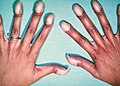
Nail clubbing, also known as digital clubbing or clubbing, is a deformity of the finger or toe nails associated with a number of diseases, anomalies and defects, some congenital, mostly of the heart and lungs. [2] [3] When it occurs together with joint effusions, joint pains, and abnormal skin and bone growth it is known as hypertrophic osteoarthropathy. [4]
Clubbing is associated with lung cancer, lung infections, interstitial lung disease, cystic fibrosis, or cardiovascular disease. [5] Clubbing may also run in families, [5] and occur unassociated with other medical problems. [6] [7]
Clubbing has been recognized as a sign of disease since the time of Hippocrates. [5]
Clubbing is associated with
Nail clubbing is not specific to chronic obstructive pulmonary disease (COPD). Therefore, in patients with COPD and significant degrees of clubbing, a search for signs of bronchogenic carcinoma (or other causes of clubbing) might still be indicated. [12] A congenital form has also been recognized. [13]
A special form of clubbing is hypertrophic pulmonary osteoarthropathy (HPOA), known in continental Europe as Pierre Marie-Bamberger syndrome. This is the combination of clubbing and thickening of periosteum (connective tissue lining of the bones) and synovium (lining of joints), and is often initially diagnosed as arthritis. It is commonly associated with lung cancer.[ citation needed ]
Primary hypertrophic osteoarthropathy is HPOA without signs of pulmonary disease. This form has a hereditary component, although subtle cardiac abnormalities can occasionally be found. It is known eponymously as the Touraine–Solente–Golé syndrome. This condition has been linked to mutations in the gene on the fourth chromosome (4q33-q34) coding for the enzyme 15-hydroxyprostaglandin dehydrogenase (HPGD); this leads to decreased breakdown of prostaglandin E2 and elevated levels of this substance. [14]
The exact cause for sporadic clubbing is unknown. Theories as to its cause include:
When clubbing is observed, pseudoclubbing should be excluded before making the diagnosis. Associated conditions may be identified by taking a detailed medical history—particular attention is paid to lung, heart, and gastrointestinal conditions—and conducting a thorough clinical examination, which may disclose associated features relevant to the underlying diagnosis. Additional studies such as a chest X-ray and a chest CT-scan may reveal otherwise asymptomatic cardiopulmonary disease. [12]
Clubbing is present in one of five stages: [12]
Schamroth's sign or Schamroth's window test (originally demonstrated by South African cardiologist Leo Schamroth on himself) [16] is a popular test for clubbing. When the distal phalanges (bones nearest the fingertips) of corresponding fingers of opposite hands are directly opposed (place fingernails of same finger on opposite hands against each other, nail to nail), a small diamond-shaped "window" is normally apparent between the nailbeds. If this window is obliterated, the test is positive and clubbing is present.
The exact frequency of clubbing in the population is not known. A 2008 study found clubbing in 1%, or 15 patients, of 1511 patients admitted to a department of internal medicine in Belgium. Of these, 40%, or 6 patients, turned out to have significant underlying disease of various causes, while 60%, or 9 patients, had no medical problems on further investigations and remained well over the subsequent year. [7]
At least since the time of Hippocrates, clubbing has been recognized as a sign of disease. [5] The phenomenon has been called "Hippocratic fingers".
The Dutch painter Dick Ket had nail clubbing as is seen from his paintings. He had an underlying disease, probably dextrocardia. [17]
Tetralogy of Fallot (TOF), formerly known as Steno-Fallot tetralogy, is a congenital heart defect characterized by four specific cardiac defects. Classically, the four defects are:
Cyanosis is the change of body tissue color to a bluish-purple hue, as a result of decrease in the amount of oxygen bound to the hemoglobin in the red blood cells of the capillary bed. Cyanosis is apparent usually in the body tissues covered with thin skin, including the mucous membranes, lips, nail beds, and ear lobes. Some medications may cause discoloration such as medications containing amiodarone or silver. Furthermore, mongolian spots, large birthmarks, and the consumption of food products with blue or purple dyes can also result in the bluish skin tissue discoloration and may be mistaken for cyanosis. Appropriate physical examination and history taking is a crucial part to diagnose cyanosis. Management of cyanosis involves treating the main cause, as cyanosis is not a disease, but rather a symptom.
A megakaryocyte is a large bone marrow cell with a lobated nucleus that produces blood platelets (thrombocytes), which are necessary for normal clotting. In humans, megakaryocytes usually account for 1 out of 10,000 bone marrow cells, but can increase in number nearly 10-fold during the course of certain diseases. Owing to variations in combining forms and spelling, synonyms include megalokaryocyte and megacaryocyte.
Pulmonary hypertension is a condition of increased blood pressure in the arteries of the lungs. Symptoms include shortness of breath, fainting, tiredness, chest pain, swelling of the legs, and a fast heartbeat. The condition may make it difficult to exercise. Onset is typically gradual. According to the definition at the 6th World Symposium of Pulmonary Hypertension in 2018, a patient is deemed to have pulmonary hypertension if the pulmonary mean arterial pressure is greater than 20mmHg at rest, revised down from a purely arbitrary 25mmHg, and pulmonary vascular resistance (PVR) greater than 3 Wood units.
A congenital heart defect (CHD), also known as a congenital heart anomaly, congenital cardiovascular malformation, and congenital heart disease, is a defect in the structure of the heart or great vessels that is present at birth. A congenital heart defect is classed as a cardiovascular disease. Signs and symptoms depend on the specific type of defect. Symptoms can vary from none to life-threatening. When present, symptoms are variable and may include rapid breathing, bluish skin (cyanosis), poor weight gain, and feeling tired. CHD does not cause chest pain. Most congenital heart defects are not associated with other diseases. A complication of CHD is heart failure.
A cyanotic heart defect is any congenital heart defect (CHD) that occurs due to deoxygenated blood bypassing the lungs and entering the systemic circulation, or a mixture of oxygenated and unoxygenated blood entering the systemic circulation. It is caused by structural defects of the heart such as right-to-left or bidirectional shunting, malposition of the great arteries, or any condition which increases pulmonary vascular resistance. The result may be the development of collateral circulation.
Interstitial lung disease (ILD), or diffuse parenchymal lung disease (DPLD), is a group of respiratory diseases affecting the interstitium and space around the alveoli of the lungs. It concerns alveolar epithelium, pulmonary capillary endothelium, basement membrane, and perivascular and perilymphatic tissues. It may occur when an injury to the lungs triggers an abnormal healing response. Ordinarily, the body generates just the right amount of tissue to repair damage, but in interstitial lung disease, the repair process is disrupted, and the tissue around the air sacs (alveoli) becomes scarred and thickened. This makes it more difficult for oxygen to pass into the bloodstream. The disease presents itself with the following symptoms: shortness of breath, nonproductive coughing, fatigue, and weight loss, which tend to develop slowly, over several months. The average rate of survival for someone with this disease is between three and five years. The term ILD is used to distinguish these diseases from obstructive airways diseases.
Primary myelofibrosis (PMF) is a rare bone marrow blood cancer. It is classified by the World Health Organization (WHO) as a type of myeloproliferative neoplasm, a group of cancers in which there is activation and growth of mutated cells in the bone marrow. This is most often associated with a somatic mutation in the JAK2, CALR, or MPL genes. In PMF, the bony aspects of bone marrow are remodeled in a process called osteosclerosis; in addition, fibroblasts secrete collagen and reticulin proteins that are collectively referred to as fibrosis. These two pathological processes compromise the normal function of bone marrow resulting in decreased production of blood cells such as erythrocytes, granulocytes and megakaryocytes, the latter cells responsible for the production of platelets.
CREST syndrome, also known as the limited cutaneous form of systemic sclerosis (lcSSc), is a multisystem connective tissue disorder. The acronym "CREST" refers to the five main features: calcinosis, Raynaud's phenomenon, esophageal dysmotility, sclerodactyly, and telangiectasia.
Hypoxemia is an abnormally low level of oxygen in the blood. More specifically, it is oxygen deficiency in arterial blood. Hypoxemia is usually caused by pulmonary disease. Sometimes the concentration of oxygen in the air is decreased leading to hypoxemia.
Respiratory diseases, or lung diseases, are pathological conditions affecting the organs and tissues that make gas exchange difficult in air-breathing animals. They include conditions of the respiratory tract including the trachea, bronchi, bronchioles, alveoli, pleurae, pleural cavity, the nerves and muscles of respiration. Respiratory diseases range from mild and self-limiting, such as the common cold, influenza, and pharyngitis to life-threatening diseases such as bacterial pneumonia, pulmonary embolism, tuberculosis, acute asthma, lung cancer, and severe acute respiratory syndromes, such as COVID-19. Respiratory diseases can be classified in many different ways, including by the organ or tissue involved, by the type and pattern of associated signs and symptoms, or by the cause of the disease.
Pachydermoperiostosis (PDP) is a rare genetic disorder that affects both bones and skin. Other names are primary hypertrophic osteoarthropathy or Touraine-Solente-Golé syndrome. It is mainly characterized by pachyderma, periostosis and finger clubbing.
Hypertrophic osteopathy is a bone disease secondary to cancer in the lungs.
In medicine, hepatopulmonary syndrome is a syndrome of shortness of breath and hypoxemia caused by vasodilation in the lungs of patients with liver disease. Dyspnea and hypoxemia are worse in the upright position.
Platelet-derived growth factor receptor beta is a protein that in humans is encoded by the PDGFRB gene. Mutations in PDGFRB are mainly associated with the clonal eosinophilia class of malignancies.
Acropachy is a dermopathy associated with Graves' disease. It is characterized by soft-tissue swelling of the hands and clubbing of the fingers. Radiographic imaging of affected extremities typically demonstrates periostitis, most commonly the metacarpal bones. The exact cause is unknown, but it is thought to be caused by stimulating auto-antibodies that are implicated in the pathophysiology of Graves' thyrotoxicosis. There is no effective treatment for acropachy.
Hypertrophic osteoarthropathy is a medical condition combining clubbing and periostitis of the small hand joints, especially the distal interphalangeal joints and the metacarpophalangeal joints. Distal expansion of the long bones as well as painful, swollen joints and synovial villous proliferation are often seen. The condition may occur alone (primary), or it may be secondary to diseases like lung cancer. Among patients with lung cancer, it is most associated with adenocarcinoma and least associated with small cell lung cancer. These patients often get clubbing and increased bone deposition on long bones. Their presenting signs and symptoms are sometimes only clubbing and painful ankles.
Shell nail syndrome is a medical condition defined by the concurrence of large, rounded fingernails and bronchiectasis. Despite the visual similarity between the two conditions, shell nail syndrome and clubbed fingernails are opposites. Shell nail syndrome results from atrophy to the nail bed, whereas clubbed fingernails results from a bulbous, hypertrophic growth of soft tissue. The concurrence of the syndrome and bronchiectasis is well-established, however the exact causes of the deformity remains unknown. The syndrome has been observed affecting both the hands and larger toenails.
Tripe palms, also known as acanthosis palmaris, is a medical sign characterized by thick ridged velvety palms, typically as part of a paraneoplastic syndrome. It resembles the lining of the stomach of some animals (tripe). Other signs that may be noted at the same time include most frequently acanthosis nigricans (AN), and less commonly finger clubbing and Leser-Trélat sign.
Pulmonary Arterial Hypertension (PAH) is a syndrome in which the blood pressure in the pulmonary arteries and pulmonary arterioles is elevated. This pre-capillary pulmonary artery pressure being elevated is essential, and by definition a mean pulmonary artery pressure greater than 20 mmHg as measured by a right heart catheterization is required for the diagnosis. This pre-capillary pulmonary hypertension is confirmed with measuring pulmonary vascular resistance being greater than 3 Woods Units. A pulmonary artery wedge pressure being less than 15 mmHg excludes post-capillary bed pulmonary hypertension. Pulmonary arterial hypertension is a subgroup of pulmonary hypertension and is categorized as World Health Organization as group 1. PAH is further subdivided into various categories based on the cause, including idiopathic, heritable, drug and toxin induced, PAH associated with specific diseases, PAH that is responsive to vasodilators, PAH with venous or capillary involvement, and persistent PAH in the newborn period.