An embolism is the lodging of an embolus, a blockage-causing piece of material, inside a blood vessel. The embolus may be a blood clot (thrombus), a fat globule, a bubble of air or other gas, amniotic fluid, or foreign material.
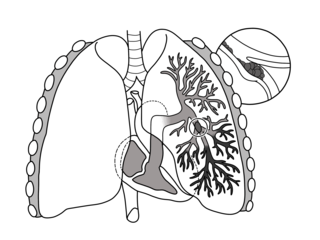
Pulmonary embolism (PE) is a blockage of an artery in the lungs by a substance that has moved from elsewhere in the body through the bloodstream (embolism). Symptoms of a PE may include shortness of breath, chest pain particularly upon breathing in, and coughing up blood. Symptoms of a blood clot in the leg may also be present, such as a red, warm, swollen, and painful leg. Signs of a PE include low blood oxygen levels, rapid breathing, rapid heart rate, and sometimes a mild fever. Severe cases can lead to passing out, abnormally low blood pressure, obstructive shock, and sudden death.

Sputum is mucus that is coughed up from the lower airways. In medicine, sputum samples are usually used for a naked eye examination, microbiological investigation of respiratory infections and cytological investigations of respiratory systems.
An air embolism, also known as a gas embolism, is a blood vessel blockage caused by one or more bubbles of air or other gas in the circulatory system. Air can be introduced into the circulation during surgical procedures, lung over-expansion injury, decompression, and a few other causes. In flora, air embolisms may also occur in the xylem of vascular plants, especially when suffering from water stress.
Decompression Illness (DCI) comprises two different conditions caused by rapid decompression of the body. These conditions present similar symptoms and require the same initial first aid. Scuba divers are trained to ascend slowly from depth to avoid DCI. Although the incidence is relatively rare, the consequences can be serious and potentially fatal, especially if untreated.
D-dimer is a dimer that is a fibrin degradation product (FDP), a small protein fragment present in the blood after a blood clot is degraded by fibrinolysis. It is so named because it contains two D fragments of the fibrin protein joined by a cross-link, hence forming a protein dimer.

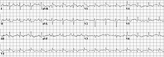
In electrocardiography, the T wave represents the repolarization of the ventricles. The interval from the beginning of the QRS complex to the apex of the T wave is referred to as the absolute refractory period. The last half of the T wave is referred to as the relative refractory period or vulnerable period. The T wave contains more information than the QT interval. The T wave can be described by its symmetry, skewness, slope of ascending and descending limbs, amplitude and subintervals like the Tpeak–Tend interval.
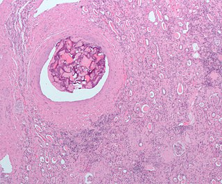
An embolus, is described as a free-floating mass, located inside blood vessels that can travel from one site in the blood stream to another. An embolus can be made up of solid, liquid, or gas. Once these masses get "stuck" in a different blood vessel, it is then known as an "embolism." An embolism can cause ischemia—damage to an organ from lack of oxygen. A paradoxical embolism is a specific type of embolism in which the embolus travels from the right side of the heart to the left side of the heart and lodges itself in a blood vessel known as an artery. It is termed "paradoxical" because venous emboli will usually be lodged in pulmonary artery in an event called pulmonary embolism, instead of systemic circulation.

A hemorrhagic infarct is determined when hemorrhage is present around an area of infarction. Simply stated, an infarction is an area of dead tissue or necrosis. When blood escapes outside of the vessel (extravasation) and re-perfuses back into the tissue surrounding the infarction, the infarction is then termed a hemorrhagic infarct (infarction). Hemorrhagic infarcts can occur in any region of the body, such as the head, trunk and abdomen-pelvic regions, typically arising from their arterial blood supply being interrupted by a blockage or compression of an artery.
In chest radiography, the Westermark sign is a sign that represents a focus of oligemia (hypovolemia) seen distal to a pulmonary embolism (PE). While the chest x-ray is normal in the majority of PE cases, the Westermark sign is seen in 2% of patients.

A ventilation/perfusion lung scan, also called a V/Q lung scan, or ventilation/perfusion scintigraphy, is a type of medical imaging using scintigraphy and medical isotopes to evaluate the circulation of air and blood within a patient's lungs, in order to determine the ventilation/perfusion ratio. The ventilation part of the test looks at the ability of air to reach all parts of the lungs, while the perfusion part evaluates how well blood circulates within the lungs. As Q in physiology is the letter used to describe bloodflow the term V/Q scan emerged.

An amniotic fluid embolism (AFE) is a life-threatening childbirth (obstetric) emergency in which amniotic fluid enters the blood stream of the mother, triggering a serious reaction which results in cardiorespiratory collapse and massive bleeding (coagulopathy). The rate at which it occurs is 1 instance per 20,000 births and it comprises 10% of all maternal deaths.

Lung infarction or pulmonary infarction occurs when an artery to the lung becomes blocked and part of the lung dies. It is most often caused by a pulmonary embolism.

A CT pulmonary angiogram (CTPA) is a medical diagnostic test that employs computed tomography (CT) angiography to obtain an image of the pulmonary arteries. Its main use is to diagnose pulmonary embolism (PE). It is a preferred choice of imaging in the diagnosis of PE due to its minimally invasive nature for the patient, whose only requirement for the scan is an intravenous line.
Michael Humpal is a former American football linebacker. He was selected by the Pittsburgh Steelers of the National Football League (NFL) in the sixth round of the 2008 NFL draft. He was part of the Steelers' victory over the Arizona Cardinals in Super Bowl XLIII. He played college football at Iowa.
Obstructive shock is one of the four types of shock, caused by a physical obstruction in the flow of blood. Obstruction can occur at the level of the great vessels or the heart itself. Causes include pulmonary embolism, cardiac tamponade, and tension pneumothorax. These are all life-threatening. Symptoms may include shortness of breath, weakness, or altered mental status. Low blood pressure and tachycardia are often seen in shock. Other symptoms depend on the underlying cause.
Embolectomy is the emergency interventional or surgical removal of emboli which are blocking blood circulation. It usually involves removal of thrombi, and is then referred to as thromboembolectomy or thrombectomy. Embolectomy is an emergency procedure often as the last resort because permanent occlusion of a significant blood flow to an organ leads to necrosis. Other involved therapeutic options are anticoagulation and thrombolysis.

Palla's sign is a clinical sign in which an enlarged right descending pulmonary artery is seen on the chest x-ray in patients with pulmonary embolism. It is of low sensitivity, and its specificity is not known. It exhibits as a "sausage" appearance on X-ray. It is named after italian radiologist Antonio Palla. In 1983, he published his observations that close to 25% of patients with pulmonary embolism had a chest x-ray sign of enlarged right descending pulmonary artery.
The 2011 Family Circle Cup was a women's tennis event on the 2011 WTA Tour. It took place from April 4 to April 10, 2011. It was the 39th edition of the tournament and one of the Premier level tournaments. The event was hosted at the Family Circle Tennis Center, on Daniel Island, Charleston, South Carolina, United States. It was the only event of the clay court season played on green clay. The total prize money offered at this tournament was US$711,000
Right heart strain is a medical finding of right ventricular dysfunction where the heart muscle of the right ventricle (RV) is deformed. Right heart strain can be caused by pulmonary hypertension, pulmonary embolism, RV infarction, chronic lung disease, pulmonic stenosis, bronchospasm, and pneumothorax.
