A CT scan or computed tomography scan makes use of computer-processed combinations of many X-ray measurements taken from different angles to produce cross-sectional (tomographic) images of specific areas of a scanned object, allowing the user to see inside the object without cutting. The 1979 Nobel Prize in Physiology or Medicine was awarded jointly to Allan M. Cormack and Godfrey N. Hounsfield "for the development of computer assisted tomography."

Radiography is an imaging technique using X-rays, gamma rays, or similar ionizing radiation and non-ionizing radiation to view the internal form of an object. Applications of radiography include medical radiography and industrial radiography. Similar techniques are used in airport security . To create an image in Conventional Radiography, a beam of X-rays is produced by an X-ray generator and is projected toward the object. A certain amount of the X-rays or other radiation is absorbed by the object, dependent on the object's density and structural composition. The X-rays that pass through the object are captured behind the object by a detector. The generation of flat two dimensional images by this technique is called projectional radiography. In computed tomography an X-ray source and its associated detectors rotate around the subject which itself moves through the conical X-ray beam produced. Any given point within the subject is crossed from many directions by many different beams at different times. Information regarding attenuation of these beams is collated and subjected to computation to generate two dimensional images in three planes which can be further processed to produce a three dimensional image.

Pulmonary embolism (PE) is a blockage of an artery in the lungs by a substance that has moved from elsewhere in the body through the bloodstream (embolism). Symptoms of a PE may include shortness of breath, chest pain particularly upon breathing in, and coughing up blood. Symptoms of a blood clot in the leg may also be present, such as a red, warm, swollen, and painful leg. Signs of a PE include low blood oxygen levels, rapid breathing, rapid heart rate, and sometimes a mild fever. Severe cases can lead to passing out, abnormally low blood pressure, and sudden death.

A pleural effusion is excess fluid that accumulates in the pleural cavity, the fluid-filled space that surrounds the lungs. This excess fluid can impair breathing by limiting the expansion of the lungs. Various kinds of pleural effusion, depending on the nature of the fluid and what caused its entry into the pleural space, are hydrothorax, hemothorax (blood), urinothorax (urine), chylothorax (chyle), or pyothorax (pus) commonly known as pleural empyema. In contrast, a pneumothorax is the accumulation of air in the pleural space, and is commonly called a "collapsed lung".

Tomography is imaging by sections or sectioning, through the use of any kind of penetrating wave. The method is used in radiology, archaeology, biology, atmospheric science, geophysics, oceanography, plasma physics, materials science, astrophysics, quantum information, and other areas of science. The word tomography is derived from Ancient Greek τόμος tomos, "slice, section" and γράφω graphō, "to write". A device used in tomography is called a tomograph, while the image produced is a tomogram.

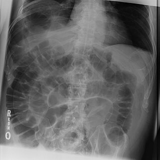
Situs inversus is a congenital condition in which the major visceral organs are reversed or mirrored from their normal positions. The normal arrangement of internal organs is known as situs solitus while situs inversus is generally the mirror image of situs solitus. Although cardiac problems are more common than in the general population, most people with situs inversus have no medical symptoms or complications resulting from the condition, and until the advent of modern medicine it was usually undiagnosed.
The Hounsfield scale, named after Sir Godfrey Hounsfield, is a quantitative scale for describing radiodensity. It is frequently used in CT scans, where its value is also termed CT number.
Manuel Dias de Abreu was a Brazilian physician and scientist, the inventor of abreugraphy, a rapid radiography of the lungs for screening tuberculosis. He is considered one of the most important Brazilian physicians, side by side with Carlos Chagas, Vital Brazil and Oswaldo Cruz.

A hemothorax is an accumulation of blood within the pleural cavity. The symptoms of a hemothorax include chest pain and difficulty breathing, while the clinical signs include reduced breath sounds on the affected side and a rapid heart rate. Hemothoraces are usually caused by an injury but may occur spontaneously: due to cancer invading the pleural cavity, as a result of a blood clotting disorder, as an unusual manifestation of endometriosis, in response to a collapsed lung, or rarely in association with other conditions.

Cryptogenic organizing pneumonia (COP), formerly known as bronchiolitis obliterans organizing pneumonia (BOOP), is an inflammation of the bronchioles (bronchiolitis) and surrounding tissue in the lungs). It should not be confused with bronchiolitis obliterans, a form of non-infectious pneumonia.

Pneumomediastinum is pneumatosis in the mediastinum. First described in 1819 by René Laennec, the condition can result from physical trauma or other situations that lead to air escaping from the lungs, airways, or bowel into the chest cavity.

In radiology, the air crescent sign is a finding on chest radiograph and computed tomography that is crescenteric and radiolucent, due to a lung cavity that is filled with air and has a round radiopaque mass. Classically, it is due to an aspergilloma, a form of aspergillosis, that occurs when the fungus Aspergillus grows in a cavity in the lung. It is also referred as Monad sign.
Willi A. Kalender is a German Medical Physicist and Professor and Chairman of the Institute of Medical Physics of the University of Erlangen-Nuremberg. He is a Fellow of the American Association of Physicists in Medicine (AAPM) and Honorary Fellow of the British Institute of Radiology (BIR) and of the Institute of Physics and Engineering in Medicine (IPEM).
In chest radiography, the Westermark sign is a sign that represents a focus of oligemia (hypovolemia) seen distal to a pulmonary embolism (PE). While the chest x-ray is normal in the majority of PE cases, the Westermark sign is seen in 2% of patients.

High-resolution computed tomography (HRCT) is a type of computed tomography (CT) with specific techniques to enhance image resolution. It is used in the diagnosis of various health problems, though most commonly for lung disease, by assessing the lung parenchyma.

Evarts Ambrose Graham (1883–1957) was an American academic, physician, and surgeon.

In radiology, ground glass opacity (GGO) is a nonspecific finding on computed tomography (CT) scans that indicates a partial filling of air spaces in the lungs by exudate or transudate, as well as interstitial thickening or partial collapse of lung alveoli.
The National Lung Screening Trial was a United States-based clinical trial which recruited research participants between 2002-2004. It was sponsored by the National Cancer Institute and conducted by the American College of Radiology Imaging Network and the Lung Screening Study Group. The major objective of the trial was to compare the efficacy of low-dose helical computed tomography and standard chest X-ray as methods of lung cancer screening. The primary study ended in 2010, and the initial findings were published in November 2010, with the main results published in 2011 in the New England Journal of Medicine.

Charles Thurstan Holland (1863–1941) was a general practitioner in Liverpool who was best known by his pioneering research in the field of Radiology. The Thurstan Holland sign is named after him.