
Croup, also known as croupy cough, is a type of respiratory infection that is usually caused by a virus. The infection leads to swelling inside the trachea, which interferes with normal breathing and produces the classic symptoms of "barking/brassy" cough, inspiratory stridor and a hoarse voice. Fever and runny nose may also be present. These symptoms may be mild, moderate, or severe. Often it starts or is worse at night and normally lasts one to two days.

Bowel obstruction, also known as intestinal obstruction, is a mechanical or functional obstruction of the intestines which prevents the normal movement of the products of digestion. Either the small bowel or large bowel may be affected. Signs and symptoms include abdominal pain, vomiting, bloating and not passing gas. Mechanical obstruction is the cause of about 5 to 15% of cases of severe abdominal pain of sudden onset requiring admission to hospital.
A steeple is a tall tower on a building, often topped by a spire.

A chest radiograph, chest X-ray (CXR), or chest film is a projection radiograph of the chest used to diagnose conditions affecting the chest, its contents, and nearby structures. Chest radiographs are the most common film taken in medicine.
Stridor is an extra-thoracic high-pitched breath sound resulting from turbulent air flow in the larynx or lower in the bronchial tree. It is different from a stertor, which is a noise originating in the pharynx.
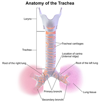
Tracheitis is an inflammation of the trachea. Although the trachea is usually considered part of the lower respiratory tract, in ICD-10 tracheitis is classified under "acute upper respiratory infections".
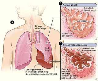
Lobar pneumonia is a form of pneumonia characterized by inflammatory exudate within the intra-alveolar space resulting in consolidation that affects a large and continuous area of the lobe of a lung.
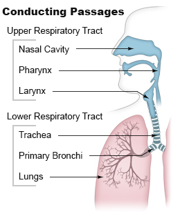
Airway obstruction is a blockage of respiration in the airway that hinders the free flow of air. Airway obstructions can occur either in the upper airway or lower airway. The upper airway consists of the nose, throat, and larynx. The lower airway comprises the trachea, bronchi, and bronchioles.
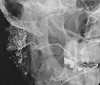
Sialography is the radiographic examination of the salivary glands. It usually involves the injection of a small amount of contrast medium into the salivary duct of a single gland, followed by routine X-ray projections.

The Stafne defect is a depression of the mandible, most commonly located on the lingual surface. The Stafne defect is thought to be a normal anatomical variant, as the depression is created by ectopic salivary gland tissue associated with the submandibular gland and does not represent a pathologic lesion as such. This cavity is commonly observed on panoramic radiograph.

An odontogenic keratocyst is a rare and benign but locally aggressive developmental cyst. It most often affects the posterior mandible and most commonly presents in the third decade of life. Odontogenic keratocysts make up around 19% of jaw cysts. Despite its more common appearance in the bone region, it can affect soft tissue.

In radiology, the thumbprint sign, or thumbprinting, is a radiologic sign found on a radiograph that suggests the diagnosis of either epiglottitis or intestinal ischemia.
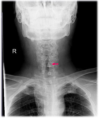
Subglottic stenosis is a congenital or acquired narrowing of the subglottic airway. It can be congenital, acquired, iatrogenic, or very rarely, idiopathic. It is defined as the narrowing of the portion of the airway that lies between the vocal cords and the lower part of the cricoid cartilage. In a normal infant, the subglottic airway is 4.5-5.5 millimeters wide, while in a premature infant, the normal width is 3.5 millimeters. Subglottic stenosis is defined as a diameter of under 4 millimeters in an infant. Acquired cases are more common than congenital cases due to prolonged intubation being introduced in the 1960s. It is most frequently caused by certain medical procedures or external trauma, although infections and systemic or autoimmune diseases can also cause it.

Projectional radiography, also known as conventional radiography, is a form of radiography and medical imaging that produces two-dimensional images by X-ray radiation. The image acquisition is generally performed by radiographers, and the images are often examined by radiologists. Both the procedure and any resultant images are often simply called 'X-ray'. Plain radiography or roentgenography generally refers to projectional radiography. Plain radiography can also refer to radiography without a radiocontrast agent or radiography that generates single static images, as contrasted to fluoroscopy, which are technically also projectional.

An abdominal x-ray is an x-ray of the abdomen. It is sometimes abbreviated to AXR, or KUB.

Retropharyngeal abscess (RPA) is an abscess located in the tissues in the back of the throat behind the posterior pharyngeal wall. Because RPAs typically occur in deep tissue, they are difficult to diagnose by physical examination alone. RPA is a relatively uncommon illness, and therefore may not receive early diagnosis in children presenting with stiff neck, malaise, difficulty swallowing, or other symptoms listed below. Early diagnosis is key, while a delay in diagnosis and treatment may lead to death. Parapharyngeal space communicates with retropharyngeal space and an infection of retropharyngeal space can pass down behind the esophagus into the mediastinum. RPAs can also occur in adults of any age.
Sialodochitis, is inflammation of the duct system of a salivary gland. This is compared to sialadenitis, which is inflammation of the gland parenchyma.

In radiology, the double bubble sign is a feature of pediatric imaging seen on radiographs or prenatal ultrasound in which two air filled bubbles are seen in the abdomen, representing two discontiguous loops of bowel in a proximal, or 'high,' small bowel obstruction. The finding is typically pathologic, and implies either duodenal atresia, duodenal web, annular pancreas, or on occasion midgut volvulus, a distinction that requires close clinical correlation and, in most cases, surgical intervention.
Spinal cord injury without radiographic abnormality (SCIWORA) is symptoms of a spinal cord injury (SCI) with no evidence of injury to the spinal column on X-rays or CT scan. Symptoms may include numbness, weakness, abnormal reflexes, or loss of bladder or bowel control. Neck or back pain is also common. Symptoms may be brief or persistent. Some do not develop symptoms until a few days after the injury.
Bat wing appearance is a radiologic sign referring to bilateral perihilar lung shadowing seen in frontal chest X-ray and in chest CT. The most common reason for bat wing appearance is the accumulation of oedema fluid in the lungs. The batwing sign is symmetrical, usually showing ground glass appearance and spares the lung cortices. This sign is seen in individuals with pneumonia, inhalation injuries, pulmonary haemorrhage, sarcoidosis, bronchoalveolar carcinoma and pulmonary alveolar proteinosis.
