Human head

Last updated
Human head
Proportions of the Head.jpg
The human head drawn by Leonardo da Vinci
Details
Identifiers
Latin caput
MeSH D006257
TA98 A01.1.00.001
TA2 98
FMA 7154
Anatomical terminology

In human anatomy, the head is at the top of the human body. It supports the face and is maintained by the skull, which itself encloses the brain.

Contents

Structure

Anatomy of the human head Head ap anatomy.jpg
Anatomy of the human head

The human head consists of a fleshy outer portion, which surrounds the bony skull. The brain is enclosed within the skull. There are 22 bones in the human head. The head rests on the neck, and the seven cervical vertebrae support it. The human head typically weighs between 2.3 and 5 kilograms (5.1 and 11.0 lb) Over 98% of humans fit into this range. There have been odd incidences where human beings have abnormally small or large heads. The Zika virus was responsible for underdeveloped heads in the early 2000s.

The face is the anterior part of the head, containing the eyes, nose, and mouth. On either side of the mouth, the cheeks provide a fleshy border to the oral cavity. The ears sit to either side of the head.

Blood supply

The head receives blood supply through the internal and external carotid arteries. These supply the area outside of the skull (external carotid artery) and inside of the skull (internal carotid artery). The area inside the skull also receives blood supply from the vertebral arteries, which travel up through the cervical vertebrae.

Nerve supply

Sensory areas of the head, showing the general distribution of the three divisions of the fifth nerve. From Gray's Anatomy 1918 Gray784.png
Sensory areas of the head, showing the general distribution of the three divisions of the fifth nerve. From Gray's Anatomy 1918

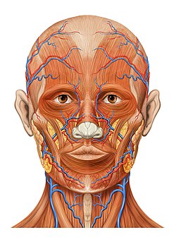
The twelve pairs of cranial nerves provide the majority of nervous control to the head. The sensation to the face is provided by the branches of the trigeminal nerve, the fifth cranial nerve. Sensation to other portions of the head is provided by the cervical nerves.

Modern texts are in agreement about which areas of the skin are served by which nerves, but there are minor variations in some of the details. The borders designated by diagrams in the 1918 edition of Gray's Anatomy are similar but not identical to those generally accepted today.

The cutaneous innervation of the head is as follows:

Function

The head contains sensory organs: two eyes, two ears, a nose and tongue inside of the mouth. It also houses the brain. Together, these organs function as a processing center for the body by relaying sensory information to the brain. Humans can process information faster by having this central nerve cluster.

Society and culture

For humans, the front of the head (the face) is the main distinguishing feature between different people due to its easily discernible features, such as eye and hair colors, shapes of the sensory organs, and the wrinkles. Humans easily differentiate between faces because of the brain's predisposition toward facial recognition. When observing a relatively unfamiliar species, all faces seem nearly identical. Human infants are biologically programmed to recognize subtle differences in anthropomorphic facial features. [1]

Dayak people were feared for their headhunting practices COLLECTIE TROPENMUSEUM Een Ibu Dajak krijger uit Long Nawan Z. en O. afdeling Borneo. TMnr 60034031.jpg
Dayak people were feared for their headhunting practices

People who have greater than average intelligence are sometimes depicted in cartoons as having bigger heads as a way of notionally indicating that they have a "larger head". Additionally, in science fiction, an extraterrestrial having a big head is often symbolic of high intelligence. Despite this depiction, advances in neurobiology have shown that the functional diversity of the brain means that a difference in overall brain size is only slightly to moderately correlated to differences in overall intelligence between two humans. [2]

The head is a source for many metaphors and metonymies in human language, including referring to things typically near the human head ( "the head of the bed"), things physically similar to the way a head is arranged spatially to a body ("the head of the table"), metaphorically ("the head of the class"), and things that represent some characteristics associated with the head, such as intelligence ("there are a lot of good heads in this company"). [3]

Ancient Greeks had a method for evaluating sexual attractiveness based on the Golden ratio, part of which included measurements of the head. [4]

Headhunting is the practice of taking and preserving a person's head after killing the person. Headhunting has been practiced across the Americas, Europe, Asia, and Oceania for millennia. [5]

Clothing

A man wearing a straw hat 0004 Madagascar (5558700320).jpg
A man wearing a straw hat

Headpieces can signify status, origin, religious/spiritual beliefs, social grouping, team affiliation, occupation, or fashion choices.

In many cultures, covering the head is seen as a sign of respect. Often, some or all of the head must be covered and veiled when entering holy places or places of prayer. For many centuries, women in Europe, the Middle East, and South Asia have covered their head hair as a sign of modesty. This trend has changed drastically in Europe in the 20th century, although is still observed in other parts of the world. In addition, a number of religions require men to wear specific head clothing—such as the Islamic taqiyah, Jewish yarmulke, or the Sikh turban. The same goes for women with the Muslim hijab or Christian nun's habit.

A hat is a head covering that can serve a variety of purposes. Hats may be worn as part of a uniform or used as a protective device, such as a hard hat, a covering for warmth, or a fashion accessory. Hats can also be indicative of social status in some areas of the world.

Anthropometry

While numerous charts detailing head sizes in infants and children exist, most do not measure average head circumference past the age of 21. Reference charts for adult head circumference also generally feature homogeneous samples and fail to take height and weight into account. [6]

One study in the United States estimated the average human head circumference to be 57 centimetres (22+12 in) in males and 55 centimetres (21+34 in) in females. [7] [ dubious ] A British study by Newcastle University showed an average size of 57.2 cm for males and 55.2 cm for females with average size varying proportionally with height [8]

Macrocephaly can be an indicator of increased risk for some types of cancer in individuals who carry the genetic mutation that causes Cowden syndrome. For adults, this refers to head sizes greater than 58 centimeters in men or greater than 57 centimeters in women. [9] [10]

Average head sizes

[ citation needed ]

Some values in the table below may not be correct. In particular, a random biocular breadth measurement showed a value of 101.6 mm for an average adult male. The first percentile value below is 110 mm for an adult male. Another user has also indicate around 100 mm being an average value for biocular breadth. Therefore, the biocular breadth values appear to be increased by around 22 mm.

MeasurementImageDescriptionSexPercentile (centimetres)
1st5th50th95th99th
Head breadth1The maximum breadth of the head, usually above and behind the ears.Men13.914.315.216.116.5
Women13.313.714.415.015.8
Interpupilliary breadth2The distance between the centres of the pupils of the eyes, while looking straight ahead.Men5.75.96.57.17.4
Women5.55.76.06.97.0
Face breadth (bizygomatic)3The breadth of the face, measured across the most lateral projections of the cheek bones (zygomatic arches).Men12.813.214.015.015.4
Women12.112.312.814.015.4
Face length (menton-sellion)4The vertical distance from the tip of the chin (menton) to the deepest point of the nasal root depression between the eyes (sellion).Men10.811.212.213.313.7
Women10.110.411.312.412.9
Biocular breadth5The distance from the outer corners of the eyes (right and left ectocanthi).Men11.011.312.213.113.6
Women10.811.111.612.913.3
Bitragion breadth6The breadth of the head from the right tragion to the left. Tragion is the cartilaginous notch at the front of the ear.Men13.113.514.515.515.9
Women12.512.813.314.315.0
Glabella to
back of head
7The horizontal distance from the most anterior point of the forehead between the brow-ridges (glabella) to the back of the head.Men18.318.820.021.121.7
Women17.518.019.120.220.7
Menton to
back of head
8The horizontal distance from the tip of the chin (menton) to the back of the head.Men15.716.518.220.020.7
Women15.215.817.318.919.6
Sellion to
top of head
9The vertical distance from the nasal root depression between the eyes (sellion) to the level of the top of the head.Men9.710.111.212.412.9
Women9.09.510.511.712.2
Stomion to top of head10The vertical distance from the midpoint of the lips (stomion) to the level of the top of the head, measured with a headboard.Men16.917.418.619.920.6
Women15.716.317.518.819.4
Sellion to back of head11The horizontal distance from the nasal root depression between the eyes (sellion), to the back of the head, measured with a headboard.Men18.018.519.720.921.4
Women17.417.818.920.020.5
Pronasale to
back of head
12The horizontal distance from the tip of the nose (pronasale) to the back of the head.Men20.020.522.023.223.9
Women19.219.721.022.222.8
Head length13The maximum length of the head; measured from the most anterior point of the forehead between the brow ridges (glabella) to the back of the head (occiput).Men18.018.519.720.921.3
Women17.217.618.719.820.2
Menton to
top of head
14The vertical distance from the bottom of the chin (menton) to the top of the head.Men21.221.823.224.725.5
Women19.820.421.823.223.8
Menton-crinion length15The vertical distance from the bottom of the chin (menton) to the midpoint of the hairline (crinion).Men16.617.419.120.921.6
Women15.516.117.719.219.9
Menton-subnasale length16The distance from the bottom of the chin (menton) to the base of the nasal septum (subnasale).Men6.16.57.38.38.7
Women5.76.06.57.88.3
Static adult human physical characteristics of the head - redrawn. AvgHeadSizes.png
Static adult human physical characteristics of the head - redrawn.

See also

Related Research Articles

<span class="mw-page-title-main">Peripheral nervous system</span> Part of the nervous system excluding the brain and spinal cord

The peripheral nervous system (PNS) is one of two components that make up the nervous system of bilateral animals, with the other part being the central nervous system (CNS). The PNS consists of nerves and ganglia, which lie outside the brain and the spinal cord. The main function of the PNS is to connect the CNS to the limbs and organs, essentially serving as a relay between the brain and spinal cord and the rest of the body. Unlike the CNS, the PNS is not protected by the vertebral column and skull, or by the blood–brain barrier, which leaves it exposed to toxins.

<span class="mw-page-title-main">Cranial nerves</span> Nerves that emerge directly from the brain and the brainstem

Cranial nerves are the nerves that emerge directly from the brain, of which there are conventionally considered twelve pairs. Cranial nerves relay information between the brain and parts of the body, primarily to and from regions of the head and neck, including the special senses of vision, taste, smell, and hearing.

<span class="mw-page-title-main">Vagus nerve</span> Main nerve of the parasympathetic nervous system

The vagus nerve, also known as the tenth cranial nerve, cranial nerve X, or simply CN X, is a cranial nerve that carries sensory fibers that create a pathway that interfaces with the parasympathetic control of the heart, lungs, and digestive tract. It comprises two nerves—the left and right vagus nerves—but they are typically referred to collectively as a single subsystem.

<span class="mw-page-title-main">Neck</span> Part of the body on many vertebrates that connects the head with the torso

The neck is the part of the body on many vertebrates that connects the head with the torso. The neck supports the weight of the head and protects the nerves that carry sensory and motor information from the brain down to the rest of the body. In addition, the neck is highly flexible and allows the head to turn and flex in all directions. The structures of the human neck are anatomically grouped into four compartments: vertebral, visceral and two vascular compartments. Within these compartments, the neck houses the cervical vertebrae and cervical part of the spinal cord, upper parts of the respiratory and digestive tracts, endocrine glands, nerves, arteries and veins. Muscles of the neck are described separately from the compartments. They bound the neck triangles.

Articles related to anatomy include:

<span class="mw-page-title-main">Parotid gland</span> Major salivary gland in many animals

The parotid gland is a major salivary gland in many animals. In humans, the two parotid glands are present on either side of the mouth and in front of both ears. They are the largest of the salivary glands. Each parotid is wrapped around the mandibular ramus, and secretes serous saliva through the parotid duct into the mouth, to facilitate mastication and swallowing and to begin the digestion of starches. There are also two other types of salivary glands; they are submandibular and sublingual glands. Sometimes accessory parotid glands are found close to the main parotid glands.

<span class="mw-page-title-main">Forehead</span> Area of the head between the eyebrows and the hairline

In human anatomy, the forehead is an area of the head bounded by three features, two of the skull and one of the scalp. The top of the forehead is marked by the hairline, the edge of the area where hair on the scalp grows. The bottom of the forehead is marked by the supraorbital ridge, the bone feature of the skull above the eyes. The two sides of the forehead are marked by the temporal ridge, a bone feature that links the supraorbital ridge to the coronal suture line and beyond. However, the eyebrows do not form part of the forehead.

<span class="mw-page-title-main">External carotid artery</span> Major artery of the head and neck

The external carotid artery is a major artery of the head and neck. It arises from the common carotid artery when it splits into the external and internal carotid artery. The external carotid artery supplies blood to the face, brain and neck.

<span class="mw-page-title-main">Internal carotid artery</span> Artery of the human brain

The internal carotid artery is an artery in the neck which supplies the anterior circulation of the brain.

<span class="mw-page-title-main">Scalp</span> Anatomical area bordered by the face at the front, and by the neck at the sides and back

The scalp is the area of the head where head hair grows. It is made up of skin, layers of connective and fibrous tissues, and the membrane of the skull. Anatomically, the scalp is part of the epicranium, a collection of structures covering the cranium. The scalp is bordered by the face at the front, and by the neck at the sides and back. The scientific study of hair and scalp is called trichology.

<span class="mw-page-title-main">Cervical vertebrae</span> Vertebrae of the neck

In tetrapods, cervical vertebrae are the vertebrae of the neck, immediately below the skull. Truncal vertebrae lie caudal of cervical vertebrae. In sauropsid species, the cervical vertebrae bear cervical ribs. In lizards and saurischian dinosaurs, the cervical ribs are large; in birds, they are small and completely fused to the vertebrae. The vertebral transverse processes of mammals are homologous to the cervical ribs of other amniotes. Most mammals have seven cervical vertebrae, with the only three known exceptions being the manatee with six, the two-toed sloth with five or six, and the three-toed sloth with nine.

<span class="mw-page-title-main">Vertebral artery</span> Major arteries of the neck

The vertebral arteries are major arteries of the neck. Typically, the vertebral arteries originate from the subclavian arteries. Each vessel courses superiorly along each side of the neck, merging within the skull to form the single, midline basilar artery. As the supplying component of the vertebrobasilar vascular system, the vertebral arteries supply blood to the upper spinal cord, brainstem, cerebellum, and posterior part of brain.

<span class="mw-page-title-main">Common carotid artery</span> One of the two arteries that supply the head and neck with blood

In anatomy, the left and right common carotid arteries (carotids) are arteries that supply the head and neck with oxygenated blood; they divide in the neck to form the external and internal carotid arteries.

<span class="mw-page-title-main">Cavernous sinus</span> Sinus in the human head

The cavernous sinus within the human head is one of the dural venous sinuses creating a cavity called the lateral sellar compartment bordered by the temporal bone of the skull and the sphenoid bone, lateral to the sella turcica.

<span class="mw-page-title-main">Petrous part of the temporal bone</span> Feature at the base of the human skull

The petrous part of the temporal bone is pyramid-shaped and is wedged in at the base of the skull between the sphenoid and occipital bones. Directed medially, forward, and a little upward, it presents a base, an apex, three surfaces, and three angles, and houses in its interior, the components of the inner ear. The petrous portion is among the most basal elements of the skull and forms part of the endocranium. Petrous comes from the Latin word petrosus, meaning "stone-like, hard". It is one of the densest bones in the body. In other mammals, it is a separate bone, the petrosal bone.

<span class="mw-page-title-main">Head and neck anatomy</span>

This article describes the anatomy of the head and neck of the human body, including the brain, bones, muscles, blood vessels, nerves, glands, nose, mouth, teeth, tongue, and throat.

<span class="mw-page-title-main">Outline of human anatomy</span> Overview of and topical guide to human anatomy

The following outline is provided as an overview of and topical guide to human anatomy:

<span class="mw-page-title-main">Parapharyngeal space</span>

The parapharyngeal space, is a potential space in the head and the neck. It has clinical importance in otolaryngology due to parapharyngeal space tumours and parapharyngeal abscess developing in this area. It is also a key anatomic landmark for localizing disease processes in the surrounding spaces of the neck; the direction of its displacement indirectly reflects the site of origin for masses or infection in adjacent areas, and consequently their appropriate differential diagnosis.

<span class="mw-page-title-main">Roots of the ciliary ganglion</span>

The ciliary ganglion is a parasympathetic ganglion located just behind the eye in the posterior orbit. Three types of axons enter the ciliary ganglion but only the preganglionic parasympathetic axons synapse there. The entering axons are arranged into three roots of the ciliary ganglion, which join enter the posterior surface of the ganglion.

References

  1. "Infants process faces long before they recognize other objects, Stanford vision researchers find". Stanford University. Retrieved 2018-11-14.
  2. Brain Size and Intelligence
  3. Lakoff and Johnson 1980, 1999
  4. Pallett PM, Link S, Lee K (January 2010). "New "golden" ratios for facial beauty". Vision Research. 50 (2): 149–54. doi:10.1016/j.visres.2009.11.003. PMC   2814183 . PMID   19896961.
  5. Christine Quigley (13 October 2005). The Corpse: A History. McFarland. pp. 249–251. ISBN   978-0-7864-2449-8.
  6. Nguyen, A.K.D (2012). "Head Circumference in Canadian Male Adults: Development of a Normalized Chart". International Journal of Morphology. 30 (4): 1474–1480. doi: 10.4067/s0717-95022012000400033 .
  7. TECHNICAL BRIEF - Relationship Between Head Mass and Circumference in Human Adults. Date: July 20, 2007. Principal Investigator: Randal P. Ching, Ph.D. Institution: University of Washington. Applied Biomechanics Laboratory.
  8. Bushby KM, Cole T, Matthews JN, Goodship JA (October 1992). "Centiles for adult head circumference". Archives of Disease in Childhood. 67 (10): 1286–7. doi:10.1136/adc.67.10.1286. PMC   1793909 . PMID   1444530.
  9. Cowden Syndrome Detection Will Allow For Early Discovery of Cancerous Polyps. Date: December 7, 2010. Principal Investigator: Charis Eng, MD, PhD. Institution: Cleveland Clinic Genomic Medicine.
  10. Mester JL, Tilot AK, Rybicki LA, Frazier TW, Eng C (July 2011). "Analysis of prevalence and degree of macrocephaly in patients with germline PTEN mutations and of brain weight in Pten knock-in murine model". European Journal of Human Genetics. 19 (7): 763–8. doi:10.1038/ejhg.2011.20. PMC   3137495 . PMID   21343951.

Further reading