This article needs additional citations for verification .(April 2014) |
Cervical plexus | |
---|---|
![]() Dermatome distribution of the trigeminal nerve (Superficial cervical plexus visible in purple, at center bottom.) | |
Details | |
From | C1-C4 |
Identifiers | |
Latin | plexus cervicalis |
MeSH | D002572 |
TA98 | A14.2.02.012 |
TA2 | 6374 |
FMA | 5904 |
Anatomical terms of neuroanatomy |
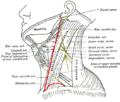
The cervical plexus is a nerve plexus of the anterior rami of the first (i.e. upper-most) four cervical spinal nerves C1-C4. [1] [2] [3] [4] The cervical plexus provides motor innervation to some muscles of the neck, and the diaphragm; it provides sensory innervation to parts of the head, neck, and chest. [1]
They are located laterally to the transverse processes between prevertebral muscles from the medial side and vertebral (m. scalenus, m. levator scapulae, m. splenius cervicis) from lateral side. There is anastomosis with accessory nerve, hypoglossal nerve and sympathetic trunk. It is located in the neck, deep to the sternocleidomastoid muscle. [5]
The branches of the cervical plexus emerge from the posterior triangle at the nerve point, a point which lies midway on the posterior border of the sternocleidomastoid.
The cervical plexus is situated deep to the sternocleidomastoid muscle, internal jugular vein, and deep cervical fascia. [1]
It is situated anterior to the middle scalene muscle, and levator scapulae muscle. [1]
The cervical plexus has two types of branches: cutaneous and muscular. [3]
{{cite book}}
: CS1 maint: location missing publisher (link)