Cutaneous innervation

Last updated

Cutaneous innervation refers to an area of the skin which is supplied by a specific cutaneous nerve.

Contents

Dermatomes are similar; however, a dermatome only specifies the area served by a spinal nerve. In some cases, the dermatome is less specific (when a spinal nerve is the source for more than one cutaneous nerve), and in other cases it is more specific (when a cutaneous nerve is derived from multiple spinal nerves.) [1]

Modern texts are in agreement about which areas of the skin are served by which nerves, but there are minor variations in some of the details. The borders designated by the diagrams in the 1918 edition of Gray's Anatomy are similar, but not identical, to those generally accepted today.

Importance of the peripheral nervous system

The peripheral nervous system (PNS) is divided into the somatic nervous system, the autonomic nervous system, and the enteric nervous system. However, it is the somatic nervous system, responsible for body movement and the reception of external stimuli, which allows one to understand how cutaneous innervation is made possible by the action of specific sensory fibers located on the skin, as well as the distinct pathways they take to the central nervous system. The skin, which is part of the integumentary system, plays an important role in the somatic nervous system because it contains a range of nerve endings that react to heat and cold, touch, pressure, vibration, and tissue injury.

Importance of the central nervous system

The central nervous system (CNS) works with the peripheral nervous system in cutaneous innervation. The CNS is responsible for processing the information it receives from the cutaneous nerves that detect a given stimulus, and then identifying the kind of sensory inputs which project to a specific region of the primary somatosensory cortex.

The role of nerve endings on the surface of the skin

Groups of nerve terminals located in the different layers of the skin are categorized depending on whether the skin is hairy, nonhairy, or an exposed mucous membrane. [2]

Hairy skin

The hairy parts of the body such as the forearm or the leg have two groups of nerve endings: those that end along with the hair follicles, and also with the arborizations of unmyelinated axons which are referred to as free nerve endings because they are served by both myelinated and unmyelinated axons. [2]

Nonhairy skin

Nonhairy skin (glabrous), such as the palms of hands and the soles of feet, has three types of nerve terminations.

The first one, Meissner's corpuscles are encapsulated nerve endings attached to the epidermis in the dermal papilli that detect changes in texture and vibrations.

Merkel's discs are arborizations of nonmyelinated axons that end in terminals on specialized tactile cells and which detect sustained touch and pressure.

Lastly, there are also free nerve endings which are similar in structure to those in hairy skin, though they are more numerous. [2]

Exposed mucous membranes

The exposed mucous membranes of the lips, the anal mucous membrane, and the external genital organs form the most densely innervated parts of the body. Though there is no specific categorization, both free nerve endings and unencapsulated nerve endings of myelinated axons are found within the dermis of those areas.

The cornea, one of the other exposed mucous membranes, contains free nerve endings served by nonmyelinated axons.

The conjunctiva contains a less dense distribution of free nerve endings that are served by both myelinated and unmyelinated axons. [2]

Distribution of sensory neurons

The distribution of the sensory neurons within the skin accounts for the large and overlapping receptive fields of the skin. [2] The size of the receptive fields in turn explains why almost any given stimulus to the human skin can potentially activate a very large number of nerve terminals. Therefore, it is more likely that a stimulus caused by the prick of a needle be detected by more than a hundred nerve endings all sharing the same receptive field, than for that same needle prick to be detected by only one nerve ending.

Types of sensory neurons

The different kinds of sensory stimuli that are picked up by sensory neurons are grouped into two categories: epicritic and protopathic. [3]

Epicritic neurons detect gentle touch such as caresses; light vibrations; the ability to recognize the shape of an object being held; and two-point discrimination, or the spacing of two points being touched simultaneously.

Protopathic neurons are responsible for detecting pain, itch, tickle, and temperature. The different types of stimuli that are detected by a given receptor allow for a relative specificity between stimuli and receptor.

Pathways to the CNS

The sensory modality that is detected by the afferent fibers is an important factor to consider because it determines the pathway that the dorsal root ganglion neurons will take within the central nervous system. The sensory neurons coming from the body synapse in the dorsal horn of the spinal cord, bringing in information about touch sensations (epicritic), or modalities of pain (protopathic). While both types of sensory neurons must first synapse in the dorsal horn of the spinal cord, the area of the dorsal horn where they synapse is different. Their pathway to the thalamus is also different.

Neurons that carry information about touch, vibration, and proprioception sensations from the lower body enter the spinal cord below spinal level T6, where they synapse in the dorsal horn to form reflex circuits, but also send axon branches through the gracile fascicle to the brainstem. Similarly, information from the upper body enters the spinal cord at level T6 and above, and ascend toward the brainstem in the Cuneate fasciculus. Together the gracile and cuneate form the dorsal column in the spine.

Neurons that carry information about pain and temperature synapse in the dorsal horn at the anterolateral fascicles.

While the neurons for touch sensations ascend ipsilaterally through the posterior column-medial lemniscus pathway to the thalamus; neurons for pain and temperature ascend contralaterally to the thalamus through the anterolateral system. [4]

When both sensory pathways reach the integrating center that is the thalamus, they make their final ascent to the somatosensory areas in the postcentral gyrus of the cerebral cortex. [5]

See also

Related Research Articles

<span class="mw-page-title-main">Central nervous system</span> Brain and spinal cord

The central nervous system (CNS) is the part of the nervous system consisting of the brain and spinal cord, the retina and optic nerve, and the olfactory nerve and epithelia. The CNS is so named because the brain integrates the received information and coordinates and influences the activity of all parts of the bodies of bilaterally symmetric and triploblastic animals—that is, all multicellular animals except sponges and diploblasts. It is a structure composed of nervous tissue positioned along the rostral to caudal axis of the body and may have an enlarged section at the rostral end which is a brain. Only arthropods, cephalopods and vertebrates have a true brain, though precursor structures exist in onychophorans, gastropods and lancelets.

In physiology, nociception, also nocioception; from Latin nocere 'to harm/hurt') is the sensory nervous system's process of encoding noxious stimuli. It deals with a series of events and processes required for an organism to receive a painful stimulus, convert it to a molecular signal, and recognize and characterize the signal to trigger an appropriate defensive response.

<span class="mw-page-title-main">Trigeminal nerve</span> Cranial nerve responsible for the faces senses and motor functions

In neuroanatomy, the trigeminal nerve (lit. triplet nerve), also known as the fifth cranial nerve, cranial nerve V, or simply CN V, is a cranial nerve responsible for sensation in the face and motor functions such as biting and chewing; it is the most complex of the cranial nerves. Its name (trigeminal, from Latin tri- 'three', and -geminus 'twin') derives from each of the two nerves (one on each side of the pons) having three major branches: the ophthalmic nerve (V1), the maxillary nerve (V2), and the mandibular nerve (V3). The ophthalmic and maxillary nerves are purely sensory, whereas the mandibular nerve supplies motor as well as sensory (or "cutaneous") functions. Adding to the complexity of this nerve is that autonomic nerve fibers as well as special sensory fibers (taste) are contained within it.

<span class="mw-page-title-main">Neural pathway</span> Connection formed between neurons that allows neurotransmission

In neuroanatomy, a neural pathway is the connection formed by axons that project from neurons to make synapses onto neurons in another location, to enable neurotransmission. Neurons are connected by a single axon, or by a bundle of axons known as a nerve tract, or fasciculus. Shorter neural pathways are found within grey matter in the brain, whereas longer projections, made up of myelinated axons, constitute white matter.

<span class="mw-page-title-main">Afferent nerve fiber</span> Axonal projections that arrive at a particular brain region

Afferent nerve fibers are axons of sensory neurons that carry sensory information from sensory receptors to the central nervous system. Many afferent projections arrive at a particular brain region.

A mechanoreceptor, also called mechanoceptor, is a sensory receptor that responds to mechanical pressure or distortion. Mechanoreceptors are innervated by sensory neurons that convert mechanical pressure into electrical signals that, in animals, are sent to the central nervous system.

<span class="mw-page-title-main">Nociceptor</span> Sensory neuron that detects pain

A nociceptor is a sensory neuron that responds to damaging or potentially damaging stimuli by sending "possible threat" signals to the spinal cord and the brain. The brain creates the sensation of pain to direct attention to the body part, so the threat can be mitigated; this process is called nociception.

<span class="mw-page-title-main">Spinothalamic tract</span> Sensory pathway from the skin to the thalamus

The spinothalamic tract is a part of the anterolateral system or the ventrolateral system, a sensory pathway to the thalamus. From the ventral posterolateral nucleus in the thalamus, sensory information is relayed upward to the somatosensory cortex of the postcentral gyrus.

<span class="mw-page-title-main">Dorsal column–medial lemniscus pathway</span> Sensory spinal pathway

The dorsal column–medial lemniscus pathway (DCML) is a sensory pathway of the central nervous system that conveys sensations of fine touch, vibration, two-point discrimination, and proprioception from the skin and joints. It transmits information from the body to the primary somatosensory cortex in the postcentral gyrus of the parietal lobe of the brain. The pathway receives information from sensory receptors throughout the body, and carries this in nerve tracts in the white matter of the dorsal column of the spinal cord to the medulla, where it is continued in the medial lemniscus, on to the thalamus and relayed from there through the internal capsule and transmitted to the somatosensory cortex. The name dorsal-column medial lemniscus comes from the two structures that carry the sensory information: the dorsal columns of the spinal cord, and the medial lemniscus in the brainstem.

<span class="mw-page-title-main">Medial lemniscus</span> Ascending bundle of axons which cross in the brainstem

In neuroanatomy, the medial lemniscus, also known as Reil's band or Reil's ribbon, is a large ascending bundle of heavily myelinated axons that decussate (cross) in the brainstem, specifically in the medulla oblongata. The medial lemniscus is formed by the crossings of the internal arcuate fibers. The internal arcuate fibers are composed of axons of nucleus gracilis and nucleus cuneatus. The axons of the nucleus gracilis and nucleus cuneatus in the medial lemniscus have cell bodies that lie contralaterally.

<span class="mw-page-title-main">Sensory neuron</span> Nerve cell that converts environmental stimuli into corresponding internal stimuli

Sensory neurons, also known as afferent neurons, are neurons in the nervous system, that convert a specific type of stimulus, via their receptors, into action potentials or graded receptor potentials. This process is called sensory transduction. The cell bodies of the sensory neurons are located in the dorsal ganglia of the spinal cord.

<span class="mw-page-title-main">Type Ia sensory fiber</span> Type of afferent nerve fiber

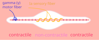
A type Ia sensory fiber, or a primary afferent fiber is a type of afferent nerve fiber. It is the sensory fiber of a stretch receptor called the muscle spindle found in muscles, which constantly monitors the rate at which a muscle stretch changes. The information carried by type Ia fibers contributes to the sense of proprioception.

The withdrawal reflex is a spinal reflex intended to protect the body from damaging stimuli. The reflex rapidly coordinates the contractions of all the flexor muscles and the relaxations of the extensors in that limb causing sudden withdrawal from the potentially damaging stimulus. Spinal reflexes are often monosynaptic and are mediated by a simple reflex arc. A withdrawal reflex is mediated by a polysynaptic reflex resulting in the stimulation of many motor neurons in order to give a quick response.

<span class="mw-page-title-main">Dorsal root ganglion</span> Cluster of neurons in a dorsal root of a spinal nerve

A dorsal root ganglion is a cluster of neurons in a dorsal root of a spinal nerve. The cell bodies of sensory neurons known as first-order neurons are located in the dorsal root ganglia.

<span class="mw-page-title-main">Pseudounipolar neuron</span>

A pseudounipolar neuron is a type of neuron which has one extension from its cell body. This type of neuron contains an axon that has split into two branches. A single process arises from the cell body and then divides into an axon and a dendrite. They develop embryologically as bipolar in shape, and are thus termed pseudounipolar instead of unipolar.

<span class="mw-page-title-main">Gate control theory</span> Theory about pain and the nervous system

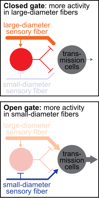
The gate control theory of pain asserts that non-painful input closes the nerve "gates" to painful input, which prevents pain sensation from traveling to the central nervous system.

<span class="mw-page-title-main">Dorsal column nuclei</span> Nuclei in the dorsal column of the brainstem

In neuroanatomy, the dorsal column nuclei are a pair of nuclei in the dorsal columns in the brainstem. The name refers collectively to the cuneate nucleus and gracile nucleus, which are situated at the lower end of the medulla oblongata. Both nuclei contain second-order neurons of the dorsal column–medial lemniscus pathway, which convey fine touch and proprioceptive information from the body to the brain. The dorsal column nuclei project to the thalamus.

<span class="mw-page-title-main">Group C nerve fiber</span> One of three classes of nerve fiber in the central nervous system and peripheral nervous system

Group C nerve fibers are one of three classes of nerve fiber in the central nervous system (CNS) and peripheral nervous system (PNS). The C group fibers are unmyelinated and have a small diameter and low conduction velocity, whereas Groups A and B are myelinated. Group C fibers include postganglionic fibers in the autonomic nervous system (ANS), and nerve fibers at the dorsal roots. These fibers carry sensory information.

Group A nerve fibers are one of the three classes of nerve fiber as generally classified by Erlanger and Gasser. The other two classes are the group B nerve fibers, and the group C nerve fibers. Group A are heavily myelinated, group B are moderately myelinated, and group C are unmyelinated.

<span class="mw-page-title-main">Outline of the human nervous system</span> Overview of and topical guide to the human nervous system

The following diagram is provided as an overview of and topical guide to the human nervous system:

References

  1. Oaklander, Anne Louise; Siegel, Sandra M. (December 2005). "Cutaneous innervation: Form and function". Journal of the American Academy of Dermatology. 53 (6): 1027–1037. doi:10.1016/j.jaad.2005.08.049. ISSN   0190-9622. PMID   16310064.
  2. 1 2 3 4 5 Weddell, G., and Miller, S. (1962) Cutaneous Sensibility. Annual Reviews 24:199-222
  3. Kandel, Eric; James Schwartz; Thomas Jessell (2000). Principles of Neural Science . McGraw-Hill. ISBN   9780838577011.
  4. Kandel, Eric; James Schwartz; Thomas Jessell (2000). Principles of Neural Science . McGraw-Hill. p.  446. ISBN   9780838577011.
  5. Kandel, Eric; James Schwartz; Thomas Jessell (2000). Principles of Neural Science . McGraw-Hill. p.  448. ISBN   9780838577011.