Posterior branches of cervical nerves

Last updated
Posterior branches of cervical nerves

Gray800.png

Posterior primary divisions of the upper three cervical nerves.
Details
From cervical nerves
Identifiers
Latin rami posteriores nervorum cervicalium

Anatomical terms of neuroanatomy

The posterior branches of cervical nerves branch from the dorsal rami of the cervical nerves.

Dorsal ramus of spinal nerve

The dorsal ramus of spinal nerve is the posterior division of a spinal nerve. The dorsal ramus is the dorsal branch of a spinal nerve that forms from the dorsal root of the nerve after it emerges from the spinal cord. The spinal nerve is formed from the dorsal and ventral rami. The dorsal ramus carries information that supplies muscles and sensation to the human back.

Contents

Branches

First

The posterior division of the first cervical or suboccipital nerve is larger than the anterior division, and emerges above the posterior arch of the atlas and beneath the vertebral artery. It enters the suboccipital triangle and supplies the muscles which bound this triangle, viz., the Rectus capitis posterior major, and the Obliqui superior and inferior; it gives branches also to the Rectus capitis posterior minor and the Semispinalis capitis. A filament from the branch to the Obliquus inferior joins the posterior division of the second cervical nerve.

Suboccipital nerve

The suboccipital nerve is the dorsal primary ramus of the first cervical nerve (C1). It exits the spinal cord between the skull and the first cervical vertebra, the atlas.

The vertebral arteries are major arteries of the neck. Typically, the vertebral arteries originate from the subclavian arteries. Each vessel courses superiorly along each side of the neck, merging within the skull to form the single, midline basilar artery. As the supplying component of the vertebrobasilar vascular system, the vertebral arteries provide supply blood to the upper spinal cord, brainstem, cerebellum, and posterior part of brain.

Suboccipital triangle

The suboccipital triangle is a region of the neck bounded by the following three muscles of the suboccipital group of muscles:

The nerve occasionally gives off a cutaneous branch which accompanies the occipital artery to the scalp, and communicates with the greater and lesser occipital nerves.

Scalp anatomical area bordered by the face anteriorly and the neck to the sides and posteriorly

The scalp is the anatomical area bordered by the human face at the front, and by the neck at the sides and back.

Second

The posterior division of the second cervical nerve is much larger than the anterior division, and is the greatest of all the cervical posterior divisions. It emerges between the posterior arch of the atlas and the lamina of the axis, below the Obliquus inferior. It supplies a twig to this muscle, receives a communicating filament from the posterior division of the first cervical, and then divides into a large medial and a small lateral branch.

Greater occipital nerve

The greater occipital nerve is a spinal nerve, specifically the medial branch of the dorsal primary ramus of cervical spinal nerve 2. This nerve arises from between the first and second cervical vertebrae, along with the lesser occipital nerve. It ascends after emerging from below the suboccipital triangle beneath the obliquus capitis inferior muscle. It then passes through the semispinalis muscle before ascending to innervate the skin along the posterior part of the scalp to the vertex. It innervates the scalp at the top of the head, over the ear and over the parotid glands.

Third

The posterior division of the third cervical is intermediate in size between those of the second and fourth.

Third occipital nerve

While under the Trapezius, the medial branch of the posterior division of the third cervical nerve gives off a branch called the third occipital nerve, which pierces the Trapezius and ends in the skin of the lower part of the back of the head.

The posterior division of the suboccipital, and the medial branches of the posterior division of the second and third cervical nerves are sometimes joined by communicating loops to form the posterior cervical plexus.

Cervical plexus

The cervical plexus is a plexus of the anterior rami of the first four cervical spinal nerves which arise from C1 to C4 cervical segment in the neck. They are located laterally to the transverse processes between prevertebral muscles from the medial side and vertebral from lateral side. There is anastomosis with accessory nerve, hypoglossal nerve and sympathetic trunk.

Fourth through eighth

The posterior divisions of the lower five cervical nerves divide into medial and lateral branches.

Additional images

Related Research Articles

Atlas (anatomy) first cervical vertebra of the spine which supports the skull

In anatomy, the atlas (C1) is the most superior (first) cervical vertebra of the spine.

Spinal nerve nerve that carries signals between the spinal chord and the body

A spinal nerve is a mixed nerve, which carries motor, sensory, and autonomic signals between the spinal cord and the body. In the human body there are 31 pairs of spinal nerves, one on each side of the vertebral column. These are grouped into the corresponding cervical, thoracic, lumbar, sacral and coccygeal regions of the spine. There are eight pairs of cervical nerves, twelve pairs of thoracic nerves, five pairs of lumbar nerves, five pairs of sacral nerves, and one pair of coccygeal nerves. The spinal nerves are part of the peripheral nervous system.

Levator scapulae muscle

The levator scapulae is a skeletal muscle situated at the back and side of the neck. As the Latin name suggests, its main function is to lift the scapula.

Obliquus capitis inferior muscle muscle

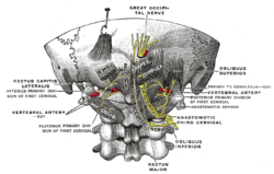
The obliquus capitis inferior muscle is the larger of the two oblique muscles of the neck. It arises from the apex of the spinous process of the axis and passes laterally and slightly upward, to be inserted into the lower and back part of the transverse process of the atlas.

Obliquus capitis superior muscle muscle

The obliquus capitis superior muscle is a small muscle in the upper back part of the neck and is one of the suboccipital muscles and part of the suboccipital triangle. It arises from the lateral mass of the atlas bone. It passes superiorly and posteriorly to insert into the lateral half of the inferior nuchal line on the external surface of the occipital bone. The muscle is innervated by the suboccipital nerve, the dorsal ramus of the first spinal nerve.

Semispinalis muscles

The semispinalis muscles are a group of three muscles belonging to the transversospinales. These are the semispinalis capitis, the semispinalis cervicis and the semispinalis thoracis.

Rectus capitis posterior minor muscle

The rectus capitis posterior minor arises by a narrow pointed tendon from the tubercle on the posterior arch of the atlas, and, widening as it ascends, is inserted into the medial part of the inferior nuchal line of the occipital bone and the surface between it and the foramen magnum, and also takes some attachment to the spinal dura mater.

Occipital artery

The occipital artery arises from the external carotid artery opposite the facial artery. Its path is below the posterior belly of digastric to the occipital region. This artery supplies blood to the back of the scalp and sterno-mastoid muscles, and deep muscles in the back and neck.

Posterior cutaneous nerve of thigh

The posterior cutaneous nerve of the thigh provides innervation to the skin of the posterior surface of the thigh and leg, as well as to the skin of the perineum.

Posterior triangle of the neck

The posterior triangle is a region of the neck.

Longissimus

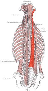
The longissimus is the muscle lateral to the semispinalis muscles. It is the longest subdivision of the erector spinae muscles that extends forward into the transverse processes of the posterior cervical vertebrae.

The posterior branches of thoracic nerves branch from the dorsal rami of the thoracic nerves.

Outline of human anatomy Overview of and topical guide to human anatomy

The following outline is provided as an overview of and topical guide to human anatomy:

Cervical spinal nerve 1

The cervical spinal nerve 1 (C1) is a spinal nerve of the cervical segment. C1 carries predominantly motor fibres, but also a small meningeal branch that supplies sensation to parts of the dura around the foramen magnum.

References

This article incorporates text in the public domain from page 921 of the 20th edition of Gray's Anatomy (1918)