Suboccipital triangle

Last updated
Suboccipital triangle

Suboccipital triangle dissection - Suboccipital triangle.png

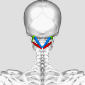
Artist depiction of the muscles that border the suboccipital triangle.

Suboccipital triangle11.png

Identifiers

Anatomical terminology

The suboccipital triangle is a region of the neck bounded by the following three muscles of the suboccipital group of muscles:

Neck part of the body, on many terrestrial or secondarily aquatic vertebrates, that distinguishes the head from the torso or trunk

The neck is the part of the body, on many vertebrates, that separates the head from the torso. It contains blood vessels and nerves that supply structures in the head to the body. These in humans include part of the esophagus, the larynx, trachea, and thyroid gland, major blood vessels including the carotid arteries and jugular veins, and the top part of the spinal cord.

Suboccipital muscles

The suboccipital muscles are a group of muscles defined by their location to the occiput. Suboccipital muscles are located below the occipital bone. These are four paired muscles on the underside of the occipital bone; the two straight muscles (rectus) and the two oblique muscles (obliquus).

Contents

(Rectus capitus posterior minor is also in this region but does not form part of the triangle)

It is covered by a layer of dense fibro-fatty tissue, situated beneath the semispinalis capitis.

The floor is formed by the posterior atlantooccipital membrane, and the posterior arch of the atlas.

Atlas (anatomy) vertebra

In anatomy, the atlas (C1) is the most superior (first) cervical vertebra of the spine.

In the deep groove on the upper surface of the posterior arch of the atlas are the vertebral artery and the first cervical or suboccipital nerve.

The vertebral arteries are major arteries of the neck. Typically, the vertebral arteries originate from the subclavian arteries. Each vessel courses superiorly along each side of the neck, merging within the skull to form the single, midline basilar artery. As the supplying component of the vertebrobasilar vascular system, the vertebral arteries provide supply blood to the upper spinal cord, brainstem, cerebellum, and posterior part of brain.

Suboccipital nerve

The suboccipital nerve is the dorsal primary ramus of the first cervical nerve (C1). It exits the spinal cord between the skull and the first cervical vertebra, the atlas.

In the past, the vertebral artery was accessed here in order to conduct angiography of the circle of Willis. In these times a formal angiography of the circle of Willis is performed via catheter angiography, with access usually being acquired at the common femoral artery. Alternatively, a computed tomographic angiogram or magnetic resonance angiogram is performed.

Angiography medical imaging technique

Angiography or arteriography is a medical imaging technique used to visualize the inside, or lumen, of blood vessels and organs of the body, with particular interest in the arteries, veins, and the heart chambers. This is traditionally done by injecting a radio-opaque contrast agent into the blood vessel and imaging using X-ray based techniques such as fluoroscopy.

Circle of Willis circulatory anastomosis that supplies blood to the brain and surrounding structures

The circle of Willis is a circulatory anastomosis that supplies blood to the brain and surrounding structures. It is named after Thomas Willis (1621–1675), an English physician.

Contents of the suboccipital triangle

1) Third part of vertebral artery
2) Dorsal ramus of nerve C1-suboccipital nerve
3) Suboccipital venous plexus

The purpose of these muscles is to provide fine motor function in movements of the head. The actions of trapezius, sternocleidomastoid and other larger muscles that move the head are refined by the relatively small suboccipital triangle muscles.

Additional images

See also

Related Research Articles

Articles related to anatomy include:

Greater occipital nerve

The greater occipital nerve is a spinal nerve, specifically the medial branch of the dorsal primary ramus of cervical spinal nerve 2. This nerve arises from between the first and second cervical vertebrae, along with the lesser occipital nerve. It ascends after emerging from below the suboccipital triangle beneath the obliquus capitis inferior muscle. It then passes through the semispinalis muscle before ascending to innervate the skin along the posterior part of the scalp to the vertex. It innervates the scalp at the top of the head, over the ear and over the parotid glands.

Obliquus capitis inferior muscle muscle

The obliquus capitis inferior muscle is the larger of the two oblique muscles of the neck. It arises from the apex of the spinous process of the axis and passes laterally and slightly upward, to be inserted into the lower and back part of the transverse process of the atlas.

Obliquus capitis superior muscle muscle

The obliquus capitis superior muscle is a small muscle in the upper back part of the neck and is one of the suboccipital muscles and part of the suboccipital triangle. It arises from the lateral mass of the atlas bone. It passes superiorly and posteriorly to insert into the lateral half of the inferior nuchal line on the external surface of the occipital bone. The muscle is innervated by the suboccipital nerve, the dorsal ramus of the first spinal nerve.

Common carotid artery

In anatomy, the left and right common carotid arteries (carotids) are arteries that supply the head and neck with oxygenated blood; they divide in the neck to form the external and internal carotid arteries.

Rectus capitis anterior muscle

The rectus capitis anterior is a short, flat muscle, situated immediately behind the upper part of the Longus capitis.

Rectus capitis posterior major muscle

The rectus capitis posterior major arises by a pointed tendon from the spinous process of the axis, and, becoming broader as it ascends, is inserted into the lateral part of the inferior nuchal line of the occipital bone and the surface of the bone immediately below the line.

Rectus capitis posterior minor muscle

The rectus capitis posterior minor arises by a narrow pointed tendon from the tubercle on the posterior arch of the atlas, and, widening as it ascends, is inserted into the medial part of the inferior nuchal line of the occipital bone and the surface between it and the foramen magnum, and also takes some attachment to the spinal dura mater.

Occipital artery

The occipital artery arises from the external carotid artery opposite the facial artery. Its path is below the posterior belly of digastric to the occipital region. This artery supplies blood to the back of the scalp and sterno-mastoid muscles, and deep muscles in the back and neck.

Posterior triangle of the neck

The posterior triangle is a region of the neck.

Deep cervical vein

The deep cervical vein accompanies its artery between the Semispinales capitis and colli.

Lateral parts of occipital bone

The lateral parts of the occipital bone are situated at the sides of the foramen magnum; on their under surfaces are the condyles for articulation with the superior facets of the atlas.

Condylar canal

The condylar canal is a canal in the condyloid fossa of the lateral parts of occipital bone behind the occipital condyle. Resection of the rectus capitis posterior major and minor muscles reveals the bony recess leading to the condylar canal, which is situated posterior and lateral to the occipital condyle. It is immediately superior to the extradural vertebral artery, which makes a loop above the posterior C1 ring to enter the foramen magnum. The anteriomedial wall of the condylar canal thickens to join the foramen magnum rim and connect to the occipital condyle.

Posterior atlantooccipital membrane

The posterior atlantooccipital membrane is a broad but thin membrane. It is connected above to the posterior margin of the foramen magnum and below to the upper border of the posterior arch of the atlas.

Posterior branches of cervical nerves

The posterior branches of cervical nerves branch from the dorsal rami of the cervical nerves.

Outline of human anatomy scientific study of the morphology of the human body

The following outline is provided as an overview of and topical guide to human anatomy:

References

This article incorporates text in the public domain from page 402 of the 20th edition of Gray's Anatomy (1918)