Submental triangle

Last updated
Submental triangle
Musculi coli base, my edits for tringles, Submental.svg
Submental triangle
Gray1210.png
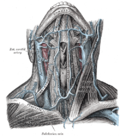
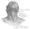
Side of neck, showing chief surface markings (nerves are yellow, arteries are red)
Details
Identifiers
Latin trigonum submentale
TA98 A01.2.02.006
TA2 233
FMA 61604
Anatomical terminology

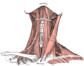
The submental triangle (or suprahyoid triangle) is a division of the anterior triangle of the neck.

Contents

Boundaries

It is limited to:

Contents

It contains one or two lymph glands, the submental lymph nodes (three or four in number) and Submental veins and commencement of anterior jugular veins.

(The contents of the triangle actually lie in the superficial fascia over the roof of submental triangle)

Additional images

See also

Related Research Articles

<span class="mw-page-title-main">Levator ani</span> Broad, thin muscle group, situated on either side of the pelvis

The levator ani is a broad, thin muscle group, situated on either side of the pelvis. It is formed from three muscle components: the pubococcygeus, the iliococcygeus, and the puborectalis.

<span class="mw-page-title-main">Femoral triangle</span> Anatomical region of the thigh

The femoral triangle is an anatomical region of the upper third of the thigh. It is a subfascial space which appears as a triangular depression below the inguinal ligament when the thigh is flexed, abducted and laterally rotated.

<span class="mw-page-title-main">Omohyoid muscle</span> Human neck muscle

The omohyoid muscle is a muscle in the neck. It is one of the infrahyoid muscles. It consists of two bellies separated by an intermediate tendon. Its inferior belly is attached to the scapula; its superior belly is attached to the hyoid bone. Its intermediate tendon is anchored to the clavicle and first rib by a fascial sling. The omohyoid is innervated by the ansa cervicalis of the cervical plexus. It acts to depress the hyoid bone.

<span class="mw-page-title-main">Digastric muscle</span> Small muscle located under the jaw in mammals

The digastric muscle is a bilaterally paired suprahyoid muscle located under the jaw. Its posterior belly is attached to the mastoid notch of temporal bone, and its anterior belly is attached to the digastric fossa of mandible; the two bellies are united by an intermediate tendon which is held in a loop that attaches to the hyoid bone. The anterior belly is innervated via the mandibular nerve, and the posterior belly is innervated via the facial nerve. It may act to depress the mandible or elevate the hyoid bone.

<span class="mw-page-title-main">Sublingual gland</span> Major salivary glands in the mouth

The sublingual gland is a seromucous polystomatic exocrine gland. Located underneath the oral diaphragm, the sublingual gland is the smallest and most diffuse of the three major salivary glands of the oral cavity, with the other two being the submandibular and parotid. The sublingual gland provides approximately 3-5% of the total salivary volume.

<span class="mw-page-title-main">Pectineus muscle</span> Adductor of the thigh

The pectineus muscle is a flat, quadrangular muscle, situated at the anterior (front) part of the upper and medial (inner) aspect of the thigh. The pectineus muscle is the most anterior adductor of the hip. The muscle's primary action is hip flexion; it also produces adduction and internal rotation of the hip.

<span class="mw-page-title-main">Superior mesenteric artery</span> Artery which supplies blood to the intestines and pancreas

In human anatomy, the superior mesenteric artery (SMA) is an artery which arises from the anterior surface of the abdominal aorta, just inferior to the origin of the celiac trunk, and supplies blood to the intestine from the lower part of the duodenum through two-thirds of the transverse colon, as well as the pancreas.

<span class="mw-page-title-main">Inferior mesenteric artery</span>

In human anatomy, the inferior mesenteric artery (IMA) is the third main branch of the abdominal aorta and arises at the level of L3, supplying the large intestine from the distal transverse colon to the upper part of the anal canal. The regions supplied by the IMA are the descending colon, the sigmoid colon, and part of the rectum.

<span class="mw-page-title-main">Medial cutaneous nerve of forearm</span>

The medial cutaneous nerve of the forearm is a sensory branch of the medial cord of the brachial plexus derived from the ventral rami of spinal nerves C8-T1. It provides sensory innervation to the skin of the medial forearm and skin overlying the olecranon. It descends through the (upper) arm within the brachial fascia alongside the basilic vein, then divides into an anterior branch and a posterior branch upon emerging from the brachial fascia; the two terminal branches travel as far distally as the wrist.

<span class="mw-page-title-main">Triangles of the neck</span>

Anatomists use the term triangles of the neck to describe the divisions created by the major muscles in the region.

<span class="mw-page-title-main">Posterior triangle of the neck</span>

The posterior triangle is a region of the neck.

<span class="mw-page-title-main">Superior thyroid artery</span>

The superior thyroid artery arises from the external carotid artery just below the level of the greater cornu of the hyoid bone and ends in the thyroid gland.

<span class="mw-page-title-main">Transverse cervical nerve</span>

The transverse cervical nerve is a cutaneous (sensory) nerve of the cervical plexus that arises from the second and third cervical spinal nerves (C2-C3). It curves around the posterior border of the sternocleidomastoideus muscle, then pierces the fascia of the neck before dividing into two branches. It provides sensory innervation to the front of the neck.

<span class="mw-page-title-main">Anterior triangle of the neck</span>

The anterior triangle is a region of the neck.

<span class="mw-page-title-main">Submandibular triangle</span>

The submandibular triangle corresponds to the region of the neck immediately beneath the body of the mandible.

<span class="mw-page-title-main">Carotid triangle</span>

The carotid triangle is a portion of the anterior triangle of the neck.

<span class="mw-page-title-main">Muscular triangle</span>

The inferior carotid triangle, is bounded, in front, by the median line of the neck from the hyoid bone to the sternum; behind, by the anterior margin of the sternocleidomastoid; above, by the superior belly of the omohyoid.

<span class="mw-page-title-main">Subclavian triangle</span>

The subclavian triangle, the smaller division of the posterior triangle, is bounded, above, by the inferior belly of the omohyoideus; below, by the clavicle; its base is formed by the posterior border of the sternocleidomastoideus.

<span class="mw-page-title-main">Anal triangle</span> Posterior part of the perineum

The anal triangle is the posterior part of the perineum. It contains the anal canal.

<span class="mw-page-title-main">Submental lymph nodes</span>

The submental lymph nodes are 2-3 lymph nodes situated in the submental triangle, between the anterior bellies of the digastric muscle and the hyoid bone.

References

PD-icon.svgThis article incorporates text in the public domain from page 565 of the 20th edition of Gray's Anatomy (1918), Page 88 of Textbook of Anatomy; head, neck and brain by Vishram Singh