Anterior jugular vein

Last updated
Anterior jugular vein
Gray558.png
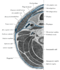
The veins of the neck, viewed from in front
(anterior jugular visible at center)
Gray557.png
Veins of the head and neck
(anterior jugular visible at bottom right)
Details
Drains to External jugular vein
Identifiers
Latin vena jugularis anterior [1]
TA98 A12.3.05.047
TA2 4959
FMA 13318
Anatomical terminology

The anterior jugular vein is a vein in the neck.

Structure

The anterior jugular vein lies lateral to the cricothyroid ligament. [2] It begins near the hyoid bone by the confluence of several superficial veins from the submandibular region. [1] Its tributaries are some laryngeal veins, and occasionally a small thyroid vein. [1] It descends between the median line and the anterior border of the sternocleidomastoid muscle, and, at the lower part of the neck, passes beneath that muscle to open into the termination of the external jugular vein, or, in some instances, into the subclavian vein. [1] Just above the sternum the two anterior jugular veins communicate by a transverse trunk, the venous jugular arch, which receive tributaries from the inferior thyroid veins; each also communicates with the internal jugular. [1]

Contents

There are no valves in this vein. [1]

The pretracheal lymph nodes follow the anterior jugular vein on each side of the midline. [3]

Variation

The anterior jugular vein varies considerably in size, bearing usually an inverse proportion to the external jugular. [1] Most frequently, there are two anterior jugulars, a right and left. [1] [4] However, there is sometimes only one. [1]

A duplicate anterior jugular vein may be present on one side, which may cross over the midline.

Clinical significance

Ultrasound

The anterior jugular vein, if present, is easily identified using ultrasound of the neck. [5]

Tracheotomy

The anterior jugular vein may be damaged during tracheotomy, causing significant bleeding. [6] [7] [8] The significant variation in vein course, such as duplicate veins, creates this risk. [4] Performing a midline incision helps to avoid the anterior jugular vein. [7] [8]

Additional images

Related Research Articles

<span class="mw-page-title-main">Subclavian artery</span> Major arteries of the upper thorax, below the clavicle

In human anatomy, the subclavian arteries are paired major arteries of the upper thorax, below the clavicle. They receive blood from the aortic arch. The left subclavian artery supplies blood to the left arm and the right subclavian artery supplies blood to the right arm, with some branches supplying the head and thorax. On the left side of the body, the subclavian comes directly off the aortic arch, while on the right side it arises from the relatively short brachiocephalic artery when it bifurcates into the subclavian and the right common carotid artery.

<span class="mw-page-title-main">Spinal canal</span> Passage through the vertebral column containing the spinal cord

In human anatomy, the spinal canal, vertebral canal or spinal cavity is an elongated body cavity enclosed within the dorsal bony arches of the vertebral column, which contains the spinal cord, spinal roots and dorsal root ganglia. It is a process of the dorsal body cavity formed by alignment of the vertebral foramina. Under the vertebral arches, the spinal canal is also covered anteriorly by the posterior longitudinal ligament and posteriorly by the ligamentum flavum. The potential space between these ligaments and the dura mater covering the spinal cord is known as the epidural space. Spinal nerves exit the spinal canal via the intervertebral foramina under the corresponding vertebral pedicles.

<span class="mw-page-title-main">Omohyoid muscle</span> Human neck muscle

The omohyoid muscle is a muscle in the neck. It is one of the infrahyoid muscles. It consists of two bellies separated by an intermediate tendon. Its inferior belly is attached to the scapula; its superior belly is attached to the hyoid bone. Its intermediate tendon is anchored to the clavicle and first rib by a fascial sling. The omohyoid is innervated by the ansa cervicalis of the cervical plexus. It acts to depress the hyoid bone.

<span class="mw-page-title-main">Celiac artery</span> First major branch of the abdominal aorta

The celiacartery, also known as the celiac trunk or truncus coeliacus, is the first major branch of the abdominal aorta. It is about 1.25 cm in length. Branching from the aorta at thoracic vertebra 12 (T12) in humans, it is one of three anterior/ midline branches of the abdominal aorta.

<span class="mw-page-title-main">Subclavian vein</span> Blood vessel that drains the arm

The subclavian vein is a paired large vein, one on either side of the body, that is responsible for draining blood from the upper extremities, allowing this blood to return to the heart. The left subclavian vein plays a key role in the absorption of lipids, by allowing products that have been carried by lymph in the thoracic duct to enter the bloodstream. The diameter of the subclavian veins is approximately 1–2 cm, depending on the individual.

<span class="mw-page-title-main">Cricothyroid ligament</span>

The cricothyroid ligament is a ligament in the neck. It connects the cricoid cartilage to the thyroid cartilage. It prevents these cartilages from moving too far apart. It is cut during an emergency cricothyrotomy to treat upper airway obstruction.

<span class="mw-page-title-main">Platysma muscle</span> Human neck muscle

The platysma muscle is a superficial muscle of the human neck that overlaps the sternocleidomastoid. It covers the anterior surface of the neck superficially. When it contracts, it produces a slight wrinkling of the neck, and a "bowstring" effect on either side of the neck.

<span class="mw-page-title-main">Psoas minor muscle</span> Long, slender skeletal muscle located anterior to the psoas major muscle

The psoas minor muscle is a long, slender skeletal muscle. When present, it is located anterior to the psoas major muscle.

<span class="mw-page-title-main">Suprasternal notch</span> Visible dip in the neck in humans

The suprasternal notch, also known as the fossa jugularis sternalis, jugular notch, or Plender gap, is a large, visible dip in between the neck in humans, between the clavicles, and above the manubrium of the sternum.

<span class="mw-page-title-main">Carotid sheath</span>

The carotid sheath is a condensation of the deep cervical fascia enveloping multiple vital neurovascular structures of the neck, including the common and internal carotid arteries, the internal jugular vein, the vagus nerve, and ansa cervicalis. The carotid sheath helps protects the structures contained therein.

Postural drainage (PD) is the drainage of lung secretions using gravity. It is used to treat a variety of conditions that cause the build-up of secretions in the lungs.

<span class="mw-page-title-main">Supraspinous ligament</span>

The supraspinous ligament, also known as the supraspinal ligament, is a ligament found along the vertebral column.

<span class="mw-page-title-main">Thyrohyoid membrane</span> Elastic membrane in the larynx

The thyrohyoid membrane is a broad, fibro-elastic sheet of the larynx. It connects the upper border of the thyroid cartilage to the hyoid bone.

<span class="mw-page-title-main">Lingual artery</span>

The lingual artery arises from the external carotid artery between the superior thyroid artery and facial artery. It can be located easily in the tongue.

<span class="mw-page-title-main">Retromandibular vein</span> Major face vein

The retromandibular vein is a major vein of the face. It is formed within the parotid gland by the confluence of the maxillary vein, and superficial temporal vein. It descends in the gland and splits into two branches upon emerging from the gland. Its anterior branch then joins the (anterior) facial vein forming the common facial vein, while its posterior branch joins the posterior auricular vein forming the external jugular vein.

<span class="mw-page-title-main">Right lymphatic duct</span> Lymphatic vessel

The right lymphatic duct is an important lymphatic vessel that drains the right upper quadrant of the human body. It forms various combinations with the right subclavian vein and right internal jugular vein.

<span class="mw-page-title-main">Radial tuberosity</span> Bony eminence on the radius

Beneath the neck of the radius, on the medial side, is an eminence, the radial tuberosity; its surface is divided into:

<span class="mw-page-title-main">Supraglenoid tubercle</span> Region of the scapula from which the long head of the biceps brachii muscle originates

The supraglenoid tubercle is a region of the scapula from which the long head of the biceps brachii muscle originates. It is a small, rough projection superior to the glenoid cavity near the base of the coracoid process. The term supraglenoid is from the Latin supra, meaning above, and glenoid, meaning socket or cavity.

<span class="mw-page-title-main">Linea semilunaris</span>

The linea semilunaris is a curved tendinous intersection found on either side of the rectus abdominis muscle.

<span class="mw-page-title-main">Pretracheal lymph nodes</span>

The pretracheal lymph nodes are lymph nodes located anterior to the trachea in the neck.

References

  1. 1 2 3 4 5 6 7 8 9 PD-icon.svgOne or more of the preceding sentences incorporates text in the public domain from page 647 of  the 20th edition of Gray's Anatomy (1918)
  2. Cattano, Davide; Cavallone, Laura F. (2013-01-01), Hagberg, Carin A. (ed.), "Chapter 30 - Percutaneous Dilational Cricothyrotomy and Tracheostomy", Benumof and Hagberg's Airway Management (Third Edition), Philadelphia: W.B. Saunders, pp. 613–639.e3, doi:10.1016/b978-1-4377-2764-7.00030-0, ISBN   978-1-4377-2764-7 , retrieved 2021-01-12
  3. Rhys, Rhian (2011-01-01), Allan, Paul L.; Baxter, Grant M.; Weston, Michael J. (eds.), "CHAPTER 46 - Cervical lymph nodes", Clinical Ultrasound (Third Edition), Edinburgh: Churchill Livingstone, pp. 920–937, ISBN   978-0-7020-3131-1 , retrieved 2021-01-12
  4. 1 2 El-Sayed, Ivan H.; Bhatki, Amol M.; Khabie, Nissim (2009-01-01), Eisele, David W.; Smith, Richard V. (eds.), "CHAPTER 34 - Complications of Tracheostomy and Tracheal Surgery", Complications in Head and Neck Surgery (Second Edition), Philadelphia: Mosby, pp. 405–424, ISBN   978-1-4160-4220-4 , retrieved 2021-01-12
  5. Rhys, Rhian (2011-01-01), Allan, Paul L.; Baxter, Grant M.; Weston, Michael J. (eds.), "CHAPTER 45 - Ultrasound of the neck", Clinical Ultrasound (Third Edition), Edinburgh: Churchill Livingstone, pp. 890–919, ISBN   978-0-7020-3131-1 , retrieved 2021-01-12
  6. Osborn, Timothy Marx; Dierks, Eric J. (2012-01-01), Bagheri, Shahrokh C.; Bell, R. Bryan; Khan, Husain Ali (eds.), "Chapter 34 - The Surgical Airway", Current Therapy In Oral and Maxillofacial Surgery, Saint Louis: W.B. Saunders, pp. 275–281, ISBN   978-1-4160-2527-6 , retrieved 2021-01-12
  7. 1 2 Gross, Ronald I.; Jacobs, Lenworth M. (2008-01-01), Asensio, JUAN A.; Trunkey, DONALD D. (eds.), "CHAPTER 17 - AIRWAY MANAGEMENT: WHAT EVERY TRAUMA SURGEON SHOULD KNOW, FROM INTUBATION TO CRICOTHYROIDOTOMY", Current Therapy of Trauma and Surgical Critical Care, Philadelphia: Mosby, pp. 95–105, ISBN   978-0-323-04418-9 , retrieved 2021-01-12
  8. 1 2 Minokadeh, Anushirvan; Wilson, William C. (2010-01-01), Jeremias, Allen; Brown, David L. (eds.), "CHAPTER 49 - Emergency Airway Management", Cardiac Intensive Care (Second Edition), Philadelphia: W.B. Saunders, pp. 598–631, ISBN   978-1-4160-3773-6 , retrieved 2021-01-12