Cerebellar veins

Last updated
Cerebellar veins
CerebellumArteries.jpg
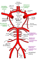
Corresponding arterial circulation of the cerebellum (AICA and PICA).
Gray565.png
Veins and plexa of the cerebellum seen.
Details
Drains from cerebellum
Drains to dural venous sinuses
Artery anterior inferior cerebellar artery (AICA), posterior inferior cerebellar artery (PICA)
Identifiers
Latin venae cerebelli
TA98 A12.3.06.056
TA2 4935
FMA 70879
Anatomical terminology

The cerebellar veins are veins which drain the cerebellum. They consist of the superior cerebellar veins and the inferior cerebellar veins (dorsal cerebellar veins). The superior cerebellar veins drain to the straight sinus and the internal cerebral veins. The inferior cerebellar veins drain to the transverse sinus, the superior petrosal sinus, and the occipital sinus.

Contents

Structure

The superior cerebellar veins pass partly forward and medialward, across the superior cerebellar vermis. They end in the straight sinus, [1] and the internal cerebral veins, partly lateralward to the transverse and superior petrosal sinuses.

The inferior cerebellar veins are larger. They end in the transverse sinus, [2] the superior petrosal sinus, and the occipital sinus.

Clinical significance

The cerebellar veins may be affected by infarction or thrombosis. [3] They may be the draining site of abnormal fistulas. [4]

Additional images

Related Research Articles

<span class="mw-page-title-main">Dura mater</span> Outermost layer of the protective tissues around the central nervous system (meninges)

In neuroanatomy, dura mater is a thick membrane made of dense irregular connective tissue that surrounds the brain and spinal cord. It is the outermost of the three layers of membrane called the meninges that protect the central nervous system. The other two meningeal layers are the arachnoid mater and the pia mater. It envelops the arachnoid mater, which is responsible for keeping in the cerebrospinal fluid. It is derived primarily from the neural crest cell population, with postnatal contributions of the paraxial mesoderm.

<span class="mw-page-title-main">Great cerebral vein</span>

The great cerebral vein is one of the large blood vessels in the skull draining the cerebrum of the brain. It is also known as the vein of Galen, named for its discoverer, the Greek physician Galen.

<span class="mw-page-title-main">Posterior cranial fossa</span> Area of the cranium containing the brainstem and cerebellum

The posterior cranial fossa is the part of the cranial cavity located between the foramen magnum, and tentorium cerebelli. It is formed by the sphenoid bones, temporal bones, and occipital bone. It lodges the cerebellum, and parts of the brainstem.

<span class="mw-page-title-main">Cavernous sinus</span> Sinus in the human head

The cavernous sinus within the human head is one of the dural venous sinuses creating a cavity called the lateral sellar compartment bordered by the temporal bone of the skull and the sphenoid bone, lateral to the sella turcica.

<span class="mw-page-title-main">Confluence of sinuses</span> Venous sinus in the skull

The confluence of sinuses, torcular Herophili, or torcula is the connecting point of the superior sagittal sinus, straight sinus, and occipital sinus. It is below the internal occipital protuberance of the skull. It drains venous blood from the brain into the transverse sinuses. It may be affected by arteriovenous fistulas, a thrombus, major trauma, or surgical damage, and may be imaged with many radiology techniques.

<span class="mw-page-title-main">Straight sinus</span> Venous sinus draining the brain

The straight sinus, also known as tentorial sinus or the sinus rectus, is an area within the skull beneath the brain. It receives blood from the inferior sagittal sinus and the great cerebral vein, and drains into the confluence of sinuses.

<span class="mw-page-title-main">Superior sagittal sinus</span> Anatomical structure of the brain

The superior sagittal sinus, within the human head, is an unpaired area along the attached margin of the falx cerebri. It allows blood to drain from the lateral aspects of anterior cerebral hemispheres to the confluence of sinuses. Cerebrospinal fluid drains through arachnoid granulations into the superior sagittal sinus and is returned to venous circulation.

<span class="mw-page-title-main">Inferior sagittal sinus</span> Anatomical structure of the brain

The inferior sagittal sinus, within the human head, is an area beneath the brain which allows blood to drain outwards posteriorly from the center of the head. It drains to the straight sinus, which connects to the transverse sinuses. See diagram : labeled in the brain as "SIN. SAGITTALIS INF.".

<span class="mw-page-title-main">Superior petrosal sinus</span> One of the dural venous sinuses located beneath the brain

The superior petrosal sinus is one of the dural venous sinuses located beneath the brain. It receives blood from the cavernous sinus and passes backward and laterally to drain into the transverse sinus. The sinus receives superior petrosal veins, some cerebellar veins, some inferior cerebral veins, and veins from the tympanic cavity. They may be affected by arteriovenous malformation or arteriovenous fistula, usually treated with surgery.

<span class="mw-page-title-main">Inferior petrosal sinus</span>

The inferior petrosal sinuses are two small sinuses situated on the inferior border of the petrous part of the temporal bone, one on each side. Each inferior petrosal sinus drains the cavernous sinus into the internal jugular vein.

<span class="mw-page-title-main">Superior ophthalmic vein</span> Vein of the orbit around the eye

The superior ophthalmic vein is a vein of the orbit that drains venous blood from structures of the upper orbit. It is formed by the union of the angular vein, and supraorbital vein. It passes backwards within the orbit alongside the ophthalmic artery, then exits the orbit through the superior orbital fissure to drain into the cavernous sinus.

<span class="mw-page-title-main">Falx cerebelli</span> Skull anatomy

The falx cerebelli is a small sickle-shaped fold of dura mater projecting forwards into the posterior cerebellar notch as well as projecting into the vallecula of the cerebellum between the two cerebellar hemispheres.

<span class="mw-page-title-main">Dural arteriovenous fistula</span> Medical condition

A dural arteriovenous fistula (DAVF) or malformation is an abnormal direct connection (fistula) between a meningeal artery and a meningeal vein or dural venous sinus.

<span class="mw-page-title-main">Sigmoid sinus</span>

The sigmoid sinuses, also known as the pars sigmoid, are paired dural venous sinuses within the skull that receive blood from posterior transverse sinuses.

<span class="mw-page-title-main">Transverse sinuses</span>

The transverse sinuses, within the human head, are two areas beneath the brain which allow blood to drain from the back of the head. They run laterally in a groove along the interior surface of the occipital bone. They drain from the confluence of sinuses to the sigmoid sinuses, which ultimately connect to the internal jugular vein. See diagram : labeled under the brain as "SIN. TRANS.".

<span class="mw-page-title-main">Angular vein</span> Vein of the face

The angular vein is a vein of the face. It is the upper part of the facial vein, above its junction with the superior labial vein. It is formed by the junction of the supratrochlear vein and supraorbital vein, and joins with the superior labial vein. It drains the medial canthus, and parts of the nose and the upper lip. It can be a route of spread of infection from the danger triangle of the face to the cavernous sinus.

<span class="mw-page-title-main">Occipital vein</span> Vein of the scalp

The occipital vein is a vein of the scalp. It originates from a plexus around the external occipital protuberance and superior nuchal line to the back part of the vertex of the skull. It usually drains into the internal jugular vein, but may also drain into the posterior auricular vein. It drains part of the scalp.

The quadrigeminal cistern is a subarachnoid cistern situated between splenium of corpus callosum, and the superior surface of the cerebellum. It contains a part of the great cerebral vein, the posterior cerebral artery, quadrigeminal artery, glossopharyngeal nerve, and the pineal gland.

<span class="mw-page-title-main">Inferior petrosal sulcus</span>

The inferior petrosal sulcus is the groove containing the inferior petrosal sinus.

References

PD-icon.svgThis article incorporates text in the public domain from the 20th edition of Gray's Anatomy (1918)

  1. Drake, Richard L. (2005). Gray's anatomy for students. Wayne Vogl, Adam W. M. Mitchell, Henry Gray. Philadelphia: Elsevier / Churchill Livingstone. p. 795. ISBN   0-443-06612-4. OCLC   55139039.{{cite book}}: CS1 maint: date and year (link)
  2. Scremin, Oscar U. (2015). "31 - Cerebral Vascular System". The Rat Nervous System (4th ed.). Academic Press. pp. 985–1011. doi:10.1016/B978-0-12-374245-2.00031-0. ISBN   978-0-12-374245-2.
  3. Bousser, Marie-Germaine; Barnett, Henry J. M. (2004). "12 - Cerebral Venous Thrombosis". Stroke - Pathophysiology, Diagnosis, and Management (4th ed.). Churchill Livingstone. pp. 301–325. doi:10.1016/B0-44-306600-0/50016-X. ISBN   978-0-443-06600-9.
  4. Cole, Tyler S.; Lawton, Michael T. (2021). "8 - Surgical management of cerebral dural arteriovenous fistulas". Cerebral Dural Arteriovenous Fistulas. Academic Press. pp. 105–124. doi:10.1016/B978-0-12-819525-3.00010-1. ISBN   978-0-12-819525-3. S2CID   242605747.