Cerebral veins

Last updated
Cerebral veins
Gray568.png
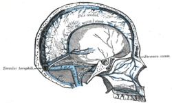
Sagittal section of the skull, showing the sinuses of the dura. (Cerebral veins labeled at center left.)
Details
Artery cerebral arteries
Identifiers
Latin venae encephali, venae cerebri
MeSH D002550
TA98 A12.3.06.001
TA2 4901
FMA 70861
Anatomical terminology
3D model of cerebral veins CNS Venography 3D SR Nevit Dilmen.stl
3D model of cerebral veins

In human anatomy, the cerebral veins are blood vessels in the cerebral circulation which drain blood from the cerebrum of the human brain. They are divisible into external (superficial cerebral veins) and internal (internal cerebral veins) groups according to the outer or inner parts of the hemispheres they drain into.

Contents

External veins

The external cerebral veins known as the superficial cerebral veins are the superior cerebral veins, inferior cerebral veins, and middle cerebral veins. The superior cerebral veins on the upper side surfaces of the hemispheres drain into the superior sagittal sinus. [1] The superior cerebral veins include the superior anastomotic vein.

Internal veins

The internal cerebral veins are also known as the deep cerebral veins and drain the deep internal parts of the hemispheres.

Related Research Articles

<span class="mw-page-title-main">Vein</span> Blood vessels that carry blood towards the heart

Veins are blood vessels in the circulatory system of humans and most other animals that carry blood towards the heart. Most veins carry deoxygenated blood from the tissues back to the heart; exceptions are those of the pulmonary and fetal circulations which carry oxygenated blood to the heart. In the systemic circulation, arteries carry oxygenated blood away from the heart, and veins return deoxygenated blood to the heart, in the deep veins.

<span class="mw-page-title-main">External carotid artery</span> Major artery of the head and neck

The external carotid artery is a major artery of the head and neck. It arises from the common carotid artery when it splits into the external and internal carotid artery. The external carotid artery supplies blood to the face, brain and neck.

<span class="mw-page-title-main">Internal carotid artery</span> Artery of the human brain

The internal carotid artery is an artery in the neck which supplies the anterior circulation of the brain.

<span class="mw-page-title-main">Great cerebral vein</span>

The great cerebral vein is one of the large blood vessels in the skull draining the cerebrum of the brain. It is also known as the vein of Galen, named for its discoverer, the Greek physician Galen.

<span class="mw-page-title-main">Cerebral circulation</span> Brain blood supply

Cerebral circulation is the movement of blood through a network of cerebral arteries and veins supplying the brain. The rate of cerebral blood flow in an adult human is typically 750 milliliters per minute, or about 15% of cardiac output. Arteries deliver oxygenated blood, glucose and other nutrients to the brain. Veins carry "used or spent" blood back to the heart, to remove carbon dioxide, lactic acid, and other metabolic products. The neurovascular unit regulates cerebral blood flow so that activated neurons can be supplied with energy in the right amount and at the right time. Because the brain would quickly suffer damage from any stoppage in blood supply, the cerebral circulatory system has safeguards including autoregulation of the blood vessels. The failure of these safeguards may result in a stroke. The volume of blood in circulation is called the cerebral blood flow. Sudden intense accelerations change the gravitational forces perceived by bodies and can severely impair cerebral circulation and normal functions to the point of becoming serious life-threatening conditions.

<span class="mw-page-title-main">External jugular vein</span> Large blood vessel

The external jugular vein receives the greater part of the blood from the exterior of the cranium and the deep parts of the face, being formed by the junction of the posterior division of the retromandibular vein with the posterior auricular vein.

<span class="mw-page-title-main">Falx cerebri</span> Anatomical structure of the brain

The falx cerebri is a large, crescent-shaped fold of dura mater that descends vertically into the longitudinal fissure between the cerebral hemispheres of the human brain, separating the two hemispheres and supporting dural sinuses that provide venous and CSF drainage to the brain. It is attached to the crista galli anteriorly, and blends with the tentorium cerebelli posteriorly.

<span class="mw-page-title-main">Common carotid artery</span> One of the two arteries that supply the head and neck with blood

In anatomy, the left and right common carotid arteries (carotids) are arteries that supply the head and neck with oxygenated blood; they divide in the neck to form the external and internal carotid arteries.

<span class="mw-page-title-main">Arachnoid mater</span> Web-like middle layer of the three meninges

The arachnoid mater is one of the three meninges, the protective membranes that cover the brain and spinal cord. It is so named because of its resemblance to a spider web. The arachnoid mater is a derivative of the neural crest mesoectoderm in the embryo.

<span class="mw-page-title-main">Cavernous sinus</span> Sinus in the human head

The cavernous sinus within the human head is one of the dural venous sinuses creating a cavity called the lateral sellar compartment bordered by the temporal bone of the skull and the sphenoid bone, lateral to the sella turcica.

<span class="mw-page-title-main">Superior sagittal sinus</span> Anatomical structure of the brain

The superior sagittal sinus, within the human head, is an unpaired area along the attached margin of the falx cerebri. It allows blood to drain from the lateral aspects of anterior cerebral hemispheres to the confluence of sinuses. Cerebrospinal fluid drains through arachnoid granulations into the superior sagittal sinus and is returned to venous circulation.

<span class="mw-page-title-main">Inferior sagittal sinus</span> Anatomical structure of the brain

The inferior sagittal sinus, within the human head, is an area beneath the brain which allows blood to drain outwards posteriorly from the center of the head. It drains to the straight sinus, which connects to the transverse sinuses. See diagram : labeled in the brain as "SIN. SAGITTALIS INF.".

<span class="mw-page-title-main">Inferior cerebral veins</span>

The inferior cerebral veins are veins that drain the undersurface of the cerebral hemispheres and empty into the cavernous and transverse sinuses.

<span class="mw-page-title-main">Superior cerebral veins</span>

The superior cerebral veins are several cerebral veins that drain the superolateral and superomedial surfaces of the cerebral hemispheres into the superior sagittal sinus. There are 8-12 cerebral veins. They are predominantly found in the sulci between the gyri, but can also be found running across the gyri.

<span class="mw-page-title-main">Internal cerebral veins</span>

The internal cerebral veins are two veins included in the group of deep cerebral veins that drain the deep parts of the hemispheres; each internal cerebral vein is formed near the interventricular foramina by the union of the superior thalamostriate vein and the superior choroid vein.

<span class="mw-page-title-main">Carotid triangle</span>

The carotid triangle is a portion of the anterior triangle of the neck.

<span class="mw-page-title-main">Dorsal veins of the penis</span>

In human male anatomy, the dorsal veins of the penis are blood vessels that drain the shaft, the skin and the glans of the human penis. They are typically located in the midline on the dorsal aspect of the penis and they comprise the superficial dorsal veinof the penis, that lies in the subcutaneous tissue of the shaft, and the deep dorsal veinof the penis, that lies beneath the deep fascia.

<span class="mw-page-title-main">Outline of human anatomy</span> Overview of and topical guide to human anatomy

The following outline is provided as an overview of and topical guide to human anatomy:

<span class="mw-page-title-main">Middle cerebral veins</span>

The middle cerebral veins - the superficial middle cerebral vein and the deep middle cerebral vein - are two veins running along the lateral sulcus. The superficial middle cerebral vein is also known as the superficial Sylvian vein, and the deep middle cerebral vein is also known as the deep Sylvian vein. The lateral sulcus is also known as the Sylvian fissure.

The inferior anastomotic vein is one of several superficial cerebral veins. It forms an anastomosis between the superficial middle cerebral vein and transverse sinus, opening into either at either end.

References

PD-icon.svgThis article incorporates text in the public domain from page 652 of the 20th edition of Gray's Anatomy (1918)

  1. Moore, Keith L. (2018). Clinically oriented anatomy (Eighth ed.). Philadelphia. p. 891. ISBN   9781496347213.{{cite book}}: CS1 maint: location missing publisher (link)